This study reports a successful method of endoscopical adipose tissue-derived stromal cell (ADSC)-sheet transplantation for esophageal stricture prevention after an extended endoscopic submucosal dissection (ESD) in a swine model.
In past years, the cell-sheet construct has spurred wide interest in regenerative medicine, especially for reconstructive surgery procedures. The development of diversified technologies combining adipose tissue-derived stromal cells (ADSCs) with various biomaterials has led to the construction of numerous types of tissue-engineered substitutes, such as bone, cartilage, and adipose tissues from rodent, porcine, or human ADSCs. Extended esophageal endoscopic submucosal dissection (ESD) is responsible for esophageal stricture formation. Stricture prevention remains challenging, with no efficient treatments available. Previous studies reported the effectiveness of mucosal cell-sheet transplantation in a canine model and in humans. ADSCs are attributed anti-inflammatory properties, local immune modulating effects, neovascularization induction, and differentiation abilities into mesenchymal and non-mesenchymal lineages. This original study describes the endoscopic transplantation of an ADSC tissue-engineered construct to prevent esophageal stricture in a swine model. The ADSC construct was composed of two allogenic ADSC sheets layered upon each other on a paper support membrane. The ADSCs were labeled with the PKH67 fluorophore to allow probe-based confocal laser endomicroscopy (pCLE) monitoring. On the day of transplantation, a 5-cm and hemi-circumferential ESD known to induce esophageal stricture was performed. Animals were immediately endoscopically transplanted with 4 ADSC constructs. The complete adhesion of the ADSC constructs was obtained after 10 min of gentle application. Animals were sacrificed on day 28. All animals were successfully transplanted. Transplantation was confirmed on day 3 with a positive pCLE evaluation. Compared to transplanted animals, control animals developed severe strictures, with major fibrotic tissue development, more frequent alimentary trouble, and reduced weight gain. In our model, the transplantation of allogenic ADSCs, organized in double cell sheets, after extended ESD was successful and strongly associated with a lower esophageal stricture rate.
The management of superficial esophageal tumors has changed with the development of new endoscopic techniques. Nowadays, endoscopic resection is the first-line treatment. Indeed, it is associated with lower morbidity and mortality rates than a surgery with equal oncological results1,2,3. Endoscopic mucosal resection (EMR) and endoscopic submucosal resection (ESD) are the most widely-used techniques. In the case of an extended superficial tumor, ESD is preferred. Compared to EMR, ESD allows en bloc resectioning, regardless of lesion size and shape4,5,6. The main delayed complication of ESD is esophageal stricture formation, which generally occurs between one and two weeks after resection. Recent published studies have shown that stricture formation is correlated to the size of the resection. The Japanese Endoscopic Society recommends avoiding ESD sizes larger than ¾ of the esophageal circumference because they are associated with stricture development in more than 90% of cases and are responsible for severe feeding troubles and major deterioration in quality of life.
The prevention of esophageal stricture remains challenging. Mechanisms involved in stricture formation are only partially known. Stricture formations seems to result from the association of two different mechanisms: (1) pro-inflammatory cellular recruitment and (2) excessive fibrosis development7. Several preventive treatments have been proposed. However, results were unsatisfactory, with little benefit and severe side effects8,9. Recently, a Japanese team, Ohki et al., proposed to transplant a single-layer cell sheet of autologous oral mucosal cells into the esophageal scar. Transplantation was performed immediately after ESD10,11. They demonstrated the effectiveness of this innovative approach, first in a canine model and then in patients.
Adipose tissue-derived stromal cells (ADSCs) are promising in regenerative medicine. Their application in several fields has shown interesting results, especially in the wound-healing process. ADSC therapy offers several advantages, because the cells are easily isolated and are associated with anti-inflammatory properties, local immunomodulating effects, neovascularization induction, and differentiation abilities into mesenchymal and non-mesenchymal lineages12,13,14.
In a previous study, our team demonstrated the effectiveness of double ADSC-sheet endoscopic transplantation for esophageal stricture prevention after extended ESD in a swine model15. In this article, reports of ADSC-sheet construction and endoscopical transplantation technique are presented.
All animals were treated according to the Animal Research Ethics Committee (guidelines of the French Ministry of Agriculture). The protocol received the approval of the local ethics committee authorized for animal experimentations at the Paris Descartes University (registered number MESR 2035.02; Faculty of Medicine Paris Descartes, Paris, France).
1. ADSC Culture and Labeling
2. Double ADSC-sheet Construct
3. Esophageal Endoscopic Submucosal Dissection (ESD)
4. Endoscopic Transplantation
5. Postoperative Evaluation and Follow-up
The culture of ADSCs and the procedure to obtain the ADSC sheet is shown in Figure 1. Figure 2 shows the construction of the graft, composed of two ADSC sheets layered upon each other on their paper support membrane. ADSCs were previously labeled with the PKH67 fluorophore to allow in vivo graft monitoring with pCLE. Figure 3 shows the different steps of extended esophageal endoscopic submucosal dissection, resulting in a 5-cm and hemi-circumferential esophageal scar. Figure 4 shows the endoscopic transplantation procedure. The transplantation was successful for all animals after 10 min of gentle application. Figure 5 shows the positive pCLE evaluation on day 3, confirming the success of the transplantation procedure.
The follow-up period allowed for multimodal stricture analysis. The different assessments are showed in the following figures and table. Compared to control animals, the transplanted animal group showed less frequent alimentary trouble and greater weight gain on day 28. One animal from the transplanted group developed a severe esophageal stricture compared to all animals in the control group. Compared to transplanted animals, control animals developed significant fibrotic tissue. Table 1 shows clinical and endoscopical findings during the follow-up period. Figure 6 shows the endoscopic and radiological findings at the end of the follow-up period in transplanted and non-transplanted animals. Figure 7 shows the different histological findings between transplanted and control animals. These data confirm the effectiveness of ADSC-sheet transplantation for esophageal stricture prevention.
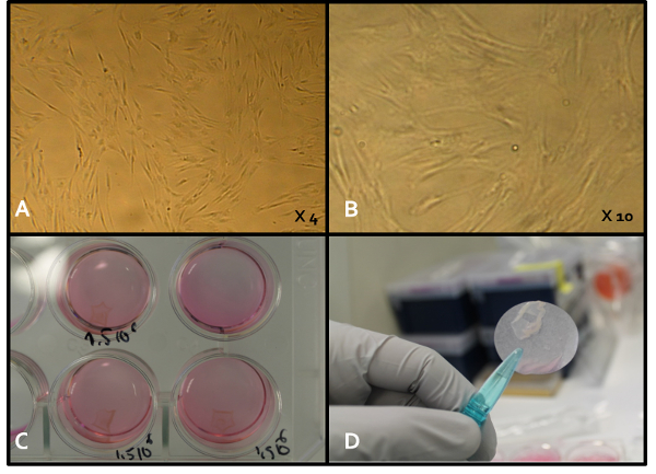
Figure 1. ADSC-sheet Construct. A and B. Early-passage ADSCs in standard culture dishes with different magnifications. ADSC sheets were obtained by culturing PKH-labeled ADSCs on commercial temperature-responsive culture dishes. C. When the incubation temperature was reduced to 20 °C (room temperature), all cells spontaneously detached from the dish surfaces as intact ADSC sheets, without the need for enzymatic treatment. D. An ADSC sheet harvested before graft construction using a transfer membrane. Please click here to view a larger version of this figure.
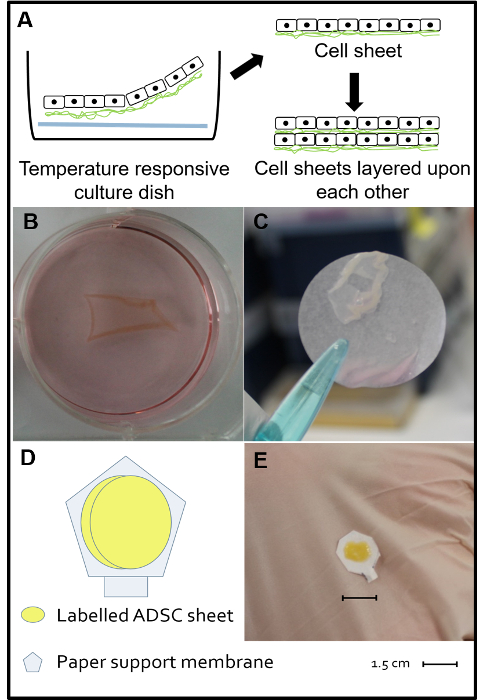
Figure 2. Double ADSC-sheet Construct. A, B, and C. The day before transplantation, PKH-labeled ADSCs were harvested at early passage number (P4 – P5), seeded on a 12-well temperature-responsive cell culture, and cultured at 37 °C and 5% CO2 with alpha minimum essential medium including 10% fetal veal serum and 1% antibiotics (penicillin and streptomycin). Each well was coated with a poly-N-isopropylacrylamide membrane (at the bottom of each well) and seeded with 1.5 x 10⁶ cells. When the incubation temperature was reduced to 20 °C (room temperature), all cells spontaneously detached as intact ADSC sheets without the need for enzymatic treatment. D. Theoretical schema of the double cell-sheet construct composed of two ADSC sheets layered upon each other and applied to a paper support membrane. E. Macroscopic view of a completed double cell-sheet construct. Modified from Reference 15. Please click here to view a larger version of this figure.
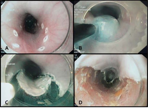
Figure 3. Endoscopic Submucosal Dissection Procedure. A. Endoscopic marks were created to delineate the resection area, ranging from 40 to 45 cm from the dental arch and involving half of the circumference. B. The submucosal injection of a glycerol solution containing indigo carmine dye. The goal of this injection was to separate the mucosal layer from the muscular layer in order to expose the submucosal layer. C. Peripheral incisions were performed externally to the endoscopic marks, revealing the injected submucosal layer. D. The endoscopic view at the end of the submucosal dissection showing a 5 cm-long and hemi-circumferential esophageal scar. Please click here to view a larger version of this figure.
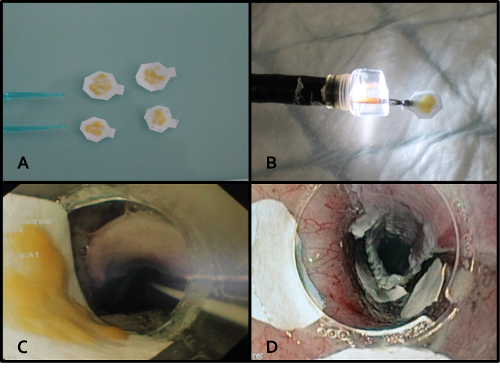
Figure 4. Double ADSC-sheet Endoscopic Transplantation Procedure. A. Four ADSC constructs ready to be transplanted. B. Protection of the ADSC construct under a large endoscopic cap before transplantation. C. Endoscopic view of the crossing of the animal pharynx, showing the ADSC construct protected under the endoscopic cap. D. 10 min of application was sufficient to obtain sheet adherence. Application was performed using the endoscopic cap or endoscopic forceps. Please click here to view a larger version of this figure.
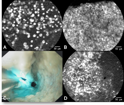
Figure 5. pCLE Findings. pCLE on day 3 shows spontaneous and intense signals similar to those obtained in vitro, compatible with a successful cell-sheet transplantation. No signal was found on days 14 and 28. A. The in vitro pCLE signal of ADSCs in suspension. B. The in vitro pCLE signal of ADSCs organized in a cell sheet. C. The in vivo pCLE probe application to the esophageal scar. D. The in vivo pCLE signal of ADSCs visualized on day 3 after transplantation. Modified from Reference 15. Please click here to view a larger version of this figure.
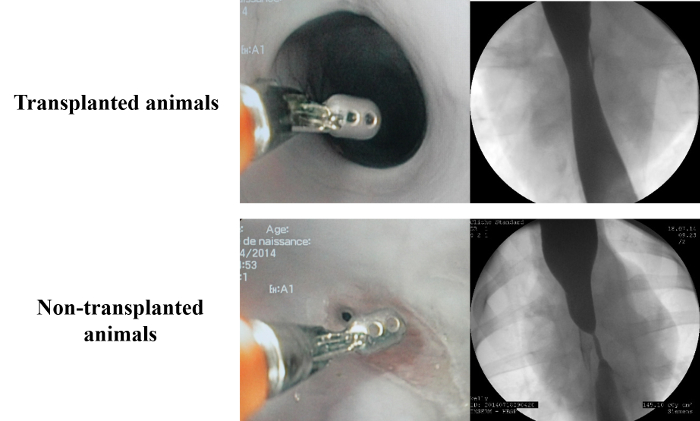
Figure 6. Results of the Morphological Analysis on Day 28. Compared to the control group, the endoscopic and radiological findings on day 28 showed a short and light stricture without upstream dilatation and a completely re-epithelialized mucosa. Modified from Reference 15. Please click here to view a larger version of this figure.
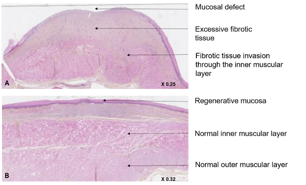
Figure 7. Histological Evaluation of the Stricture Areas. The histological analysis was performed after HES-labeling and slide digitalization. In transplanted animals (A), the healing process was improved, with increased re-epithelialization and decreased fibrosis development, compared to non-transplanted animals (B). Intense fibrotic tissue development was observed due to muscularis mucosae destruction, fibrotic inner muscularis layer invasion, and large mucosal defects. Modified from Reference 15. Please click here to view a larger version of this figure.
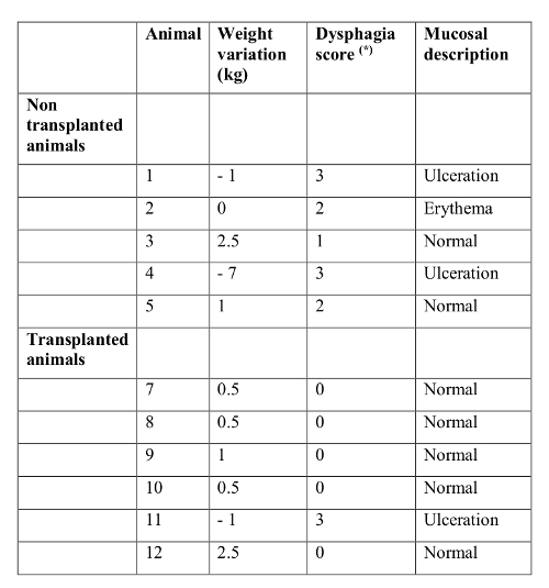
Table 1. Clinical Assessments during the Follow-up Period. Clinical assessments were performed using a daily clinical evaluation according to the Mellow Pinkas score and with weight variation. (*) Mellow Pinkas score16. Modified from Reference 15.
In this pig model, the ADSC-sheet transplantation was technically successful, and the in vivo pCLE evaluation allowed for cell engraftment monitoring. Clinical, endoscopical, radiological, and histological evaluations demonstrated the effectiveness of the endoscopic ADSC sheet at esophageal stricture prevention after extended ESD.
The endoscopic transplantation of an ADSC glycerol solution containing an indigo carmine dye sheet is an innovative approach in regenerative medicine. Ohki et al. first described the endoscopic transplantation procedure. In their studies, cell-sheet constructs were composed of oral mucosal epithelial cell sheets layered on a thin sheet of polyvinylidene difluoride, used as a support membrane10,11,19. The support membrane was grasped with endoscopic forceps and carefully placed on the ulcer bed. Cell-sheet adherence was obtained after 10 min of gentle application. This procedure has not yet been reproduced. In this study, double ADSC-sheet constructs were transplanted for multiple reasons. The choice of ADSC was based on several studies that demonstrated their anti-inflammatory abilities, especially in the skin-healing process. ADSCs are known to modulate keratinocyte and fibroblast interactions7. The mechanism of fibrosis development is complex and only partially understood. The paracrine effect seems to play a major role in ADSC action, including anti-inflammatory factor production, such as TGFβ and pro-angiogenic factors20.
Endoscopic ADSC-sheet transplantation has several limitations. First, cell sheets were applied for 10 min to the esophageal scar to obtain a stable adhesion. This part of the procedure was challenging, technically difficult, and had to be performed by an experienced endoscopist. Nevertheless, all animals were successfully transplanted, confirming the feasibility of the endoscopic procedure. Recently, the use of a 3D-printed device to improve the success of cell-sheet transplantation was reported21. Second, this technique is associated with an unknown survival rate. An esophageal scar is an aggressive environment for cells. A temperature-responsive dish allows for the production of the cell-sheet construct through the extracellular matrix and for the formation of cell connections. Thus, to improve the survival rate and the ADSC interactions, a double ADSC-sheet construct was chosen22,23. This study was not designed to assess either the cell survival rate or the dose effect of transplantation on esophageal stricture prevention, which are both important questions. Third, as in Ohki et al., the use of a support membrane was necessary to carry the ADSC-sheet construct. After transplantation, the paper support membrane could not be removed without construct destruction. This intra-esophageal foreign material was responsible for the increased inflammation. An optimized technique would allow for the removal of the paper support membrane.
Transplantation monitoring with pCLE was also challenging. This new endoscopic imaging technique allows in vivo histological analysis, including visualization of the glandular pattern, micro-vascularization, and cellular alteration. pCLE was evaluated in many digestive diseases, such as inflammatory bowel disease, Barrett's esophagus, and mucinous pancreas cysts24,25. The pCLE-compatible PKH67 fluorophore was used for cell labeling. A fluorescent signal compatible with the presence of the ADSC sheet was observed in 4 animals on day 3. This positive result reflected the success of the ADSC-sheet transplantation. However, because PKH67 has a decreased signal over time and after cell division, pCLE evaluation was possible only a few days after transplantation26. Ohki et al. and Kanai et al. monitored cell sheet transplantation through immunostaining, and they confirmed the presence of cells until day 8 after transplantation. Mature cells were used in this study, which have the advantage of specific immunostaining.
Esophageal stricture is one of the most serious adverse events following ESD, thereby limiting technique development. Optimal techniques to prevent stricture formation are still lacking. In clinical practice, patients with extended esophageal ESD are prescribed different preventive treatments, such as corticosteroids, endoscopic balloon dilatations, or esophageal stenting27. Local application of biomaterial has been conducted, with varying results28,29. The aim of regenerative medicine is to reconstruct a tissue or organ using various approaches, such as tissue replacement or local immunomodulation. For tissue replacement, the use of biological scaffolds with or without epithelial cells was evaluated in the literature30,31. A precocious and increased re-epithelialization was observed in the esophageal scar. For local immunomodulation, little data are available in the literature about ADSC injection or amniotic membrane application32,33. A delayed stricture formation and a decreased stricture rate were observed.
In this pig model, the endoscopic cell-sheet transplantation procedure was successfully performed. Transplanted ADSCs organized in double cell sheets demonstrated their ability to prevent esophageal stricture formation after extended ESD. Mechanisms involved in the ADSC healing process are poorly understood and must be investigated further in future studies. Thus, in view of these promising results, a clinical trial should be conducted to assess the effectiveness of the ADSC-sheet construct after extended ESD in patients.
The authors have nothing to disclose.
This study was supported and funded by the Avenir Foundation (Fondation de l’Avenir, 255 rue de Vaugirard, 75719 Paris cedex 15, Paris, France). This study would never have been conducted without the precious help of the veterinary team of the Laboratory of Biosurgical Research from the Alain Carpentier Foundation.
Transparent endoscopic cap Q180 compatible | Olympus Optical Co | ||
GIF-Q180 gastroscope | Olympus Optical Co | ||
Videoscope System Exera II | Olympus Optical Co | ||
Injection needle 18 G | Olympus Optical Co | ||
Electrosurgery unit ERBE ICC 350 | ERBE Technology | ||
Indigo carmin 1% | Life | ||
Endoscopic hybrid knife | Life | ||
Minisonde Z pCLE green probe | Mauna Kea Technology | You must learn how tu use the probe. The manipulation could be difficult | |
Fetal bovine serum | Sigma Aldrich | 12105C | |
Trypsin | Sigma Aldrich | T146 | |
Alpha minimum essential medium | Thermo Fisher | 22561-021 | |
Phosphate-Buffered Salines | Thermo Fisher | 10010-023 | |
PKH67 dye kit | Sigma Aldrich | Mini67-KT | |
12-well temperature responsive cell culture dish | Upcell Thermo Scientific | 174900 | Feel the weel with 4 mL standard medium culture 30 minutes before seeding cells |
Esomeprazole 40 mg | Biogaran | ||
Moprhine sulfate 50 mg/ mL | Lavoisier | ||
Amoxicilline 1 g | Biogaran | ||
Ketamine 250 mg/5 mL | Panpharma | ||
Propofol 10 mg/ mL | Fresenius | ||
Hydrophobic paper | Carrefour |