Here we describe and validate a method to consistently generate robust human induced pluripotent stem cell-derived cardiomyocytes and characterize their function. These techniques may help in developing mechanistic insight into signaling pathways, provide a platform for large-scale drug screening, and reliably model cardiac diseases.
Human induced pluripotent stem cell-derived cardiomyocytes (iPSC-CMs) provide a valuable human source for studying the basic science of calcium (Ca2+) handling and signaling pathways as well as high-throughput drug screening and toxicity assays. Herein, we provide a detailed description of the methodologies used to generate high-quality iPSC-CMs that can consistently reproduce molecular and functional characteristics across different cell lines. Additionally, a method is described to reliably assess their functional characterization through the evaluation of Ca2+ handling properties. Low oxygen (O2) conditions, lactate selection, and prolonged time in culture produce high-purity and high-quality ventricular-like cardiomyocytes. Similar to isolated adult rat cardiomyocytes (ARCMs), 3-month-old iPSC-CMs exhibit higher Ca2+ amplitude, faster rate of Ca2+ reuptake (decay-tau), and a positive lusitropic response to β-adrenergic stimulation compared to day 30 iPSC-CMs. The strategy is technically simple, cost-effective, and reproducible. It provides a robust platform to model cardiac disease and for the large-scale drug screening to target Ca2+ handling proteins.
Human induced pluripotent stem cell-derived cardiomyocytes (iPSC-CMs) are an attractive human-based platform to model a large variety of cardiac diseases in vitro1,2,3,4,5,6,7,8. Moreover, iPSC-CMs can be used for the prediction of patient responses to novel or existing drugs, to screen hit compounds, and develop new personalized drugs9,10. However, despite significant progress, several limitations and challenges need to be considered when using iPSC-CMs11. Consequently, methods to improve cardiac differentiation protocols, to enhance iPSC-CMs efficiency and maturation, and to generate specific cardiomyocyte subtypes (ventricular, atrial, and nodal) have been intensely studied and already led to numerous culture strategies to overcome these hurdles12,13,14,15.
Notwithstanding the robustness of these protocols, a major concern for the use of iPSC-CMs is the reproducibility of long and complex procedures to obtain high-quality cardiomyocytes that can ensure the same performance and reproducible results. Reproducibility is critical not only when comparing cell lines with different genetic backgrounds, but also when repeating cellular and molecular comparisons of the same cell line. Cell variability, such as well-to-well differences in iPSCs density, may affect cardiac differentiation, generating a low yield and poor-quality cardiomyocytes. These cells could still be used to perform experiments that do not require a pure population of CMs (e.g., when performing Ca2+ transient measurements). Indeed, when performing electrophysiological analysis, the non-CMs will not beat, neither spontaneously nor under electrical stimulation, so it will be easy to exclude them from the analysis. However, because of the poor quality, iPSC-CMs can show altered electrophysiological characteristics (e.g., irregular Ca2+ transient, low Ca2+ amplitude) which are not due to their genetic makeup. Therefore, especially when using iPSC-CMs to model cardiac disease, it is important not to confuse results from a poor-quality CM with the disease phenotype. Careful screening and exclusion processes are required prior to proceeding to electrophysiological studies.
This method includes optimized protocols to generate high-purity and high-quality cardiomyocytes and to assess their function by performing Ca2+ transient measurements using a calcium and contractility acquisition and analysis system. This technique is a simple, yet powerful, way to distinguish between high efficiency and low efficiency iPSC-CM preparations and provide a more physiologically relevant characterization of human iPSC-CMs.
The experiments using adult rat cardiomyocytes in this study were conducted with approved Institutional Animal Care and Use Committee (IACUC) protocols of Icahn School of Medicine at Mount Sinai. The adult rat cardiomyocytes were isolated from Sprague Dawley rat hearts by the Langendorff-based method as previously described16.
1. Preparation of Media
2. Preparation of Human Embryonic Stem Cell (hESC)-qualified Matrix Coated Plates and Coverslips
NOTE: Perform all the steps under a sterilized tissue culture hood.
3. Preparation of Small Molecules
NOTE: Reconstitute all small molecules and Wnt modulators in DMSO unless otherwise stated.
4. Maintenance and Passaging of iPSCs
NOTE: Perform all of the following steps under a sterile tissue culture hood.
5. Cardiomyocyte Differentiation
6. Selection Procedure and iPSC-CM Dissociation
7. Preparation of iPSC-CMs for Flow Cytometry
8. Plating Cardiomyocytes onto Glass Coverslips
NOTE: Perform all steps in a sterile environment.
9. Fixing Cells
10. Immunofluorescence Staining
11. Assessment of Intracellular Ca2+ Transients
The protocol described in Figure 1A generated highly pure cardiomyocytes that acquire a ventricular/adult-like phenotype with time in culture. As assessed by immunofluorescence staining for the atrial and ventricular myosin regulatory light chain 2 isoforms (MLC2A and MLC2V, respectively), the majority of the cells generated by this protocol were MLC2A-positive at day 30 after induction of cardiac differentiation, while MLC2V was expressed in much lower amounts at the same time point (Figure 2A, top panels). As the time in culture increased (day 60 and 90), a complete switch of MLC2 isoforms (MLC2A to MLC2V) was observed (Figure 2A, bottom panels). In order to quantify the MLC2A and MLC2V-positive cells, flow cytometry analysis was performed. In accordance with the immunofluorescence results, the flow cytometry data demonstrated early stage (day 30) iPSC-CMs mostly expressing MLC2A (29.8% MLC2A + vs. 1.9% MLC2V +) (see Figure 2B, left panel), as compared to late stage (day 90) iPSC-CMs, which mostly expressed MLC2V (41.3% MLC2V + vs. 16.7% MLC2A +) (see Figure 2B, right panel). Because the expression pattern of MLC2A and MLC2V is known to be a hallmark of cardiac differentiation and maturation, these results suggest that prolonged culture time increases maturation of iPSC-CMs and that the majority of cells appear to be committed to the ventricular phenotype. We then assessed the time dependence of Ca2+ handling maturation. Ca2+ transient was measured in iPSC-CMs at the three differentiation times (day 30, 60, and 90), and compared to isolated adult rat cardiomyocytes (ARCMs). The Ca2+ amplitude was significantly increased in the iPSC-CMs at day 90 and was similar to the ARCMs (Figure 3A,B). The rate of Ca2+ reuptake (decay-tau) at day 90 was significantly faster compared to day 30 iPSC-CMs, and closer to ARCMs (Figure 3C). The effect of β-adrenergic stimulation on Ca2+ transients was further evaluated by treating the cells with 10 nM of isoproterenol (ISO) for 10 min at 37 °C. As observed in ARCMs, ISO significantly increased Ca2+ transient and accelerated the rate of Ca2+ reuptake in day 90 iPSC-CMs. No changes were observed in day 30 iPSC-CMs (Figure 3D-F). Interestingly, day 90 iPSC-CMs were able to follow increasing electrical stimulation (from 0.5−3 Hz), in a similar manner to that observed in ARCMs (Figure 4B). Taken together, these results show that iPSC-CMs derived from this method have similar characteristics to those seen in native CMs, specifically ventricular-like phenotypes, mature Ca2+ handling properties, and positive β-adrenergic responses.
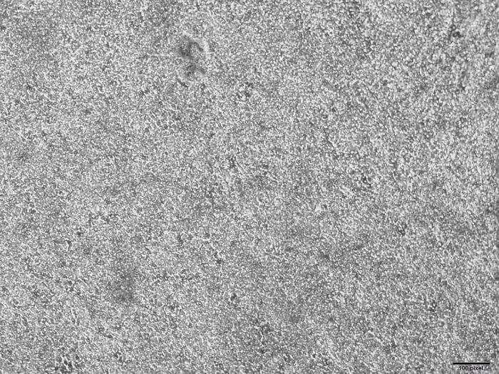
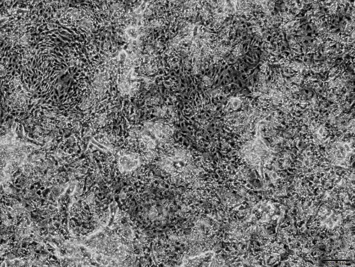
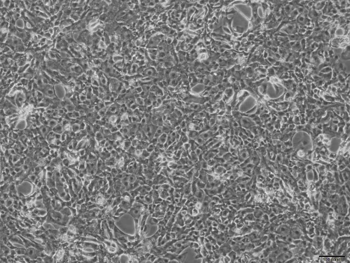
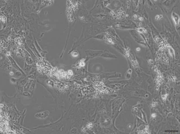
Contaminating noncardiac cells may influence the functional maturation of human iPSC-CMs during differentiation17. Figure 4A shows a comparison between 3 month-old cells obtained from high efficiency differentiations (top panels, Video 1), robustly expressing the specific ventricular marker (MLC2V), and 3-month-old cells obtained from low efficiency differentiations (bottom panels, Video 2), showing iPSC-CMs mixed with alpha-smooth muscle actin (αSMA)-positive cells. Interestingly, functional analysis demonstrated altered Ca2+ transients in the mixed iPSC-CMs/non-CMs preparation compared to the iPSC-CMs from the high efficiency differentiation. In particular, the cells obtained from low efficiency differentiations exhibited very small Ca2+ amplitude and automaticity (Figure 4B, middle panel) compared to pure iPSC-CMs (Figure 4B, top panel) and ARVCs (Figure 4B, bottom panel). In addition, cells from low efficiency differentiations presented arrhythmic patterns (Figure 4C).
It is important to note that the iPSC-CMs shown in the top panel of Figure 4A also underwent metabolic selection, which is an important step to further enrich a population of iPSC-CMs that is already derived from a highly efficient differentiation. These data indicate that high efficiency cardiac differentiation protocols that generate high-quality CMs are necessary to accurately recapitulate a cardiac phenotype in vitro.
Figure 5 shows the Ca2+ amplitude variability throughout different areas of the CMs monolayer, plotted over a time course of 1,200 s. The Ca2+ amplitudes measured at the beginning of a stimulation frequency of 1 Hz (200 s) were highly variable and became more consistent as the stimulation continued (200−900 s). However, after a time period of 900 s the Ca2+ amplitude was considerably reduced. These data indicate that, when recording Ca2+ transients in iPSC-CMs, it is necessary to let the cells stabilize at 37 °C under constant stimulation for at least 200 s. Additionally, recordings have to be restricted to a specific time window (200−900 s) to ensure reproducible results.
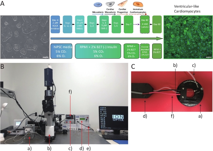
Figure 1: Overall schematic of iPCS-CMs preparation and Ca2+ transient instruments. (A) Schematic of cardiac differentiation protocol showing the developmental stages of differentiating iPSCs. Scale bar = 50 μm. (B) The calcium and contractility acquisition and analysis system. a) Peristalitic pump b) Inverted microscope c) Power source for the digital camera d) Electrical stimulator e) Fluorescence system interface f) Filter wheel controller (C) System chamber. a) System chamber body. b) Fluid inlet. c) Fluid outlet. d) Fluidic inline solution heater. f) Electrodes connecting the system chamber to the electrical stimulator. Please click here to view a larger version of this figure.
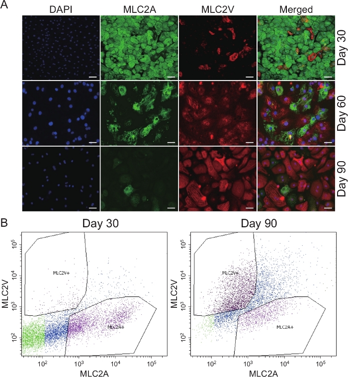
Figure 2: Comparison of MLC2A and MLC2V expression in iPSC-CMs at different days of differentiation. (A) Immunofluorescence staining of iPSC-CMs at day 30, 60, and 90 after differentiation induction for MLC2A and MLC2V. Scale bar = 20 μm. (B) Flow cytometry analysis of iPSC-CMs for MLC2V and MLC2A at 1 month and 3 months post differentiation. Please click here to view a larger version of this figure.
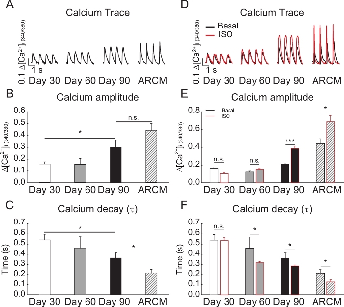
Figure 3: Comparison of iPSC-CMs properties at different days of differentiation. (A) Representative Ca2+ transient traces at different days of differentiation. (B–C) Average Ca2+ amplitude and Ca2+ decay elicited during stimulation at 1 Hz. (D–F) Effect of isoproterenol (ISO, 10 nM) treatment in iPSC-CMs at different days of differentiation. ARCMs indicate isolated rat adult cardiomyocytes. N = 70−100 areas; *p < 0.05; ***p < 0.001, as determined by Student's t-test. Data are represented as Mean ± S.E.M. Please click here to view a larger version of this figure.
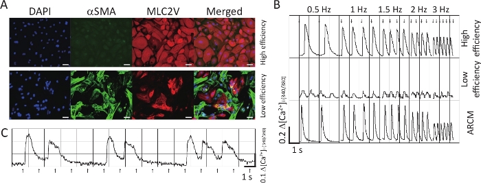
Figure 4: Comparison between high efficiency and low efficiency differentiations of iPSC-CMs. (A) Immunofluorescence staining of iPSC-CMs for αSMA and MLC2V. Scale bar = 20 μm. (B) Representative traces showing Ca2+ transients from high efficiency (above) and low efficiency (middle) differentiations, and isolated rat adult cardiomyocytes (below). The myocytes were stimulated at 0.5 Hz, 1 Hz, 1.5 Hz, 2 Hz, and 3 Hz. (C) Representative trace showing an arrhythmic pattern from a bad differentiation. Arrows indicate point pacing. ARCMs indicates isolated rat adult cardiomyocytes. Please click here to view a larger version of this figure.
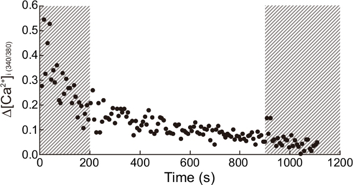
Figure 5: Time-dependent Ca2+ amplitude from a single coverslip. Ca2+ amplitudes from different areas in a coverslip were plotted in a time-dependent manner. Non-recommended time periods to measure Ca2+ transient are indicated in the dashed areas (<200 s or >900 s). Please click here to view a larger version of this figure.
Video 1: Representative video showing an example of a high efficiency differentiation. Please click here to download this video.
Video 2: Representative video showing an example of a low efficiency differentiation. Please click here to download this video.
Video 3: Representative video showing an example of a homogeneous monolayer distribution of iPSC-CMs on a glass coverslip. This video shows the recommended cell density to be used for functional analysis. Please click here to download this video.
Video 4: Representative video showing an example of cells from a low efficiency differentiation plated onto a glass coverslip. The cells in the video were obtained from a low efficiency differentiation. Cells from such differentiations are usually not distributed homogeneously across the coverslip and it is difficult to consistently find beating cells. Please click here to download this video.
Critical steps for using human iPSC-CMs as experimental models are: 1) generating high-quality cardiomyocytes (CMs) that can ensure the consistent performance and reproducible results; 2) allowing the cells to mature in culture for at least 90 days to adequately assess their phenotype; 3) performing electrophysiological studies, e.g. calcium (Ca2+) transient measurements, to provide a physiologically relevant functional characterization of human iPSC-CMs. We developed a monolayer-based differentiation method that produces high-quality ventricular-like iPSC-CMs. Our method relies on several crucial factors and is a variant of existing protocols14,18. Unlike other protocols, this one uses low oxygen conditions (5% O2) for iPSCs maintenance as well as during the first week of their differentiation into CMs. Low levels of oxygen mimic the environmental condition during embryonic and fetal heart development19. Hypoxic conditions have also been shown to increase iPSCs proliferation and their subsequent differentiation to CMs20. Seeding density and proper iPSCs passaging are also critical factors for a successful cardiac differentiation. High differentiation efficiency is observed when 70−80% confluent iPSCs are dissociated into small cell aggregates using an enzyme-free solution, and seeded at a split ratio of 1:6. Cardiac differentiation can be started when the cells reach a confluency of 70−80%, expected about 4 days after splitting. CHIR99021 (10 μM) treatment at the beginning of the differentiation will cause significant cell death. However, cell proliferation is observed when CHIR99021 is removed from culture the following day. A correct balance between cell death and proliferation is essential for preserving a monolayer culture throughout the differentiation, which is another important factor for a successful differentiation. If the iPSC density is either too low or too high, the subsequent differentiation will be a low efficiency cardiac differentiation (Figure 4A, Video 2). Figure 4A shows such an example of a low efficiency differentiation, where the cardiomyocytes derived from a healthy donor are mixed with other cell types, such as α-smooth muscle actin-positive cells. Importantly, Ca2+ transients recorded from such preparations showed altered characteristics, such as low Ca2+ amplitude and arrhythmic patterns, which could easily be misinterpreted as biological variation. A successful and high efficiency differentiation from the same cell line, instead, generated a pure population of ventricular-like cardiomyocytes (Figure 4A, Video 1) along with regular Ca2+ transients (Figure 4B, top panel). Thus, the quality of the differentiation plays an important role in the overall magnitude, shape, regularity, and frequency of a Ca2+ transient.
Another important aspect of a successful and efficient differentiation is obtaining an enriched population of cardiomyocytes. While undifferentiated stem cells and other iPSC-derived cell types use glucose as an energy source, CMs can use lactate efficiently for ATP and glutamate production21. Thus, in order to obtain an even purer population of CMs, the cells are cultured in lactate-supplemented and glucose-depleted culture medium. Culturing iPSC-CMs in this selecting media for a long time, however, may lead to functional impairment. Therefore, to purify iPSC-CMs and still preserve their function, metabolic selection is performed only for 10 days, from days 80−90 since differentiation induction. After this period, the selection media is replaced, and the cells are maintained in RPMI/B27 media. While the selection protocol can be implemented much earlier22 (e.g., around day 15), it is advisable to wait until day 80, as doing so can prevent residual iPSCs or other cell types from proliferating by the time the cells are ready for functional studies (day 90). It is vital therefore, that for a successful, efficient, and robust cardiac differentiation, all of the above-mentioned factors are taken into consideration.
In addition to the robustness and efficiency of cardiac differentiation protocols, maturation and cell type specification (e.g., atrial and ventricular cell types) also pose a major challenge for using iPSC-CMs to model cardiac disease. Over the last few years, many promising maturation strategies have been reported, including electrical stimulation, mechanical stretch, and substrate stiffness23. However, prolonged in vitro culture of iPSC-CMs continues to be a simple and practical approach to generate adult-like cardiomyocytes24,25.
Consistent with other published reports12, iPSC-CMs generated by this in vitro differentiation protocol show robust expression of ventricle-specific MLC2V and much lower levels of MLC2A at day 90 (Figure 2A,B). While fetal CMs contain both MLC2A and MLC2V isoforms, adult ventricular CMs only contain MLC2V. This switch in MLC isoform prevalence occurs during the neonatal stage26.
Cardiomyocytes generated through this protocol express ventricular specific marker, such as MLC2V, which progressively increases in abundance as the cells are kept in culture longer (Figure 2A,B). This indicates that a prolonged in vitro culture enhances the maturation of CMs that appear to be committed to the ventricular phenotype.
Time in culture also greatly affects Ca2+ handling maturation24,25. Previous reports described that after 20 days of the differentiation induction, there were no major changes in the calc Ca2+ transients as the cells grew older25. However, the cardiomyocytes generated from the protocol detailed here show progressive changes in Ca2+ amplitude and Ca2+ decay over the three time points evaluated (30, 60, and 90 days after induction of cardiac differentiation) (Figure 3A-C). Interestingly, these data show that Ca2+ transient parameters, such as Ca2+ amplitude and Ca2+ decay, measured in day 90 iPSC-CMs were similar to those measured in isolated rat adult cardiomyocytes (ARCMs). Moreover, the 90 days old iPSC-CMs were also able to follow various electrical stimulations ranging from 0.5 Hz to 3 Hz consistently, similar to ARCMs (Figure 4B). In future work, it would be interesting to see how an even longer time in culture affects the functional characteristics of these iPSC-CMs as compared to ARCMs.
Additionally, since β-adrenergic receptor signaling shows a distinct time-dependent maturational pattern after cardiac induction27, the effect of isoproterenol on Ca2+ transient in iPSC-CMs generated through this protocol was tested. Stimulation with isoproterenol led to a positive lusitropic response in day 90 iPSC-CMs, similar to what is observed in ARCMs (Figure 3D-F).
The functional assessments of iPSC-CMs performed in this study have some differences and limitations as compared to isolated adult CMs. The adult CMs have well-developed and well-arranged sarcomeres. This allows for contractility measurements through detection of sarcomere movement. Because none of the current protocols generate fully matured, adult-like iPSC-CMs, contractility measurements through sarcomere detection are not feasible. While it is possible to detect the movement of the cell edges when the cell contracts, it is significantly more challenging to track those movements in a precise manner in iPSC-CMs. As of now, technological advancements are needed to allow for accurate assessments of contractility in these cells. On the other hand, it is possible to measure Ca2+ transients of iPSC-CMs using a Ca2+ indicator like Fura-2. Unlike adult CMs, however, iPSC-CMs are clustered and do not have a well-defined border. Therefore, the recorded Ca2+ transients usually do not represent a single cell, but rather a group of cells in a specific area. While it is possible to adjust the area size, recording Ca2+ transients from a single cell is incredibly challenging. It is critical that when CMs are plated onto the coverslips, the density is such that they achieve a homogenous monolayer distribution (Video 3), which allows for consistently finding areas with cells. If the cells are not distributed as a monolayer on the coverslip (Video 4), there will be areas without cells, and if the coverslip is screened specifically for areas with beating cells to perform the measurements, this can lead to a "selection bias". Instead of selecting a specific area of beating cells, it is recommended to collect data from multiple areas throughout the coverslip, which is only possible when the cells are distributed as a homogenous monolayer. Measurements from 100 such different areas, which take about 10 min, are sufficient to provide reliable functional properties of the cells. Lastly, as shown in Figure 5, iPSC-CMs need time to adjust to the measurement conditions. For reliable measurements, it is recommended that the cells should be stabilized at the measurement conditions for at least 3 min.
The authors have nothing to disclose.
This research was supported by AHA Scientist Development Grant 17SDG33700093 (F.S.); Mount Sinai KL2 Scholars Award for Clinical and Translational Research Career Development KL2TR001435 (F.S.); NIH R00 HL116645 and AHA 18TPA34170460 (C.K.).
Anti-Actin, α-Smooth Muscle antibody, Mouse monoclonal | Sigma Aldrich | A5228 | |
Alexa Fluor 488 goat anti mouse | Invitrogen | A11001 | |
Alexa Fluor 555 goat anti rabbit | Invitrogen | A21428 | |
B27 Supplement | Gibco | 17504-044 | |
B27(-) insulin Supplement | Gibco | A18956-01 | |
CHIR-99021 | Selleckchem | S2924 | |
DAPI nuclear stain | ThermoFisher | D1306 | |
DMEM/F12 (1:1) (1X) + L- Glutamine + 15mM Hepes | Gibco | 11330-032 | |
Double Ended Cell lifter, Flat blade and J-Hook | Celltreat | 229306 | |
Falcon Multiwell Tissue Culture Plate, 6 well | Corning | 353046 | |
Fluidic inline heater | Live Cell Instrument | IL-H-10 | |
Fura-2, AM | Invitrogen | F1221 | |
hESC-qualified matrix | Corning | 354277 | Matrigel Matrix |
hPSC media | Gibco | A33493-01 | StemFlex Basal Medium |
IWR-1 | Sigma Aldrich | I0161 | |
Live cell imaging chamber | Live Cell Instrument | EC-B25 | |
MLC-2A, Monoclonal Mouse Antibody | Synaptic Systems | 311011 | |
Myocyte calcium and contractility system | Ionoptix | ISW-400 | |
Myosin Light Chain 2 Antibody, Rabbit Polyclonal (MLC2V) | Proteintech | 10906-1-AP | |
Nalgene Rapid Flow Sterile Disposable Filter units with PES Membrane | ThermoFisher | 124-0045 | |
PBS with Calcium and Magnesium | Corning | 21-030-CV | |
PBS without Calcium and Magensium | Corning | 21-031-CV | |
Premium Glass Cover Slips | Lab Scientific | 7807 | |
RPMI medium 1640 (-) D-glucose (1X) | Gibco | 11879-020 | |
RPMI medium 1640 (1X) | Gibco | 11875-093 | |
Sodium L-lactate | Sigma Aldrich | L7022 | |
StemFlex Supplement | Gibco | A33492-01 | |
Thiazovivin | Tocris | 3845 | |
Trypsin-EDTA (0.25%) | ThermoFisher | 25200056 | |
Tyrode's solution | Boston Bioproducts | BSS-355w | Adjust pH at 7.2. Add 1.2mM Calcium Chloride |