The protocol presented here is intended to demonstrate the occurrence of heterologous interactions between Golgi-resident type III membrane proteins with cytoplasmically exposed N- and/or C-termini in live mammalian cells using the most recent variant of the split luciferase complementation assay.
The goal of this protocol is to explore the applicability of the most recent variant of split luciferase complementation for demonstrating heterologous complexes formed by nucleotide sugar transporters (NSTs). These ER- and Golgi-resident multitransmembrane proteins carry the cytoplasmically synthesized nucleotide sugars across organelle membranes to supply enzymes that mediate glycosylation with their substrates. NSTs exist as dimers and/or higher oligomers. Heterologous interactions between different NSTs have also been reported. To verify whether the technique is suitable for studying the phenomenon of NST heteromerization, we tested it against a combination of the two Golgi-resident NSTs that have been previously shown to associate by several other means. The luciferase complementation assay appears to be particularly suitable for studying interactions between Golgi-resident membrane proteins, as it does not require high expression levels, which often trigger protein mislocalization and increase the risk of false positives.
This manuscript describes a step-by-step protocol to check for the presence of heterologous interactions between Golgi-resident type III membrane proteins in transiently transfected human cells using the most recent variant of the split luciferase complementation assay. The procedure has been most extensively tested against nucleotide sugar transporters (NSTs) but we were also able to obtain positive results for other Golgi-resident type III membrane proteins whose N- and/or C-termini are facing the cytoplasm.
Our research group explores the role of NSTs in glycosylation of macromolecules. NSTs are Golgi- and/or ER-resident type III membrane proteins with N- and C-termini facing the cytoplasmic side of the organellar membrane1. NSTs are thought to carry nucleotide-activated sugars across organelle membranes to supply glycosyltransferases with their substrates. NSTs form dimers and/or higher oligomers2,3,4,5,6,7,8,9,10. Moreover, heterologous interactions between different NSTs have also been reported6,11. NSTs were also demonstrated to form complexes with functionally related glycosylation enzymes12,13,14. We sought for an alternative to the presently used technique, fluorescence lifetime imaging (FLIM)-based FRET approach, for studying interactions of NSTs and functionally related Golgi-resident proteins, so we decided to test the split luciferase complementation assay. It allowed us to identify a novel interaction between an NST and a functionally related glycosylation enzyme9.
The most recent modification of the split luciferase complementation assay, NanoBiT, is used in the protocol presented here15. It relies on the reconstitution of the luciferase enzyme (e.g., NanoLuc) from the two fragments – the large one, termed as large BiT or LgBiT, a 17.6 kDa protein, and the small one, composed of only 11 amino acids, termed as small BiT or SmBiT. The two proteins of interest are fused with the complementary fragments and transiently expressed in a human cell line. If the two fusion proteins interact, a luminescence is produced in situ upon addition of a cell-permeable substrate. These two fragments have been optimized so that they associate with minimum affinity unless being brought together by an interaction between the proteins of interest they are fused to.
In general, bioluminescence-based methods have some advantages over the ones based on fluorescence. Bioluminescent signals have a higher signal-to-noise ratio because the background luminescence is negligible compared to the luciferase-derived signal16. In contrast, fluorescence-based approaches usually suffer from a relatively high background caused by the phenomenon of autofluorescence. Besides, bioluminescence is less detrimental to the analyzed cells than fluorescence, as in the former case there is no need to excite the sample. For those reasons bioluminescent approaches to studying PPIs in vivo outcompete the commonly used fluorescent methods like Förster Resonance Energy Transfer (FRET) or Bimolecular Fluorescence Complementation (BiFC).
Our protocol relies on referring luminescence obtained for the protein combination of interest to luminescence obtained for the control combination. The latter includes the one of the tested proteins which is fused with a larger fragment and a control protein (e.g., HaloTag), fused with a smaller fragment. The latter is a protein of bacterial origin that is not expected to interact with any of mammalian proteins. Using this protein as a control poses limitations to the topology of the Golgi-resident pairs of proteins to be analyzed. Since in mammalian cells this protein is synthesized in the cytoplasm, both proteins of interest should have at least one cytoplasmic tail.
This approach can be particularly useful for initial screening of PPIs. It may become the method of choice when the fusion proteins of interest are expressed at levels that are simply insufficient for other approaches to be applied. Similarly, the split luciferase complementation assay can be the best option if the proteins of interest are expressed at high levels, but this adversely affects their subcellular localization or is known to force non-specific interactions. Since the smaller fragment has only 11 amino acids, the split luciferase complementation assay can be applied when using larger tags is impossible. Finally, it can be employed to further confirm data obtained using other techniques, as in the case presented here.
1. Generation of expression plasmids
2. Transient transfection of the expression plasmids into the cells
3. Medium exchange
4. Preparation of furimazine working solution
5. Measuring luminescence
6. Data analysis
To obtain the most reliable data in this approach all the possible combinations should be tested (see Figure 1). In parallel, positive and negative controls should be included. The positive control should consist of the two proteins that are known to interact, of which one is fused with the larger fragment and the other is fused with the smaller fragment. The negative control ideally should consist of the two non-interacting type III membrane proteins tagged likewise. However, establishing such a control may be challenging, as the lack of interaction of the two control proteins should be thoroughly confirmed using several alternative approaches. Therefore, one might employ a cytosolic protein of non-human origin that is not expected to interact with any human protein (e.g., HaloTag) as a negative control, if N- and/or C-termini of the proteins, whose interaction is to be determined, are cytoplasmically exposed as in the case presented here. We highly recommend an additional verification of the results obtained using this type of control by co-expression of the untagged variants of either of the proteins of interest combined with titration of the corresponding plasmids. If the two proteins specifically interact, an extra copy of either of them lacking the luciferase fragment should result in a specific decrease of luminescence.
Relative luminescence units (RLU) values typical of positive and negative combinations are listed in Table 1. The results were obtained for the two NSTs, SLC35A2 and SLC35A3, that were shown to associate by co-immunoprecipitation and FLIM-FRET6 as well as in situ proximity ligation assay11. Both SLC35A2 and SLC35A3 are Golgi-resident type III membrane proteins with N- and C-termini facing the cytoplasm (Figure 1A). Therefore, there are eight possible tagging options and the resulting fusion proteins can be set together also in eight possible ways (Figure 1B). Mean RLU values corresponding to all the tested and control combinations are depicted in Figure 2A. The positive control is also included. An initial requirement for a selected result to be considered indicative of an interaction is that the RLU value obtained for the combination of interest is statistically significantly higher than the RLU value obtained for the corresponding control combination. In Figure 2A three such combinations are seen (LgBiT-SLC35A2 + SLC35A3-SmBiT, SmBiT-SLC35A2 + LgBiT-SLC35A3 and SLC35A2-SmBiT + LgBiT-SLC35A3). The next step in the analysis of results involves obtaining ratio values for the combinations of interest by dividing the corresponding RLU values by RLU values obtained for the respective controls. The results of such analysis are shown in Figure 2B. According to the manufacturer’s suggestions, ratios between 10 and 1000 are highly indicative of specific interactions. There are two combinations that meet these criteria (SmBiT-SLC35A2 + LgBiT-SLC35A3 and SLC35A2-SmBiT + LgBiT-SLC35A3) and support the idea that SLC35A2 and SLC35A3 interact. However, the fact that the other six combinations are negative does not mean that there are no interactions at all between the respective fusion proteins, but rather suggests that the tagging strategy was not optimal in these cases. This example shows how important it is to test all the possible combinations.
Data presented in Figure 2 were additionally confirmed by the co-expression of HA-tagged SLC35A2 or SLC35A3 with the combination that resulted in the highest relative luminescence, namely SLC35A2-LgBiT + SmBiT-SLC35A3. Varying amounts of the plasmids encoding HA-tagged NST variants were used for co-transfection. This resulted in a statistically significant, dose-dependent decrease in RLU values (Figure 3). Specific and dose-dependent disruption of the interaction between SLC35A2-LgBiT and SmBiT-SLC35A3 by the simultaneous co-expression of HA-tagged NST variants is shown in Table 2.
The most common problem associated with the method presented here is the poor efficiency of transfection. To verify whether this is the cause of suboptimal results, include positive control in all experiments. The positive control we employed to obtain the data presented here consists of the two interacting fusion proteins: cAPM-dependent protein kinase catalytic subunit alpha (PRKACA) fused with the smaller fragment and cAPM-dependent protein kinase type II-alpha regulatory subunit (PRKAR2A) fused with the larger fragment. In our hands, the corresponding RLU value reached ~6 x 10⁵. A significant (2-3 orders of magnitude) drop in this number might be indicative of the suboptimal efficiency of the performed transfection. In such a case we recommend checking on the well-being of the cultured cells and making sure that the appropriate number of cells is being plated for transfection.
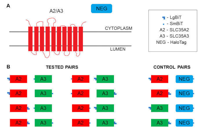
Figure 1: Schematics of membrane topology and tagging. (A) Membrane topology of SLC35A2 and SLC35A3 proteins. (B) Possibilities of tagging SLC35A2 and SLC35A3 proteins with the split luciferase complementation assay fragments and combining the resulting fusion proteins for the assay. Please click here to view a larger version of this figure.
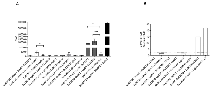
Figure 2: Results of the split luciferase complementation assay performed in HEK293T cells for the selected protein combinations and the corresponding negative controls (processed data). (A) RLU values obtained for the selected protein combinations and the corresponding negative controls. Negative, HaloTag tagged with SmBiT; PRKAR2A, cAPM-dependent protein kinase type II-alpha regulatory subunit; PRKACA, cAPM-dependent protein kinase catalytic subunit alpha. Data were analyzed using one-way ANOVA with multiple comparisons and are presented as a mean ± standard deviation (SD) from three technical replicates. p < 0.1 *, p < 0.05 **, p < 0.01 ***. (B) Fold changes calculated by dividing a mean luminescence obtained for the tested combination (RLU SAMPLE) by a mean luminescence obtained for the corresponding negative control (RLU CONTROL). Threshold value considered indicative of an interaction was set at 10. Please click here to view a larger version of this figure.
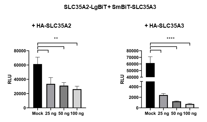
Figure 3: Specific and dose-dependent disruption of the interaction between SLC35A2-LgBiT and SmBiT-SLC35A3 by simultaneous co-expression of HA-tagged NST variants. HEK293T cells were transfected with plasmids encoding SLC35A2-LgBiT and SmBiT-SLC35A3 and, additionally, either with an empty pSelect plasmid (mock) or with increasing amounts of pSelect plasmids encoding HA-tagged SLC35A2 (left panel) and SLC35A3 (right panel). Data were analyzed using one-way ANOVA with multiple comparisons and are presented as a mean ± standard deviation (SD) from three technical replicates. p < 0.05 **, p < 0.001 ****. Please click here to view a larger version of this figure.
Table 1: Results of the assay performed in HEK293T cells for the selected protein combinations and the corresponding negative controls (raw data). Unprocessed RLU values obtained for the selected protein combinations and the corresponding negative controls are shown. Negative, HaloTag tagged with SmBiT; PRKAR2A, cAPM-dependent protein kinase type II-alpha regulatory subunit; PRKACA, cAPM-dependent protein kinase catalytic subunit alpha, TR, technical replicate. Please click here to download this table.
Table 2: Specific and dose-dependent disruption of the interaction between SLC35A2-LgBiT and SmBiT-SLC35A3 by simultaneous co-expression of HA-tagged NST variants (raw data). Unprocessed RLU values obtained for the selected protein combinations are shown. Mock, cells expressing SLC35A2-LgBiT and SmBiT-SLC35A3 co-transfected with an empty pSelect vector; HA-SLC35A2, cells expressing SLC35A2-LgBiT and SmBiT-SLC35A3 co-transfected with the indicated amounts of a pSelect plasmid encoding SLC35A2 tagged with the HA epitope at the N-terminus; HA-SLC35A3, cells expressing SLC35A2-LgBiT and SmBiT-SLC35A3 co-transfected with the indicated amounts of a pSelect plasmid encoding SLC35A3 tagged with the HA epitope at the N-terminus; TR, technical replicate. Please click here to download this table.
Here we provide a detailed protocol enabling the demonstration of heterologous complexes formed between Golgi-resident type III membrane proteins, such as NSTs, using the split luciferase complementation assay. The proposed approach to data analysis and interpretation involves relating the luminescence obtained for the protein combination of interest to the luminescence obtained for the corresponding control combination, which is composed of one of the proteins of interest fused with the larger fragment and the control protein of a bacterial origin fused with the smaller fragment. The latter is expressed in the cytoplasm of mammalian cells; therefore, using it as a reference requires that N- and/or C-termini of the proteins, whose interaction is to be analyzed, are on the cytoplasmic side of the Golgi membrane.
The critical steps in the protocol are plasmid design, plating the cells to be transfected, transfection itself, the medium exchange, preparation of the furimazine working solution and adding it to the cells. The plasmid design should be made in such a way that both luciferase fragments are facing the cytoplasm, otherwise control combinations will not work and the reference RLU values will be missing. Cells to be transfected should be plated as specified in the protocol, otherwise the transfection efficiency might be suboptimal. We recommend using multiwell plates with the poly-L-lysine-coated surface to support cell attachment while the medium is being exchanged. Finally, the furimazine working solution should be freshly prepared and immediately added to the cells. Once it is added, the readout should be performed as soon as possible.
To avoid inconclusive results, positive and negative controls should always be included. The positive control should be always employed so that the transfection efficiency is monitored. It is very important that all the possible fusion proteins that are topologically compatible with each other as well as with the HaloTag-based negative control are generated and combined. If possible, the negative control comprising of a membrane protein non-interacting with any of the proteins of interest should be used. Here, we did not employ such a control, as its development is still on the way. Instead, we forced a specific disassembly of the complexes of interest by co-expressing an extra, untagged copy of one of the analyzed proteins. The expression vectors we used carry the relatively weak herpes simplex virus-thymidine kinase (HSV-TK) promoter, which ensures low expression levels that are optimal for obtaining specific outcome. However, in the case of suboptimal results it might be beneficial to use the CMV-based vectors or, alternatively, optimizing the amount of plasmids used for transfection.
The presented method, although powerful and convenient, has some limitations. First, this method does not allow to conclude whether the identified complexes correspond to dimers or higher-order oligomers. Besides, the subcellular localization of the interacting proteins cannot be monitored. This is, however, possible upon the extension of the basic protocol with bioluminescent imaging, although spatial resolution of such images would be significantly lower when compared with fluorescence-based approaches.
The presented method is very fast and efficient (the measurement takes up to several minutes and the data are obtained from thousands of cells). Data processing and interpretation is also relatively straightforward. Little or no optimization of the basic protocol is needed. The only specific equipment required is a luminometer. The split luciferase complementation assay and BiFC work on the same essential principle, i.e., reconstitution of a functional protein from its non-functional fragments. General advantages of bioluminescence-based approaches over the ones based on fluorescence were already listed in the Introduction. A specific advantage of the split luciferase complementation assay over BiFC is that the former is fully reversible15. In BiFC, once a fluorescent protein is reconstituted, it would not dissociate back into the corresponding non-fluorescent fragments17. In contrast, the assembly of the NanoLuc subunits is reversible, which creates a unique opportunity to study the dynamics of PPIs. Finally, the exceptional sensitivity of the presented method allows to assume that this approach should work even with the cell lines that are difficult to transfect.
The protocol presented here allows to determine whether the two Golgi-resident type III membrane proteins interact. As already mentioned, the basic setup of the method can be coupled with bioluminescence imaging to confirm the subcellular localization of the PPI of interest. The reversibility of this split luciferase complementation assay allows to study the dynamics of PPIs in real time. The luminescent signal derived from furimazine is sustained for about 2 hours. However, substrates for the split luciferase complementation assay ensuring a substantially longer (hours to days) duration of the resulting signal are also available. This method allows for the identification of the factors that trigger or prevent the PPI of interest. Some of the most recent examples of its application include studies on interactions between G proteins and G-protein-coupled receptors18, protein conformational changes19, protein ubiquitination20, internalization of cell surface receptors21, and identification of factors that modulate PPIs22,23. Therefore, this method appears to be a versatile tool with a high potential to fulfill even very challenging experimental goals.
The authors have nothing to disclose.
This work was supported by grant no. 2016/23/D/NZ3/01314 from the National Science Centre (NCN), Krakow, Poland.
0.25% trypsin-EDTA solution | |||
Adherent mammalian cell line | |||
BioCoat Poly-D-Lysine 96-well White/Clear Flat Bottom TC-treated Microplate, with Lid | Corning | 356651 | |
Cell culture centrifuge | |||
Cell culture supplements (heat-inactivated fetal bovine serum, L-glutamine, penicillin, streptamycin) | |||
CO2 incubator | |||
Expression plasmids encoding protein(s) of interest not tagged with NanoBiT fragments | |||
FuGENE HD Transfection Reagent | Promega | E2311 | |
GloMax Discover Microplate Reader (or a different luminescence microplate reader) | Promega | GM3000 | |
Growth medium dedicated to the cell line used | |||
Materials and reagents for standard molecular cloning (bacteria, thermostable polymerase, restriction enzymes, DNA ligase, materials and reagents for nucleic acid purification) | |||
NanoBiT MCS Starter System | Promega | N2014 | This kit contains vectors enabling tagging of the proteins of interest with NanoBiT fragments at different orientations as well as the control plasmid encoding HaloTag protein fused with SmBiT and a positive control plasmid pair. |
Nano-Glo Live Cell Assay System | Promega | N2011 | This kit contains furimazine, which is a substrate enabling detection of the NanoLuc activity in living cells, and a dedicated dilution buffer. |
Opti-MEM I Reduced Serum Medium, no phenol red | Gibco | 11058021 | |
Oribital shaker | |||
Software for data analysis (e.g. GraphPad Prism) | |||
Thermocycler |