This protocol outlines methodologies behind the Ramsay assay, ion-selective microelectrodes, Scanning Ion-selective Electrode Technique (SIET) and in vitro contraction assays, applied to study the adult mosquito excretory system, comprised of the Malpighian tubules and hindgut, to collectively measure ion and fluid secretion rates, contractile activity, and transepithelial ion transport.
Studies of insect physiology, particularly in those species that are vectors of pathogens causing disease in humans and other vertebrates, provide the foundation to develop novel strategies for pest control. Here, a series of methods are described that are routinely utilized to determine the functional roles of neuropeptides and other neuronal factors (i.e., biogenic amines) on the excretory system of the mosquito, Aedes aegypti. The Malpighian tubules (MTs), responsible for primary urine formation, can continue functioning for hours when removed from the mosquito, allowing for fluid secretion measurements following hormone treatments. As such, the Ramsay assay is a useful technique to measure secretion rates from isolated MTs. Ion-selective microelectrodes (ISME) can sequentially be used to measure ion concentrations (i.e., Na+ and K+) in the secreted fluid. This assay allows for the measurement of several MTs at a given time, determining the effects of various hormones and drugs. The Scanning Ion-selective Electrode Technique uses ISME to measure voltage representative of ionic activity in the unstirred layer adjacent to the surface of ion transporting organs to determine transepithelial transport of ions in near real time. This method can be used to understand the role of hormones and other regulators on ion absorption or secretion across epithelia. Hindgut contraction assays are also a useful tool to characterize myoactive neuropeptides, that may enhance or reduce the ability of this organ to remove excess fluid and waste. Collectively, these methods provide insight into how the excretory system is regulated in adult mosquitoes. This is important because functional coordination of the excretory organs is crucial in overcoming challenges such as desiccation stress after eclosion and before finding a suitable vertebrate host to obtain a bloodmeal.
Maintenance of salt and water levels in insects allows them to succeed in many ecological and environmental niches, utilizing a variety of feeding strategies1. Most insects have evolved mechanisms to regulate the composition of their haemolymph within narrow limits in order to withstand the different challenges associated with their particular environment2. Terrestrial insects are often faced with the challenge of conserving water and the excretory system undergoes anti-diuresis to prevent loss of water and some essential salts, therefore, avoiding desiccation. In contrast, diuresis occurs when the insect feeds and is challenged with excess water and potentially salts3,4. Through their specialized and highly active excretory system, insects have evolved regulatory mechanisms acting to counter their osmoregulatory challenges. In adult Aedes aegypti mosquitoes, the excretory system is comprised of the Malpighian tubules (MTs) and hindgut, the latter of which is made up of the anterior ileum and posterior rectum5. MTs are responsible for generating primary urine, usually rich in NaCl and/or KCl. The primary urine is then modified through secretory and reabsorptive processes as it travels downstream of the tubule and enters the hindgut5. The final excreta can be hyper- or hypoosmotic to the haemolymph, depending on feeding/environmental conditions, and is enriched in toxic and nitrogenous wastes2.
MTs are ideal for studying many features of epithelial fluid and solute transport as they carry out a great variety of transport and excretory functions2,6. Through hormonal regulation2, MTs function by secreting ions and other solutes from the blood into the tubule lumen7, providing an osmotic gradient allowing water to be transported by aquaporins8,9, which collectively creates the primary urine, before traveling toward the reabsorptive hindgut2. Thus, by collecting the secreted fluid from isolated MTs, one can continuously monitor transepithelial transport of fluid and ions. Measuring secretion rate and urine composition provides insight on mechanisms responsible for transepithelial ion and fluid transport. A popular method for studying fluid secretion rates is the Ramsay assay, which was first introduced by Ramsay in 195310. In this method, the distal (closed) end of the tubule is treated with a hormone (or other test compound/drug), while the proximal (open) end is wrapped around a pin in water-saturated paraffin oil, which secretes the primary urine, accumulating as a droplet on the tip of the pin. Isolated MTs are able to survive and function for long periods (up to 24 h) under optimized in vitro conditions, which make them suitable and efficient models for fluid secretion measurement. Insects have open circulatory systems, thus the MTs are easily dissected and removed as they are usually freely floating in the haemolymph6. Additionally, with the exception of aphids—which lack MTs11—the number of MTs in a given insect species can vary considerably from four to hundreds (five in Aedes mosquitoes) allowing for multiple measurements from one insect.
The MTs in Aedes mosquitoes, in common with other endopterygote insects, are composed of two cell types forming a simple epithelium2,12; large principal cells, which facilitate active transport of cations (i.e., Na+ and K+) into the lumen, and thin stellate cells, which aid in transepithelial Cl– secretion13. The MTs are not innervated2, and instead are regulated by several hormones including both diuretic and anti-diuretic factors, allowing for the control of ion transport (mainly Na+, K+, and Cl–) and osmotically-obliged water2. Numerous studies have examined the hormonal regulation of Aedes MTs to understand the role of endocrine factors on transepithelial transport14,15,16,17,18. As shown in the representative results, the protocols herein demonstrate the effects of different hormonal factors on isolated MTs from adult female A. aegypti mosquitoes, including both diuretic and anti-diuretic control (Figure 1). The Ramsay assay is used to demonstrate how an anti-diuretic hormone, AedaeCAPA-1, inhibits fluid secretion of MTs stimulated by diuretic hormone 31 (DH31) (Figure 1).
The smaller size of insects has required the development of micro methods for measuring ionic activity and concentrations in fluid samples, or near the surface of isolated tissues such as the MTs and gut. Varying methods have been implemented, including the use of radioisotopes of ions19, which requires collection of the secreted fluid drops for measurement of ion concentrations20. Stimulated Aedes tubules in vitro typically secrete ~0.5 nL/min21, thus handling of such small volumes can pose a challenge and potentially introduce error upon transfer. As a result, ion-selective microelectrodes (ISMEs) have been extensively used to measure ion concentrations in secreted droplets of MTs in vitro. In this method, a reference electrode and ISME, filled with the appropriate backfill solution and ionophore, are positioned into the secreted urine droplet to determine ion concentrations22. Adapted from Donini and colleagues23, this current protocol uses a Na+-selective ionophore to measure ion activity in secreted droplets from stimulated MTs in adult Aedes mosquitoes. Since ion-selective microelectrodes measure ion activity, this data can be expressed as ion concentrations following the assumption that the calibration solutions and experimental samples share the same ion activity coefficient21 (Figure 1B,C).
The Scanning Ion-selective Electrode Technique (SIET) also makes use of ISMEs to measure ion concentration gradients in the unstirred layer adjacent to organs, tissues, or cells that are transporting ions. The ISMEs measure voltage gradients which can then be used to calculate the ion concentration gradients and direction and magnitude of ion flux across the organ, tissue, or cell20. In this technique, the ISME is mounted to a three axes manipulator controlled by computerized micro-stepper motors so that its 3D position is controlled to the micrometer level20. Voltages are measured at two points within the unstirred layer using a sampling protocol programmed into and controlled by computer software. The two points are typically separated by a distance of 20–100 µm with one point within 5–10 µm of the surface of the organ, tissue, or cell and the second point a further 20–100 µm away. The difference in magnitude of voltages between the two points is calculated to obtain a voltage gradient24,25,26, which is then used to calculate the concentration gradient and subsequently the net flux using Fick’s Law24,27. This method is useful for assessing the transport of specific ions across different regions of the insect gut and MTs, or at specific timepoints following a bloodmeal or treatment exposure. For instance, the SIET can be used to understand how absorptive and secretory processes in the mosquito excretory system are regulated by hormones28 as well as different feeding behaviors and rearing conditions25. Previous work utilizing the SIET revealed sites involved in ion transport along the anal papillae and rectum of larval and adult mosquitoes24,28. The current protocol, described previously by Paluzzi and colleagues26, measures Na+ flux across the rectal pad epithelia of the adult female rectum (Figure 2).
The final segment of the mosquito excretory system requires coordinated muscular movement to help mix food and secrete waste26. Non-absorbable products of digestion from the midgut, along with primary urine secreted by the MTs, are passed through the pyloric valve and delivered to the hindgut2. Spontaneous hindgut contractions begin at the pyloric valve and occur in peristaltic waves, which are relayed over the ileum through the coordinated contraction of circular and longitudinal muscles surrounding the basal surface of epithelial cells26. Finally, the muscles within the rectum help to propel and eliminate waste through the anal canal. Although insect hindgut motility is myogenic, requiring extracellular Ca2+ to produce spontaneous contractions, these processes can also be regulated neuronally26,29,30. This exogenous regulation by the nervous system is important after feeding, as the animal must expel wastes from the gut and restore haemolymph balance31. As a result, performing in vitro bioassays to identify myostimulatory or myoinhibitory neuropeptides is useful in assessing how neurochemicals influence hindgut motility. The current protocol, performed by Lajevardi and Paluzzi28, uses video recordings to examine ileal motility in response to neuropeptides (Figure 3). Similarly, a force transducer or impedance converter may also be used to observe traces of contractions through a data acquisition software32,33. However, using video technology allows us to visually assess the organ and further analyze using a subset of parameters to identify the role of hormones on hindgut motility.
Using these techniques can help characterize factors that regulate and coordinate fluid and ion transport along the excretory system along with hindgut motility. Importantly, a functional link between the diuretic response by the MTs and hindgut motility is supported, as diuretic hormones, such as DH31 and 5HT, characterized by their ability to stimulate fluid secretion by the MTs, have also been found to exhibit myotropic actions along the mosquito hindgut21,34,35. These findings highlight the importance of stringent coordination between the MTs and hindgut during events such as post-prandial diuresis in insects requiring rapid waste elimination.
Herein, the detailed approach behind the Ramsay assay technique to measure fluid secretion rate in the mosquito, A. aegypti, and the use of ion-selective microelectrodes to determine Na+ concentrations within the secreted fluid of the MTs are described, which when combined allows for transepithelial ion transport rates to be determined. Additionally, the Scanning Ion-selective Electrode Technique and hindgut contraction assays are described to measure ion flux and motility, respectively, which helps to elucidate hormonal regulation of the hindgut (Figure 4).
1. Making silicone-lined dishes
NOTE: This step should be done prior to the experiments. These dishes will be made to prepare the assay dish for dissections, and for contraction assay experiments.
2. Making the Ramsay assay dish
NOTE: The dish can be re-used from experiment to experiment, thus, repeat this step only if the dish is damaged or breaks. A separate dish is used for dissections.
3. Making the poly-L-lysine-coated SIET dish
NOTE: This step is important for the organ to adhere to the bottom of the dish during SIET measurements ensuring the site of measurement remains the same for each sample. Preparation of these dishes should be done at least 2 days prior to the experiments. Each dish should only be used once when applying a specific treatment. Dispose following every sample or if the poly-L-lysine coat is scratched off or damaged.
4. Preparing the Ramsay and contraction assay dishes for experiments
NOTE: The dish can be re-used (provided appropriate washing to remove previously used saline/treatments), thus only repeat this step if the plate is damaged or breaks. A separate dish is used for dissections. This step is performed on the day of the experiment.
5. Preparing solutions
6. Mosquito MTs and hindgut dissections
7. Setting up the Ramsay assay
8. ISME setup
9. Preparing the microelectrodes for ISME and SIET
10. Preparing the backfill syringes
NOTE: This step is done to create fine-tipped syringes to backfill electrodes for ISME.
11. Filling the Ion-Selective and Reference Microelectrode for ISME and SIET
NOTE: The microelectrode may be re-used as long as it is still working (calibrate before each experiment). For ISME, this step can be done while the MTs are incubated in the Ramsay assay.
12. Calibrating electrodes for ISME
NOTE: This step is performed right before taking measurements of the secreted fluid (~10–15 min before). Calibrations should be done every ~5–6 measurements to ensure that the slope is consistent.
13. ISME recordings and calculations
14. SIET setup
NOTE: The SIET system has been described previously27,39. To reduce background noise, a Faraday cage is installed around the light microscope and headstage. Experiments presented in this paper use the following settings on the Automated Scanning Electrode Technique (ASET) software 2.0: a 4 second wait period to allow ion gradients to fully re-establish following microelectrode movements, with voltage being recorded for 0.5 s following the wait period, an excursion distance of 100 µm and three repetitions for every recording. Certain settings can be user-modified in ASET, as needed.
15. Calibrating electrodes for SIET
16. SIET measurements
NOTE: The motor switch on the Computer Motion Control unit should always be switched to Disable except when manipulating the electrode using computer keys through ASET, or during measurement recordings (at which point the key should be switched to Enable).
17. Hindgut contraction assays
Application of DH31 against unstimulated MTs results in a significant increase in fluid secretion rate, confirming its role as a diuretic hormone in Aedes mosquitoes (Figure 1A). When tubules are treated with AedaeCAPA-1, a reduction in secretion rate is observed in DH31-stimulated MTs. Figure 1B demonstrates the use of ion-selective electrodes to measure Na+ concentrations in the secreted droplets. Treatment of DH31 on the MTs had no effect on the Na+ concentration in the secreted droplet; however, with the application of AedaeCAPA-1, the Na+ concentration in the secreted fluid was significantly increased. Additionally, compared with unstimulated controls, DH31 led to a significantly greater Na+ transport rate, whereas AedaeCAPA-1 abolished this increase in DH31-stimulated tubules (Figure 1C). Together, these sample results demonstrate how fluid secretion rates following application of diuretic and anti-diuretic hormones can be measured as well as the ion concentration within the secreted droplets. Three to six-day old females were dissected and between 23–35 MTs were analyzed for the experiment. Differences were denoted as statistically significant if p < 0.05 using a one-way ANOVA and unpaired t-tests.
The SIET was used to assess changes in Na+ transport along the rectal pad epithelia of adult female mosquitoes. Most unstimulated recta examined exhibited haemolymph-directed Na+ transport (absorption), and so only these preparations were examined. Due to variability in baseline activity, the difference in ion flux, following either saline or peptide application, relative to initial transport activity in saline was calculated. These values yield a negative change in ion flux (ion flux following treatment – ion flux in baseline conditions), although all organs remained absorptive following treatments. A leucokinin analog (Drosokinin) was used to examine changes in Na+ absorption, which resulted in a four-fold decrease in Na+ absorption compared to saline control (Figure 2).
To assess the role of a neuropeptide, pyrokinin-2 (PK2), on ileal motility, a Rhodnius prolixus analog was used, which was previously shown to activate the A. aegypti PK2 receptor, enriched in the mosquito ileum28. Relative to baseline levels, PK2 significantly inhibits ileal contractions (Figure 3).
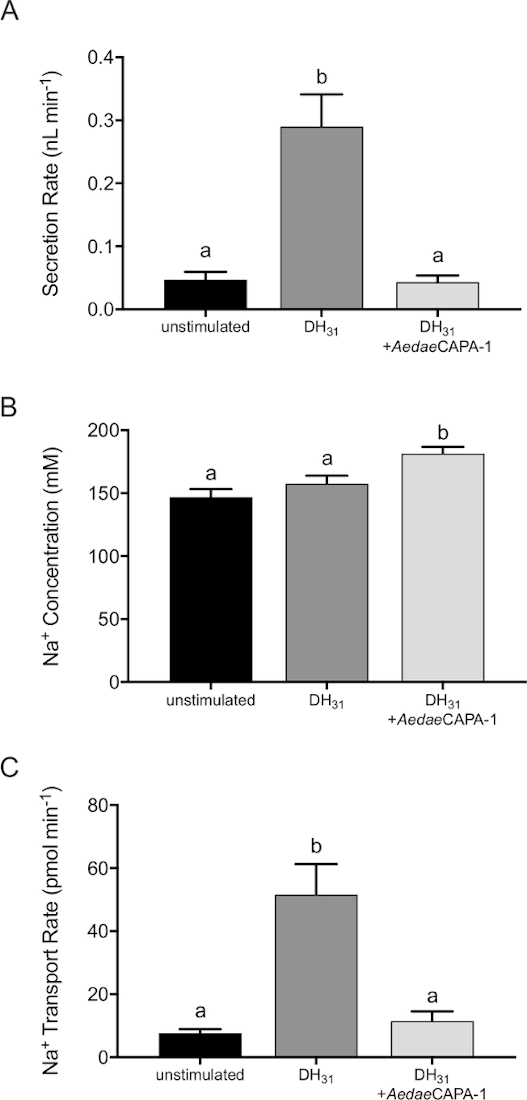
Figure 1: Effect of diuretic and anti-diuretic neuropeptides on in vitro fluid secretion rate, Na+ concentration and transport rate by adult female A. aegypti MTs. AedaeCAPA-1 (0.1 fM) was applied to MTs stimulated with DromeDH31 (25 nM). (A) MTs were incubated for 60 min with neuropeptides and 120 min for unstimulated controls. (B) Na+ concentrations in the secreted droplet were measured using ISME and values were used to calculate transport rate (C). Columns that are significantly different from controls are denoted with the same letter, as determined by a one-way ANOVA and Bonferroni post-test (n = 23–35). Please click here to view a larger version of this figure.
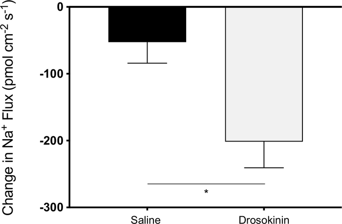
Figure 2: Changes in Na+ transport across female rectal pad epithelia measured using the SIET. Na+ absorption was recorded in unstimulated baseline saline conditions, followed by treatment with either additional saline (vehicle control) or 1 µM Drosokinin (n = 10–12). Changes in haemolymph-directed Na+ flux were measured for each treatment. There was a four-fold decrease in Na+ absorption in response to Drosokinin, as determined by an unpaired two-tailed t-test. Please click here to view a larger version of this figure.
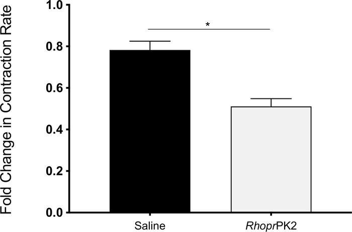
Figure 3: Change in ileal motility of female mosquitoes following saline (vehicle control) and RhoprPK2 application. The change in contraction frequency following both treatments was recorded for every sample relative to baseline conditions (n = 32). RhoprPK2 decreased ileal motility, as determined by a paired two-tailed t-test. Please click here to view a larger version of this figure.
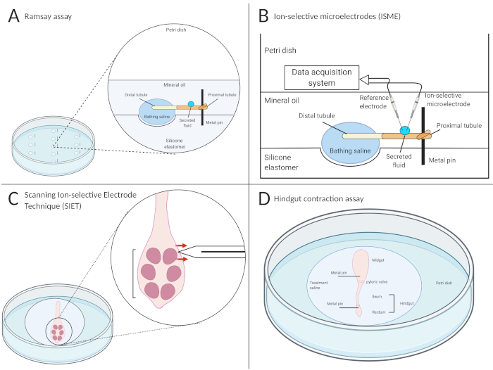
Figure 4: Schematic of the Ramsay assay, use of ion-selective microelectrodes (ISME), the scanning ion-selective electrode technique (SIET), and hindgut contraction assays. Illustrations of the four methods utilized to determine the functional roles of endocrine factors on the excretory system of the mosquito, Aedes aegypti. (A) The Ramsay assay is used to measure fluid secretion rates in isolated Malpighian tubules. The proximal (open) end of the tubule is wrapped around a pin, surrounded by mineral oil, while the distal (closed) end is bathed in the treatment saline. After a set time period, the proximal end will secrete the primary urine, accumulating as droplets on the pin. (B) A reference electrode and ISME are placed in the secreted droplet, connected to a data acquisition system, to measure ion concentration. (C) The SIET measures ion transport using an ion-selective microelectrode perpendicularly placed ~2 µm away from the organ adhered to the bottom of a poly-L-lysine dish. The direction of the red arrows in this schematic indicate that Na+ is being absorbed into the saline, indicated by a greater voltage recording along the epithelium relative to the region away from organ surface. (D) Hindgut contraction assays are conducted using video recordings to assess changes in contraction frequency, length, and relaxation duration. In this schematic, the organs are placed inside a well, and a metal pin is inserted directly into the midgut and rectum to allow the ileum to freely float and contract. Treatments are added into the well to assess changes in motility both qualitatively and quantitatively. Created using BioRender. Please click here to view a larger version of this figure.
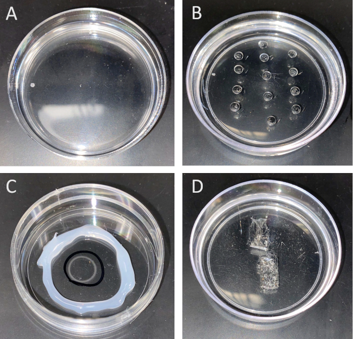
Figure 5: Dishes used for (A) dissecting mosquitoes, (B) Ramsay assay, (C) SIET and (D) contraction assay measurements. A, B and D are completely coated with silicone, whereas C is coated with poly-L-lysine in the center. To prepare the dish used for the Ramsay assay (B), a small silicone-coated Petri dish is utilized, and small ~4–5 mm wells are carved 1 cm apart (around ~20 wells can be carved/dish). Minutien metal pins are placed beside each well to hold onto the isolated tubule to measure fluid secretion rate. The poly-L-lysine dish (C) allows the organ to adhere to the bottom during SIET measurements. The wells in the contraction assay dish (D) are filled with saline and any following treatments. The contracting organ placed inside the well is observed using a video camera. Please click here to view a larger version of this figure.
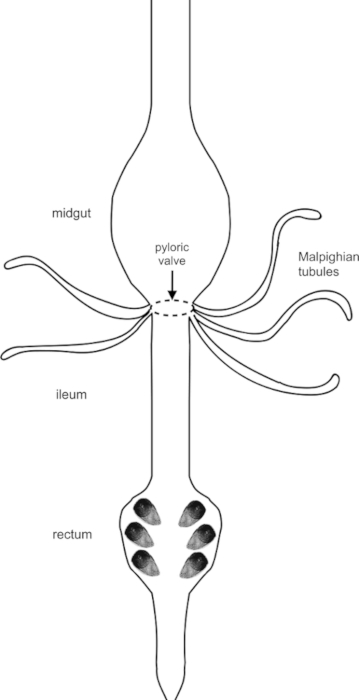
Figure 6: The excretory system of mosquito, Aedes aegypti. After a bloodmeal, solutes, water, and nutrients are passed along the alimentary canal, which includes (but is not limited to) the midgut, pyloric valve, MTs, and hindgut. The midgut is the main site for food digestion and nutrient absorption. The five MTs transport water, solutes, and waste from the surrounding haemolymph and secrete this primary urine into the hindgut, made up of an anterior ileum and posterior rectum, the latter of which includes four (male) or six (female) rectal pads. The hindgut acts as the final reabsorption site before waste is excreted. Please click here to view a larger version of this figure.
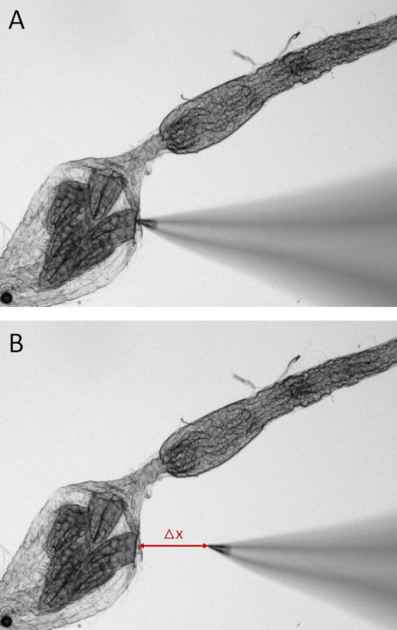
Figure 7: Still images of a dissected mosquito hindgut during SIET measurements. To measure ion activity along the epithelia of one of the six rectal pads, the Na+-selective microelectrode is placed 2 µm away from the tissue (A). Voltage is recorded at this site before the microelectrode tip is moved at an excursion distance (Δx) of 100 µm away from this point (B). The voltage gradient between these two points is used to calculate the concentration gradient and ion flux. Please click here to view a larger version of this figure.
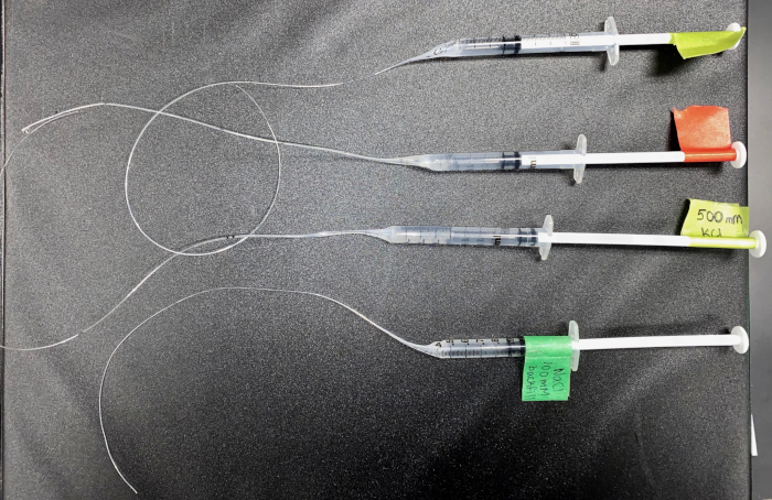
Figure 8: The backfill syringes for ISME and SIET. A picture of the syringes used to backfill NaCl and KCl solutions into the microelectrodes. This shows four 1 mL slip-tip syringes with disposable needles, with a modified stretched tip to allow for insertion into an electrode. Please click here to view a larger version of this figure.
2X Aedes aegypti saline | ||
For 500 mL: | ||
In a beaker, add 400 mL of ddH2O, then add the following solutes: | ||
Component | Weight (g) | Final concentration (mM) |
NaCl | 8.766 | 150 |
HEPES | 5.957 | 25 |
KCl | 0.253 | 3.4 |
NaOH | 0.3 | 7.5 |
NaHCO3 | 0.151 | 1.8 |
MgSO4 | 0.12 | 1 |
CaCl2-2H20 | 0.249 | 1.7 |
Stir, and adjust pH to 7.1 | ||
Add ddH2O to final volume of 500 mL to make 2x Aedes saline | ||
10X glucose | ||
For 500 mL: | ||
In a beaker, add 450 mL of ddH2O, then add: | ||
Component | Weight (g) | Final concentration (mM) |
Glucose | 4.5 | 5 |
Stir and add ddH2O to final volume of 500 mL | ||
To make 1X Aedes aegypti saline: | ||
For 100 mL: | ||
In a beaker, add 40 mL of ddH2O, then add: | ||
Component | Volume (mL) | |
2x Aedes saline | 10 | |
Stir, adjust pH to 7.1 | ||
Filter sterilize |
Table 1: Making Aedes aegypti saline. To make Aedes saline, separately prepare 2X Aedes saline and 10x glucose, store in 4 °C fridge. Use these two solutions to prepare working stocks (1x Aedes saline), used for dissecting tissues, Ramsay assays, and contraction assays.
Calcium-free Aedes aegypti Saline + NMDG | ||
For 500 mL: | ||
In a beaker, add 400 mL of ddH2O, then add the following solutes: | ||
Component | Weight (g) | Final concentration (mM) |
NaCl | 1.17 | 20 |
NMDG | 25.38 | 130 |
HEPES | 5.957 | 25 |
KCl | 0.253 | 3.4 |
NaHCO3 | 0.151 | 1.8 |
MgSO4 | 0.12 | 1 |
Glucose | 0.9 | 5 |
Stir, and adjust pH to 7.1 | ||
Add ddH2O to final volume of 500 mL |
Table 2: Modified Aedes aegypti saline used for SIET measurements. This Ca2+-free saline is used to prevent spontaneous hindgut contractions during SIET measurements. This saline is specific for measuring Na+ transport, as it consists of reduced Na+ (20 mM) made up by equimolar substitution with N-methyl-D-glucamine (NMDG) to reduce background noise.
DH31-stimulated MTs | Droplet diameter (units) | Droplet diameter (um) | Droplet volume (um3) | Secretion Rate (nl/min) |
n | Treatment #1 (60 min) | Treatment #1 (60 min) | Treatment #1 (60 min) | Treatment #1 (60 min) |
1 | 16 | 313.73 | 16167692.56 | 0.27 |
2 | 14 | 274.51 | 10831090.91 | 0.18 |
3 | 17 | 333.33 | 19392547.25 | 0.32 |
4 | 13 | 254.90 | 8671977.67 | 0.14 |
5 | 14 | 274.51 | 10831090.91 | 0.18 |
6 | 22 | 431.37 | 42029685.15 | 0.70 |
7 | 15 | 294.12 | 13321768.16 | 0.22 |
8 | 16 | 313.73 | 16167692.56 | 0.27 |
9 | 20 | 392.16 | 31577524.53 | 0.53 |
Mean | 16.33333333 | 320.26 | 18776785.52 | 0.31 |
STD ERR | 0.06 |
Table 3: Sample Ramsay assay data. Table showing sample data collected from DH31-stimulated MTs from adult female mosquitoes. When measuring the diameter of the secreted droplet, first measure with the stereomicroscope ocular micrometer and note down the value. Next, multiply the ocular unit diameter with the calibration conversion conducted before the experiment to get the diameter of the droplet in µm. Calculate the volume of the equation next, using the equation noted in step 7.7, followed by the secretion rate, using the equation noted in step 7.8.
When ingesting a blood meal, haematophagus insects face the challenge of excess solutes and water in their haemolymph2. To cope with this, they have a specialized excretory system, which is tightly controlled by hormonal factors, allowing the insects to rapidly initiate post-prandial diuresis. The Ramsay assay and use of ion-selective microelectrodes allows for measurement of fluid secretion rates along with ion concentrations and transport rates in isolated insect MTs. Critical steps within these approaches include ensuring proper dissection of the insect MTs. Individually removing each tubule from the insect must be done carefully, as any damage to the tubule will cause the fluid to be secreted from the tear or lead to a non-functional preparation. Aedes MTs do not secrete readily in unstimulated controls, thus hormones and drugs may be applied to stimulate secretion. However, even with diuretic application or careful dissections, some MTs may fail to secrete if damaged (even if not at all obvious with visual inspection), thus multiple rounds of the assay may have to be conducted. For proper use of ion-selective electrodes during both ISME and SIET measurements, ensure that the slope of the standards is within the normal range reported for the ionophore, which will give the most accurate readings. However, if the readings are unstable, examine the electrode under the microscope to confirm the ionophore has been taken up and is not leaking. A new electrode should be prepared if there are air bubbles present, the tip is broken, or the ionophore has not been taken up properly. If the ionophore/backfill border is convex, the electrodes are under-silanized and more dichlorodimethylsilane should be used. If the border is concave, the electrodes are over-silanized, and less dichlorodimethylsilane should be used. Additionally, some electrodes may be less stable than others, such as the Na+-selective electrode, whereby the slope deteriorates quicker compared to K+ or H+.
These approaches provide unlimited opportunities to studying the mechanisms behind fluid secretion by the MTs. Due to recent advances in genomic editing, reverse genetic strategies such as CRISPR-Cas9 gene-editing and RNA interference may be used to investigate the regulatory and/or functional role of specific genes expressed in excretory organs. Studies have used such tools to understand the hormonal regulation in Aedes40 and Drosophila41 MTs. Additionally, the bathing droplet may be modified to look at the specific roles of hormones14,15,18,21,42, second-messenger systems, and drugs on secretion rate and ion concentrations. These methodologies have been applied in various other insects such as Rhodnius prolixus38, larval and adult mosquito species, Drosophila41, and mayfly species (Hexagenia rigida)43. Similarly, different ions may be examined, such as Cl–, K+, H+ and NH4+.
During SIET measurements, there may be variability between every sample during baseline conditions. Differences could still arise when controlling the age, sex, and feeding state of the mosquito, particularly when examining different regions of the gut (i.e. certain distances anterior or posterior from the ileum-rectum junction). These differences can be accounted for by expressing ion flux following treatment as a change relative to baseline conditions. Over time, ion activity may also fluctuate and therefore it is important to assess changes in comparison to a vehicle control (saline), as shown in the results presented here. If voltage readings begin to fluctuate drastically, prepare a new ion-selective microelectrode and re-calibrate. Using a third calibration solution (diluted 10-fold from the lowest calibration solution concentration) can further help establish a more accurate Nernst slope. Since Na+ activity is typically highly susceptible to background noise, a modified Aedes saline consisting of less Na+ is used (Table 2). It is also important to keep a distance from the Faraday cage during measurements to avoid interference and reduce any background. Additionally, if the organ still produces spontaneous contractions when adhered to the poly-L-lysine at the bottom of the dish, adding a calcium chelator to the saline will further help to prevent this. If examining ion transport along the rectal pad epithelia, as shown in the results presented here, it is important to identify the “hotspot” site exhibiting greatest ion activity by moving the microelectrode tip in small increments along the haemolymph-facing side of the rectal pad. If unable to detect any large changes in ion activity, the organ may have been damaged or the microelectrode tip is not in the same three dimensional plane as the rectal pad opening, and thus another mosquito should be dissected.
The SIET can be used for various experiments, such as examining ion activity in mosquitoes reared in different conditions25 or fed different diets (i.e., sucrose-fed vs. blood-fed vs. unfed), which can be useful to identify ion transport mechanisms critical following a bloodmeal. It is therefore important to ensure that measurements are obtained from the same site between samples. For instance, measurements can be taken from the ileum-rectum junction or at specific distances located anteriorly from this site (along the ileum) or posteriorly (along the rectal epithelium). These measurements can also be obtained along the rectal pad epithelium that requires different positioning of the organ in the dish. Additionally, it is important to note that the SIET is not restricted to measuring ion activity solely along the hindgut; other organs, including the midgut and MTs can also be examined. Similarly, K+, H+, NH4+, and Cl– flux can be measured using the SIET25,44,45 and measurements can be taken at set time intervals following a treatment44. This technique has been used on both larval and adult mosquitoes26,44, as well as various other insects, including Chironomus riparius46, Trichoplusia ni45,47 and Hexagenia rigida43. These experiments can be used together with the Ramsay assay and ISME to gain a better understanding of the role of different treatments and environmental factors on fluid secretion, ion concentration, and ion transport.
Lastly, contraction assays are useful to identify neuroendocrine factors that influence gut motility. It is crucial that the organ under study is not damaged since this assay relies on movements generated by musculature. Therefore, forceps should only be used to grab surrounding structures when transferring the organs to the assay dish. Gripping these adjacent organs with a pin inside the well also prevents them from moving and influencing movement of the isolated organ being examined. If no spontaneous contractions are observed, the tissue may have been damaged, and a new dissection is required. Other methods commonly used to examine insect gut contractile activity include the use of a force transducer32 or impedance monitor33. These methods can also be used to measure contraction rate in the mosquito hindgut; however, if the goal is to visually examine these changes through video recordings, the methods described in this protocol will be beneficial and can be used for further analysis48.
The four techniques outlined in this protocol have helped deduce the role of a number of neuropeptides in the excretory system of various insects. Using these methods in conjunction with additional techniques utilized by insect physiologists, such as quantitative PCR and immunohistochemistry, can provide a more comprehensive understanding of underlying pathways and signaling systems, which can serve as novel potential targets for vector control.
The authors have nothing to disclose.
This research was funded by Natural Sciences and Engineering Research Council of Canada (NSERC) Discovery Grants to AD and J-PP. AL and FS received NSERC CGS-M awards in support of their graduate research.
1 mL syringes | Fisher Scientific | 14955456 | |
35 mm Petri dishes | Corning Falcon (Fisher Scientific) | C351008 | |
Borosillicate glass capillary filamented tubes (OD 1 mm, ID 0.58 mm, length 100 mm) |
World Precision Instruments | 1B100F-4 | used for ISME reference electrodes |
Borosillicate glass capillary filamented tubes (OD 2 mm, ID 1.12 mm, length 102 mm) |
World Precision Instruments | 1B200F-4 | used for SIET reference electrodes |
Borosillicate glass capillary unfilamented tubes (OD 1.5 mm, ID 1.12 mm, length 100 mm) |
World Precision Instruments | TW150-4 | used for ISME and SIET electrodes |
CO2 pad | Diamed | GEN59-114 | |
Dimethyltrimethylsilylamine solution | Sigma-Aldrich | 41716 | |
Faraday cage | Custom | Can be fabricated by local machine shop | |
Ferric chloride | Sigma-Aldrich | 157740 | |
Forceps (Dumont #5) | Fine Science Tools | 91150-20 | |
Glass Petri dish | Fisher Scientific | 08-748A | |
Hydrated mineral oil | Fisher Scientific | 8042-47-5 | Specific brand is not important |
INFINITY1-2CB video camera | Luminera | INFINITY1-2CB | |
Micromanipulators (left and right handed) | World Precision Instruments | MMJL and MMJR | Specific brand is not important so long as high quality manipulator |
Mineral Oil, Light | Fisher Scientific | 0121-4 | |
Minutien pins (0.1 mm stainless steel) | Fine Science Tools | 26002-10 | |
Non-hardening modeling clay | Sargent Art | Specific brand is not important | |
Olympus light microscope (FOR SIET) | Olympus | customized system | |
Plastic Pasteur (transfer) pipette | Fisher Scientific | 13-711-7M | |
Poly-L-lysine solution (0.1 mg/mL) | Sigma-Aldrich | A-005-M | 84 kDa |
Polyvinyl chloride (PVC) | Sigma-Aldrich | 81395 | |
Scalpel Blade | Fine Science Tools | 10050-00 | |
Scalpel Handle | Fine Science Tools | 10053-09 | |
Schneider's Drosophila medium | Sigma-Aldrich | S0146 | |
SIET system | Applicable Electronics | customized system | Details available at: http://www.applicableelectronics.com/overview |
Silver wire | World Precision Instruments | AGW1010 | |
Sodium ionophore II cocktail A | Fluka | 99357 | |
Standard polystyrene Petri (culture) dishes | Fisherbrand | FB012921 | Any size would work, but 60 mm dishes are good for both dissections and assay |
Stereomicroscope with ocular micrometer | Nikon | SMZ800 | |
Sutter P-97 Flaming Brown Pipette puller | Sutter Instruments | FGPN7 | |
Sylgard 184 Silicone Elastomer Kit | Dow Chemical Company | NC9285739 | |
Tetrahydrofuran | Sigma-Aldrich | 401757 | |
VWR advanced hotplate stirrer – aluminum | VWR | 9578 | Specific brand is not important |