Here, we present optimizations to a rat lung transplantation model that serve to improve outcomes. We provide a size guide for cuffs based on body weight, a measurement strategy to ascertain the 4th intercostal space, and methods of wound closure and BAL (bronchoalveolar lavage) fluid and tissue collection.
From our experience with rat lung transplantation, we have found several areas for improvement. Information in the existing literature regarding methods for choosing appropriate cuff sizes for the pulmonary vein (PV), pulmonary artery (PA), or bronchus (Br) are varied, thus making the determination of proper cuff size during rat lung transplantation an exercise of trial and error. By standardizing the cuffing technique to use the smallest effective cuff appropriate for the size of the vessel or bronchus, one can make the transplantation procedure safer, faster, and more successful. Since diameters of the PV, PA, and Br are related to the body weight of the rat, we present a strategy to choosing an appropriate size using a weight-based guide. Since lung volume is also related to body weight, we recommend that this relationship should also be considered when choosing the proper volume of air for donor lung inflation during warm ischemia as well as for the proper volume of PBS to be instilled during bronchoalveolar lavage (BAL) fluid collection. We also describe methods for 4th intercostal space dissection, wound closure, and sample collection from both the native and transplanted lobes.
For over three decades, researchers have been modifying and improving rat lung transplantation models so that the data generated are more consistent and more reflective of the actual clinical condition. In our laboratory's time performing this model, we have determined four areas of improvement: cuffing techniques for anastomoses, identification of the recipient's 4th intercostal space, lung inflation and wound closure during the recipient's procedure, and the harvesting of samples for analysis.
Cuffing technique modifications for anastomoses can improve the entire transplantation procedure by shortening handling time of the donor lung1,2,3,4,5,6 and making the anastomosis procedure faster and technically easier for the microsurgeon. While it is critical to use the proper sized cuffs to supply the necessary blood and airflow to the transplanted lung, there is limited guidance regarding how one should choose the size of cuffs for the pulmonary vein (PV), pulmonary artery (PA), or bronchus (Br)5,7,8,9. Since the diameters of the PV, PA, and Br are related to the body weight of the donor and recipient rats, we propose that the cuff size be based on body weight. This report provides a size guide for cuffs based on a rat's body weight (180 g to over 270 g) that serves to optimize blood and air supply to the transplanted lung (Table 1).
While a newer microsurgeon can successfully and easily procure a donor lung during the donor procedure, transplanting the lung during the recipient's procedure is more complicated and is dependent on the microsurgeon's experience. Attempts to find the 4th intercostal space to access the recipient's left lung is one of the more difficult steps that holds some subjectivity and can increase the procedure time. Therefore, we introduce a simple and objective method to assist in the identification of the 4th intercostal space location by using chest measurements and the palpitations of the heart to find the correct area chest wall to dissect4,5,6,10,11,12.
We also propose an improvement to donor lung inflation, which is a potential source of injury to the organ. The donor lung is deflated until reperfusion starts. While suturing the 4th intercostal space, the donor lung is commonly inflated by increasing PEEP from 2 cmH2O to 6 cmH2O. In order to minimize lung injury from overinflation, we propose a technique where three 6-0 nylon sutures are placed around the 4th rib inferior to the 5th rib with simple double knots. When it is time for wound closure, the ends of the three sutures are held with hemostats in both hands, the wound is closed all at once by pulling up on each side, and PEEP is immediately reduced to 2 cmH2O. In this way, the lung is allowed to expand for the shortest time possible10.
At the conclusion of an experiment, the researcher often wants to collect many types of samples for many types of analysis from each transplant. For example, snap frozen tissue, formalin fixed tissue, tissue for wet-to-dry weight ratio to determine pulmonary edema, and bronchoalvelolar lavage (BAL) fluid can all be used to assess how well the transplant went. The traditional method of collecting BAL fluid allows for a mixed pooled sample from both the recipient's native lobes and the donor's transplanted lobe13,14,15. To overcome this, we present a method of clamping the hilar areas that can yield more precise insight into the transplanted and native lungs' condition. Additionally, the volume of PBS used to collect BAL fluid from each side of the lungs is important to consider because BAL fluid contains numerous soluble factors such as cytokines and chemokines that are measured by concentration. Normalizing the volume of the fluid instilled to the estimated volume of lung capacity can help with comparison. With four lobes on the right side and one lobe on the left side, each of the rat's five lobes has a different volume and surface area16. According to a previous study on volume measurement of lung lobes by Backer et al., of the total volume of the whole lung the volume of the right lobes is 63% (4400 mm3) and the left lobe is 37% (2500 mm3). Therefore, we recommend that the volume of PBS used to collect BAL fluid should be calculated as twice the tidal volume (7.2 mL/kg) multiplied by 63% for the right lung and 37% for the left lung. By using this approach, one can better control for variables like body weight and timing10,16.
In all, in this report we will demonstrate a few modifications to the standard experimental model of rat lung transplantation that can make the procedure more efficient and increase the capability of generating more accurate and plentiful data from each experiment.
Male Sprague-Dawley rats (180-270 g body weight) were purchased commercially (e.g., Envigo) and were housed under pathogen-free conditions at The Ohio State University Animal Facility. All procedures were humanely performed according to the NIH and the National Research Council's Guide for the Humane Care and Use of Laboratory Animals and with the approval of The Ohio State University Institutional Animal Care and Use Committee (IACUC Protocol # 2012A00000135-R2).
1. Initial setup
2. Donor rat preparation
3. Donor lung warm ischemia and procurement
4. Recipient rat preparation
5. Anastomoses
6. Reperfusion
7. Collection of experimental specimens (plasma, lung tissue)
In order to measure pulmonary edema, the wet-to-dry weight ratio was calculated. The donor's native lobe, the transplanted lobe, and the recipient's native lobe were collected as described in the protocol and weighed immediately for wet weight, dried at 60 °C for 48 h, and then weighed again for the dry weight. An increased wet-to-dry weight ratio would be indicative of pulmonary edema. Our results indicate that the transplanted lobe did have a significant increase in wet-to-dry weight ratio compared to the donor's or recipient's native lobe (p=0.0050, n=6/group; Figure 11).
Angio-catheter size for cuffs | |||
Rat Body Weight (g) | PA | Br | PV |
180-200 | 20 G | 18 G | 16 G |
200-230 | 18 G | 16 G | 14 G |
230-250 | 18 G | 14 G | 14 G |
250-270 | 18 G | 14 G | 12 – 14 G |
Over 270 | 16 G | 14 G | 12 G |
Table 1. Size Guide for Cuffs. The size of the pulmonary artery (PA), bronchus (Br), or pulmonary vein (PV) is related to body weight. Depending on the body weight and which type of cuff you are making, the recommended angio-catheter size is given.
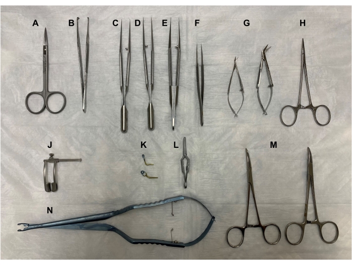
Figure 1. Surgical tools. (A) Fine scissors, (B) forceps, (C-E) microsurgical forceps, (F) Dumont #5 fine forceps, (G) Vannas-Tubingen spring scissors and Castroviejo micro dissecting scissors, (H) Halsted-mosquito hemostat, (J) retractor, (K) Yasargil clamps, (L) Dieffenbach bulldog clamp, (M) curved hemostats, and (N) Yasargil clamp applicator. All tools should be autoclaved at 121 °C for 30 min. Please click here to view a larger version of this figure.
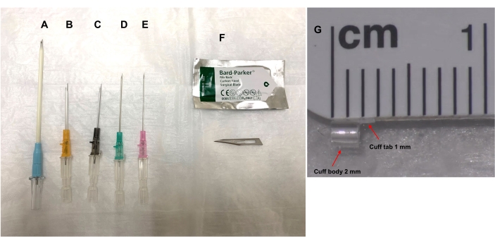
Figure 2. Cuff preparation with various sizes of angio-catheter and rib-back surgical blade #11. The size of the angio-catheter chosen for the cuff is determined by the cuff size guide (Table 1) that takes into account the rat's body weight and whether the cuff is for the pulmonary artery (PA), bronchus (Br), or pulmonary vein (PV). The angio-catheters (A) 20 G, (B) 18 G, (C) 16 G, (D) 14 G, or (E) 12 G are cut with a (F) rib-back surgical blade #11 as described in the protocol, and stored in saline until needed. (G) The cuff body length is 2 mm and a 1 mm X 1 mm tab (width x height) is left at the top of the cuff body for handling of the cuff body. Please click here to view a larger version of this figure.
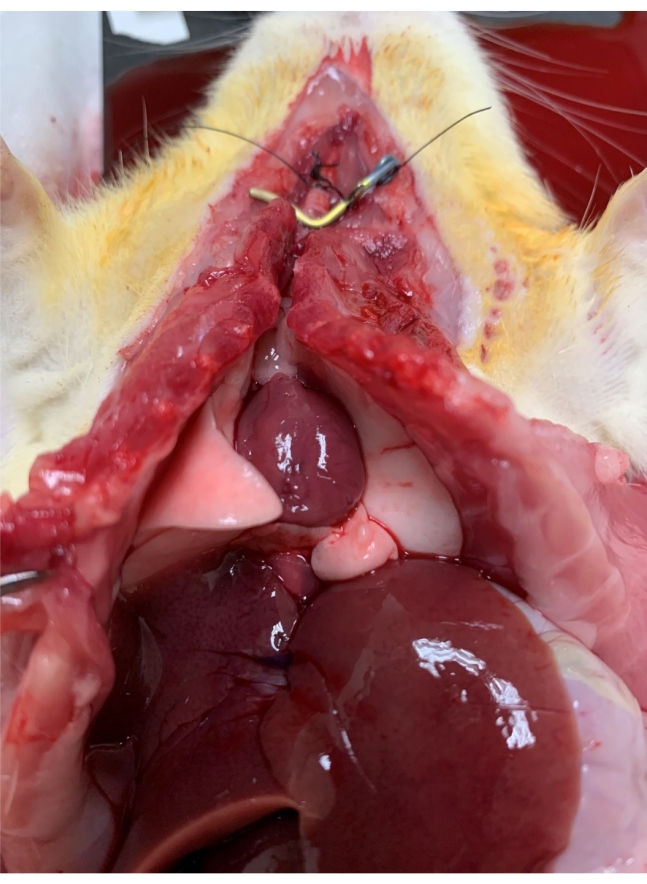
Figure 3. Warm ischemia. Lungs are flushed with the preservation solution through the pulmonary artery, inflated with twice the tidal volume of air, and then wrapped with an under pad and kept on the surgery warming board to keep the rat at normal body temperature for 1 h. Please click here to view a larger version of this figure.
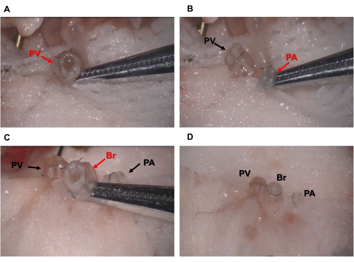
Figure 4. Cuffing of donor lung PV, PA, and Br. (A) Pulmonary vein, PV (B) pulmonary artery, PA, or (C) bronchus, Br, is inserted through a properly sized cuff, everted, secured with 8-0 nylon suture, and (D) then stored on sterile gauze dampened with ice-cold saline on a sterile Petri dish on ice. Please click here to view a larger version of this figure.
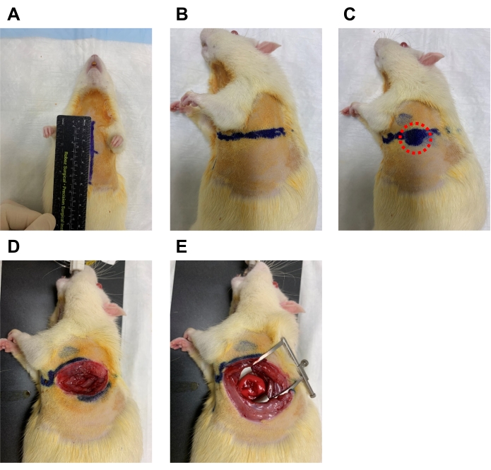
Figure 5. Measuring and dissecting at the 4th intercostal space. (A) The recipient rat is laid supine, and the chest is measured from the suprasternal notch to the xiphoid process and a line is drawn. (B) At the midpoint of this line, another line to the left side is drawn at half the length. (C) Along this line, the microsurgeon should feel for an area where the cardiac impulse is strongest to ensure the proper location of the 4th intercostal space (red circle). (D) The skin and muscle are then dissected with fine scissors. (E) The retractor is then used to open the space widely. Please click here to view a larger version of this figure.
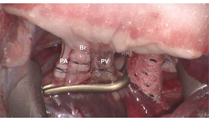
Figure 6. Anastomosis. The donor's cuffed (A) PV (B) Br, or (C) PA is inserted into the recipient's PV, Br, or PA, and then secured with 7-0 nylon suture. Please click here to view a larger version of this figure.
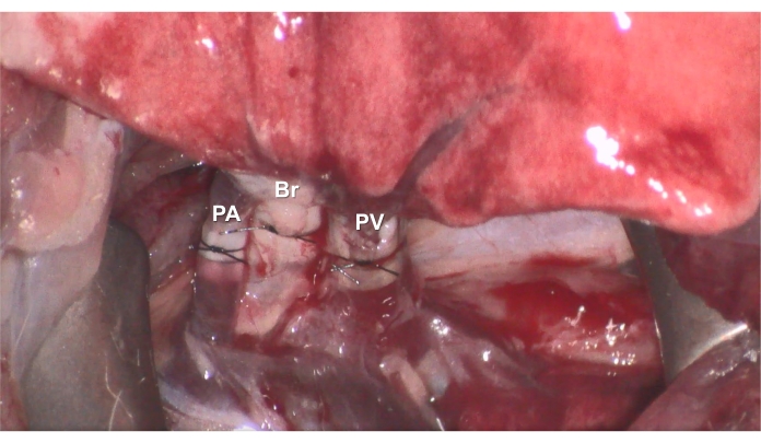
Figure 7. Reperfusion. After the anastomoses are complete, reperfusion can be started by removing the clamp, and the recipient rat is survived for 3 h under ventilation and anesthesia. Please click here to view a larger version of this figure.
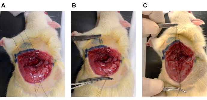
Figure 8. Wound closure. (A) Three 6-0 nylon sutures with simple double knots are placed around the ribs superior to the 4th rib and inferior to the 5th rib. (B) Use hemostats in both hands to gather the three sutures together and increase the PEEP to 6 cmH2O in the ventilation settings. (C) Tie together all three knots at the same time by pulling up and away to close the wound, decrease PEEP to 2 cmH2O immediately, and close skin with 6-0 nylon suture. Please click here to view a larger version of this figure.
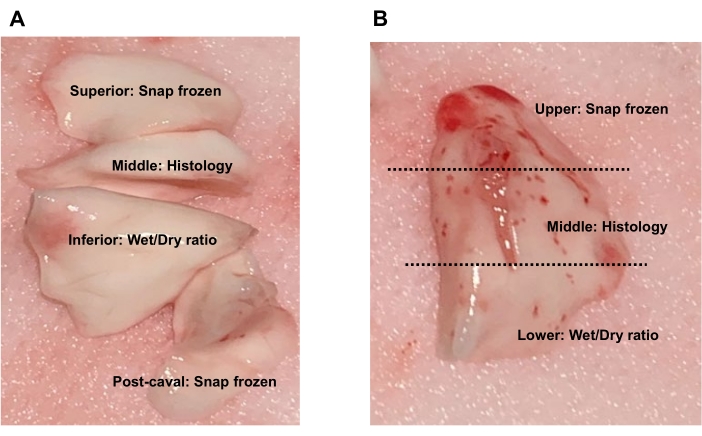
Figure 9. Lung tissue collection. (A) For the donor's or recipient's native lobes, the superior lobe and post-caval lobe can be snap frozen for protein or RNA expression analyses, the middle lobe can be preserved for histology, and the inferior lobe can be used for wet-to-dry weight ratio. (B) For the transplanted lobe, collect the upper region for snap frozen, middle region for histology, or lower region for wet-to-dry weight ratio. Please click here to view a larger version of this figure.
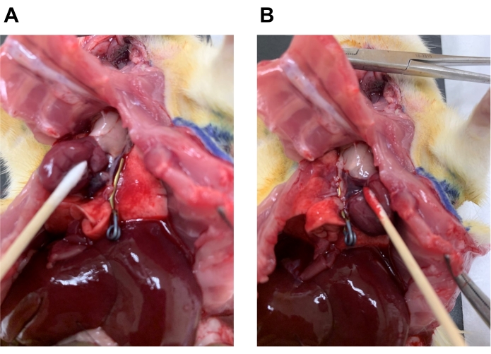
Figure 10. Application of the clamp for selective BAL fluid collection. To avoid a pooled sample, BAL fluid can be collected from either the right (native) or left (transplanted) lung. (A) The left lung hilar area can be clamped to collect BAL fluid from the right lobes. (B) The right lung hilar area can be clamped to collect BAL fluid from the left lobe. Please click here to view a larger version of this figure.
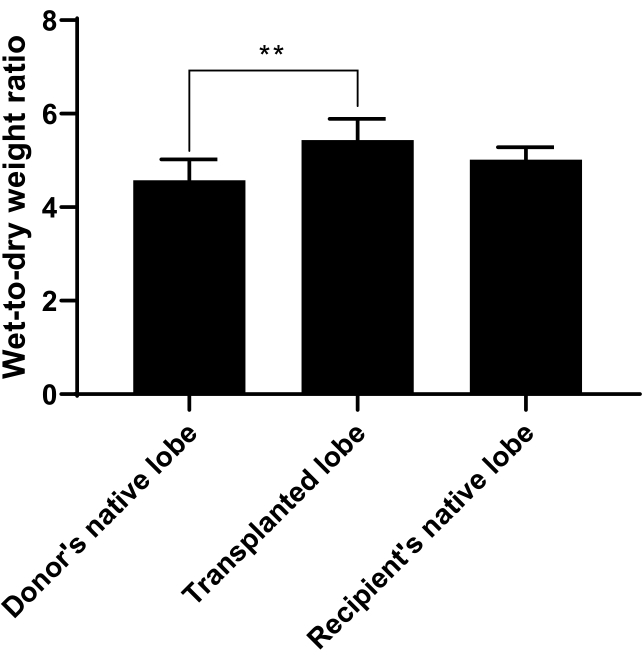
Figure 11. Wet-to-Dry Weight Ratio. Wet-to-dry weight ratio was calculated to measure pulmonary edema and can be used to indicate how well the transplant went. The donor's native lobe, the transplanted lobe, or recipient's native lobe was collected as described in the protocol and weighed immediately for wet weight, dried at 60 °C for 48 h, and then weighed again for the dry weight. A ratio of wet weight to dry weight was taken. The ratio for the transplanted lobe was significantly increased compared to the donor or recipient's native lobes. n=6 rats/group and bars represent mean ± SD. Statistical analysis was performed by using ANOVA with Tukey's post-hoc analysis. ** p<0.01. Please click here to view a larger version of this figure.
In this report, we have intervened at several critical steps in a rat lung transplantation protocol to optimize the procedure. While various cuffing techniques for rat lung transplantation has been reported1,2,3,4,5,6,7,8,9,15, the process can still be subjective and hard for microsurgeons to apply. We have emphasized that the proper size of cuffs for PV and PA to provide blood or Br to provide air into the transplanted lung should be used, and we have provided a more objective way to determine optimal cuff size based on the rat's body weight. To more consistently induce warm ischemia of the lungs, we have given recommendations of lung inflation, clamping, and how to keep the rat warm during the 1 h warm ischemia time. During the recipient's procedure, it can be difficult to locate the 4th intercostal space. We have recommended that one can more accurately locate this position by employing a method of measurement and feeling for cardiac impulse. At the time of reperfusion, to better close the wound, we also showed a technique of handling sutures and adjusting PEEP that can more quickly close the wound and prevent overinflation and injury of the lung. Finally, we have presented strategies for tissue and BAL fluid collection that allow for a more objective harvesting of samples that can be compared across experiments and different body weights.
The most significant limitation of the described techniques is the rather steep learning curve of the transplant operation in general. The learning curve is one that can be reduced with consistent surgical practice and from reviewing troubleshooting techniques in the literature. Another limitation is that this model studies the acute phase of IRI and transplantation where biochemical changes are first occurring, which is the focus of our laboratory. Future studies should test bronchial and vascular patency at longer timelines post-transplantation.
Overall, a viable small animal transplant model is critical for evaluating therapeutic interventions for transplantation and ischemia-reperfusion injury (IRI). The rat lung transplantation model, in particular, is useful as an adjunct to studying small animal ex vivo lung perfusion (EVLP)17. It is also a more viable alternative to murine lung transplantation, because the larger anatomic size allows the researcher to obtain sufficient tissue for more in-depth analyses18. Additionally, the model is essential for determining the viability of donor lung allografts after therapeutic intervention with novel small molecule and protein therapeutics19,20,21,22 either to the donor, to the donor organ through EVLP17, or to the recipient and provides a powerful avenue to gather in vivo data.The optimizations that we describe in this report are important for microsurgeons who aim to lessen their learning curve and to remove some subjectivity. By utilizing a standardized lung cuffing size algorithm, the surgical approach can be streamlined and more objective. At the conclusion of the transplant reperfusion period, the described structured approach to BAL fluid and tissue collection also provides responsible use of animals and efficient use of the microsurgeon's time by maximizing the impact of data collected per experiment.
The authors have nothing to disclose.
None.
12 Gauge angio-catheter | BD | 382277 | |
14 Gauge angio-catheter | B. Braun | 4251717-02 | |
16 Gauge angio-catheter | B. Braun | 4252586-02 | |
18 Gauge angio-catheter | B. Braun | 4251679-02 | |
20 Gauge angio-catheter | B. Braun | 4252527-02 | |
4-0 silk suture | Surgical Specialties Corp. | SP116 | |
6-0 nylon suture | AD Surgical | S-N618R13 | |
7-0 nylon suture | AD Surgical | S-N718SP13 | |
8-0 nylon suture | AD Surgical | XXS-N807T6 | |
Betadine Spray | Avrio Health L.P | UPC 367618160039 | |
Clippers | VWR | MSPP-023326 | |
Castroviejo micro dissecting spring scissors | Roboz Surgical Instrument Co | RS-5668 | |
Dumont #5 – Fine Forceps | Fine Science Tools | 11254-20 | |
Electrocautery | Macan | MV-7A | |
Endotracheal intubation kit | Kent Scientific | ETI-MSE | |
Forceps | Fine Science Tools | 11027-12 | |
Halsted-mosquito hemostat | Roboz Surgical Instrument Co | RS-7112 | |
Heparin | Fresnius Medical Care | C504701 | |
Insulin syringe | Life Technologies | B328446 | |
Isoflurane | Piramal Critical Care | NDC 66794-017-25 | |
Isopropyl Alcohol Swabs | BD | 326895 | |
Ketamine | Hikma Pharmaceuticals PLC | NDC 0413-9505-10 | |
Dieffenbach Bulldog Clamp | World Precision Instruments | WPI14117 | |
Needle holder/Forceps, Curved | Micrins | MI1542 | |
Needle holder/Forceps, Straight | Micrins | MI1540 | |
Perfadex Plus (Organ Preservation Solution) | XVIVO Perfusion AB | REF# 19950 | |
PhysioSuite | Kent Scientific | PS-MSTAT-RT | Used to check SpO2 and heartbeat |
Retractor | Roboz Surgical Instrument Co | RS-6560 | |
Saline | PP Pharmaceuticals LLC | NDC 63323-186-10 | |
Scissors | Fine Science Tools | 14090-11 | |
SomnoSuite Small Animal Anesthesia System | Kent Scientific | SS-MVG-Module | |
Sterile Cotton Gauze Pad | Fisherbrand | 22-415-469 | |
Surgical Microscope | Leica | M500-N w/ OHS | |
Syringe 5mL | BD | 309646 | |
Vannas-Tubingen Spring Scissors | Fine Science Tools | 15008-08 | |
Xylazine | Korn Pharmaceuticals Corp | NDC 59399-110-20 | |
Yasargil Clamp | Aesculap, Inc | FT351T | Used to clamp bronchus |
Yasargil Clamp | Aesculap, Inc | FT261T | Used to clamp hilum |
Yasargil Clamp Applicator | Aesculap, Inc | FT484T |