This protocol is intended to be a tool to study steatosis and the molecular, biochemical, cellular changes produced by the over exposure of hepatocytes to lipids in vitro.
Metabolic dysfunction-associated fatty liver disease (MAFLD), previously known as non-alcoholic fatty liver disease (NAFLD), is the most prevalent liver disease worldwide due to its relationship with obesity, diabetes type 2, and dyslipidemia. Hepatic steatosis, the accumulation of lipid droplets in the liver parenchyma, is a key feature of the disease preceding the inflammation observed in steatohepatitis, fibrosis, and end-stage liver disease. Lipid accumulation in hepatocytes might interfere with proper metabolism of xenobiotics and endogenous molecules, as well as to induce cellular processes leading to the advance of the disease. Although the experimental study of steatosis can be performed in vivo, in vitro approaches to the study of steatosis are complementary tools with different advantages. Hepatocyte culture in lipid overload-conditioned medium is an excellent reproducible option for the study of hepatic steatosis allowing the identification of cellular processes related to lipid accumulation, such as oxidative and reticular stresses, autophagia, proliferation, cell death, etcetera, as well as other testing including drug effectiveness, and toxicological testing, among many other possible applications. Here, it was aimed to describe the methodology of hepatocyte cell culture in lipid overload-conditioned medium. HepG2 cells were cultured in RMPI 1640 medium conditioned with sodium palmitate and sodium oleate. Importantly, the ratio of these two lipids is crucial to favor lipid droplet accumulation, while maintaining cell proliferation and a moderate mortality rate, as occurs in the liver during the disease. The methodology, from the preparation of the lipid solution stocks, mixture, addition to the medium, and hepatocyte culture is shown. With this approach, it is possible to identify lipid droplets in the hepatocytes that are readily observable by Oil-red O staining, as well as curves of proliferation/mortality rates.
Fatty liver associated with metabolic dysfunction is highly prevalent worldwide1,2; it is estimated that up to 25% of the population is affected3. This disease previously known as non-alcoholic fatty liver disease (NAFLD), has updated its nomenclature to metabolic dysfunction associated fatty liver disease (MAFLD) to accurately reflect the pathogenesis related with obesity, insulin resistance, diabetes type 2, and dyslipidemia, as well as the possible managements of the disease3,4.
Regardless of the name, the disease includes a wide spectrum of histopathological changes characterized by abnormally high accumulation of lipids in the liver (>5% of fat in the hepatocytes5) and might progress through the lipid accumulation typically found in simple steatosis to steatohepatitis, which in turn might lead to the development of fibrosis, cirrhosis, hepatocellular carcinoma, and liver failure5,6,7,8. Due to its increasing prevalence, MAFLD is expected to become the first indication of liver transplantation and the leading cause of hepatocellular carcinoma9.
Although it has been considered as a benign or mild form of fatty liver disease, hepatic steatosis is in fact the metabolic key in MAFLD10. Different metabolic pathways are affected by lipid accumulation in the liver, including but not limited to lipid synthesis, exportation, and metabolism10. Insulin resistance, oxidative stress, reticular stress, and cellular dysfunction are strongly associated to hepatic lipotoxicity11,12. On the other hand, fatty hepatocytes are the target of reactive oxygen species, rendering metabolites as lipid peroxides, protein carbonyls, and adducts of nucleic acids13. At the cellular level, fatty hepatocytes might undergo mitochondrial damage14, cellular senescence15, apoptosis16, pyroptosis12, and autophagia17, among other events.
Hepatocytes are highly responsible for metabolism, detoxification, and synthesis of a wide range of molecules. Many of these functions might be compromised by the lipid accumulation observed in steatosis. Therefore, it is of great importance to have reproducible tools that allow an accurate evaluation of steatosis. In this sense, in vitro models are readily applicable and highly reproducible. Steatosis in vitro has been used with different goals16,18,19. The HepG2 cells are widely used as hepatocyte cell line. It has advantages such as being easy to culture and well characterized. Perhaps, the only disadvantage of HepG2 cells is the fact that it is a carcinogenic cell line, so this must be considered when analyzing the outcomes. Here, the application of a mixture of fatty acids widely used in cell culture: palmitic acid (PA) and oleic acid (OA) is shown. Both PA and OA offer different outcomes in culture20. PA (C 16:0) is the most common saturated fatty acid obtained from the diet16. PA is considered as a biomarker of de-novo lipogenesis, a crucial step in the development of NAFLD21. PA is shown to be highly toxic22; therefore, it might not be recommended to induce steatosis in vitro. OA (C 18:1) is a monounsaturated fatty acid. In contrast to PA, OA has been suggested to possess anti-inflammatory and anti-oxidant properties, being able to counteract PA12. Both PA and OA are the main fatty acids present in the triglycerides, regardless of the condition of health or disease16. Table 1 provides examples of the hepatocyte culture with PA, OA, and their mixture, as well as the outcomes reported12,23,24,25,26,27. Other fatty acids have also been used in hepatocyte culture, including stearic acid (C 18:0)28,29,30, linoleic acid (C 18:1)28,30,31 and its conjugates (CLA)28,32, palmitoleic acid (C 16:1)29. However, their use is least frequently reported in the literature, perhaps because their hepatic abundance is lower than PA and OA16.
In conjunction, both fatty acids resemble steatosis in vitro, providing proliferating cells, with increased cell death and lower viability compared with control conditions. It is worth mentioning that the respective salts of these fatty acids are available and can be used as well. One of the main problems when assessing lipid overload in hepatocyte cell culture is given in the differentiation between toxicological models and a model that best represent steatosis. Many models can be accounted in the first case. In fact, the use of PA alone might be considered among them, and the high mortality is the most evident outcome12,16,23,24,25,26,27. The use of high doses even in the case of OA can also be considered as a toxicologic model. The protocol shown here is in higher accordance with steatosis development since it shows low mortality compared with that observed in other models and allows it to be followed during several days with progressive lipid accumulation as it occurs in NAFLD. The possibility to assess mild and severe steatosis through experimental conditions is considered another advantage.
Fatty acids | Conditions | Outcomes | Reference | ||
PA | Concentration: 200 μM | Lipid accumulation | Yan et al, 201925. | ||
Time exposure: 24 h | Hepatocyte damage | ||||
Transaminases elevation | |||||
PA | Concentration: 50, 100 and 200 μM | Lipid accumulation | Xing et al, 201924. | ||
Time exposure: 24 h | |||||
PA | Concentration: 250 μM , 500 μM , 750 μM and 1,000 μM | Lipid accumulation | Wang et al, 202026. | ||
Time exposure: 24 h | Progressive reduction of cell viability | ||||
Mix of OA/PA | Concentration: 1 mM | Lipid accumulation | Xiao et al, 202027. | ||
Time exposure: 24 h | Does not report lipotoxicity | ||||
Rate: 2OA:1PA | |||||
Mix of OA/PA | First stimulation with 200 μM and 400 μM of PA and then second stimulation with 200 μM of OA | Lipid accumulation. | Zeng et al, 202012. | ||
Concentration:400 μM PA: 200 μM OA | Evidence of lipotoxicity induced by PA was reduced by stimulation of OA. | ||||
Rate: 2PA:1OA | |||||
Time exposure: 24 h | |||||
Mix of OA/PA | Concentration: 400 μM PA: 200 μM OA | Lipid accumulation | Chen et al, 201823. | ||
Rate: 2PA:1OA | |||||
Time exposure: 24 h | |||||
Mix of OA/PA | Concentration :50 and 500 μM | Generation of two types of steatosis: mild steatosis and severe steatosis. |
Campos and Guzmán 2021 | ||
Rate: 2PA:1OA | Simulates chronic exposition of lipid overload | ||||
Time exposure: 24 h, 2 days,3 days and 4 days. |
Table 1. Hepatocyte culture in steatogenic conditions. The table presents the type of fatty acid used, the conditions maintained, and the observed outcomes in hepatocyte culture. PA: Palmitic acid. OA: Oleic acid.
Finally, this model is applicable not only to the study of steatosis and fatty liver, but also to the hepatic metabolic, synthetic, and detoxification pathways in the context of steatosis. Also, in vitro induced steatosis might provide evidence for the identification of potential markers of the disease as well as therapeutic targets.
1. Standard and conditioned medium preparation
2. Pre-culture
3. Steatogenic culture
4. Viability and mortality assessment
5. Lipid staining with Oil-Red O
6. Morphometric assessment of lipid contents
Hepatocytes cultured in the steatogenic medium display growth all over the surface of the well; however, fatty hepatocytes show lower growth rate compared with cells cultured in control medium. The proposed ratio and concentration of OA and PA, guarantee cell survival during culture. Seeding 1 x 10⁵ cells per well in 24-well plates provides optimum confluence as shown in Figure 1.
Viability in cultured cells was lower in the steatogenic groups, Mild and Severe, compared with the control conditions. In fact, viability progressively diminished as time of the culture increased, reaching the lowest of 60% at 4 days in severe steatosis (Figure 2A). Accordingly, the mortality rate was higher in hepatocytes cultured in the steatogenic conditions, and it progressively increased with the time of exposure to lipids (Figure 2B). Cell numbers progressively increased as a result of proliferation (Figure 2C). However, proliferation rate was lower in Mild steatosis at 3 days and 4 days. In contrast, Severe steatosis was associated with lower proliferation from 24 h.
HepG2 cells cultured in the proposed protocol show the most important feature of steatosis, intracellular lipid accumulation. Staining cells with Oil Red O allowed to observe at least a two-fold increase of lipid droplets in cells cultured under steatogenic conditions as shown in Figure 3 and Figure 4. Intracellular fat increased according to the time of exposure of culture in the steatogenic medium (Figure 3). In Mild steatosis, lipid contents were increased from day 2, whereas in Severe steatosis, they were significantly high from 24 h.
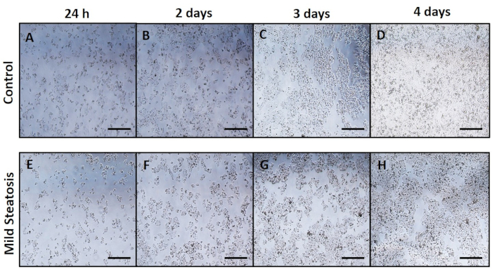
Figure 1: Cell growth. HepG2 hepatocytes culture in control (Figure 1A–D) and mild steatogenic conditions (Figure 1E–H). Photographs show growth from 1-4 days of culture. Scale bar = 500 µm. Please click here to view a larger version of this figure.
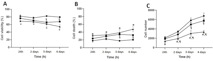
Figure 2: Viability and mortality rates. (A) Viability. (B) Mortality. (C) Cell number. HepG2 hepatocytes culture in control and steatogenic conditions were assessed for viability and mortality rates by trypan blue staining. Mean ± SD. Two independent experiments in triplicate per time of culture. Circles: control conditions; Squares: mild steatosis; Triangles: severe steatosis. One-way ANOVA was used to compare among conditions and time of culture for the same condition. p < 0.05 was considered significant: "*"- control vs severe steatosis; "§"- control vs mild steatosis; "¶"- mild vs severe steatosis. Please click here to view a larger version of this figure.
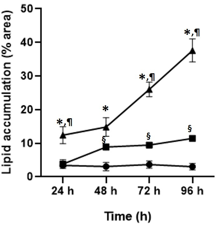
Figure 3: Lipid accumulation. HepG2 hepatocytes culture in control and steatogenic conditions were assessed for lipid contents by oil red O staining followed by a morphometric analysis using ImageJ software (NIH, USA). Percentage of lipids refers to the percentage of area stained by Oil-Red O (lipids) considering 100% as the complete area of each optical field analyzed. Mean ± SD. Two independent experiments in triplicate per time of culture. Circles: control conditions; Squares: mild steatosis; Triangles: severe steatosis. One-way ANOVA was used to compare among conditions and time of culture for the same condition. p < 0.05 was considered significant: "*"- control vs severe steatosis; "§"- control vs mild steatosis; "¶"- mild vs severe steatosis. Please click here to view a larger version of this figure.
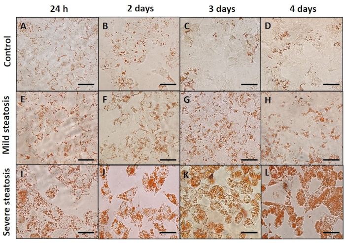
Figure 4: Steatosis in vitro. HepG2 hepatocytes culture in control (Figure 4A–D), mild steatogenic (Figure 4E–H), and severe steatogenic (Figure 4I–L) conditions were assessed for lipid contents by oil red O staining. Photographs show hepatocyte lipid droplets from 24 h to 4 days. Scale bar = 50 µm. Please click here to view a larger version of this figure.
This protocol is intended to provide a strategy to study steatosis in vitro. Cell culture is a powerful tool to study cellular, molecular, biochemical, and toxicological aspects of the cells exposed to different conditions. With this approach, steatosis can be visualized not only as a stage of the complex disease that is MAFLD, but also as the hepatocyte overexposure to lipids and the possible outcomes resulting from such exposure. Therefore, its application is not restricted to the physiopathology of MAFLD, but to the fact that patients with fatty liver are exposed to therapeutic drugs, contaminants, among other conditions that might be affected by steatosis. Thus, this protocol has potential applications in toxicology, pharmacology, and the identification of therapeutic targets for treatment of the disease.
On development of this protocol, one of the most critical steps is the preparation of the steatogenic mix: 1 part of palmitate: 2 parts of oleate, which induces steatosis from the day 2 (Figure 3, Figure 4), allowing hepatocytes to proliferate-despite a modest decrease in viability rate and increased mortality rate (Figure 2). However, decreased viability should not exceed 30%-40%, since that might represent a toxic effect rather than one that might be followed in the long-term. Hepatic steatosis is the result of a long-term overexposure to lipids. In this sense, lipids accumulate, at first, with mild affections on the hepatocytes, as observed in this model. Another feature is the lipid droplet profile. In mild steatosis, an increased size in lipid droplets is observed during the progression of the culture (Figure 4E–H). In severe steatosis, droplet size is considerably higher compared to mild steatosis (Figure 4I–L), whereas controls do not show changes in lipid droplet size (Figure 4A–D).
It is preferable to store both the mild and severe steatogenic media at 4 °C for up to a week. Afterward, it is recommended to prepare fresh steatogenic medium. However, the OA stock can be preserved at -20 °C for up to a month, whereas the PA stock can be stored at 4 °C for up to a month. Using these stock solutions after the suggested time might represent a risk of degradation of the fatty acids. To ensure the proper concentration of the solutions before every use, it is recommended to measure fatty acid concentrations by a non-esterified fatty acids (NEFA) assay kit.
PA and OA, as well as their respective salts, have been used separately to induce lipid accumulation; however, differences are observed for every fatty acid16,20. On one hand, palmitate used alone is a good inductor of steatosis. It induces cell death, hepatic insulin resistance, mitochondrial dysfunction, reticular stress16,34,35,36. However, palmitate is highly toxic16,34, and the outcomes expected of using it alone in culture include lower viability and higher mortality compared with the mixture of PA and OA16,20. On the other hand, oleate also induces steatosis. It induces de novo lipogenesis, insulin resistance37,38, and hyperproliferation38. However, the outcomes observed with oleate are often milder compared with palmitate and the mixture16,20. This might be related to its protective role. Oleate is the major component in olive oil, a key ingredient in the Mediterranean diet, one of the well-known successful strategies against MAFLD39.
This protocol might be considered as a nice tool to study steatosis due to its reproducibility and the short time it takes to obtain results. This is in comparison with using experimental models of MAFLD, adding the fact that it does not imply the ethical issues inherent to using rodent models. This protocol allows being followed for several days, provided that steatogenic medium is refreshed every 24 h. This model is also cost-effective and possesses a wide range of applications. It can also be adjusted to other cell lines, not only hepatocytes, but a wide range of cells affected by lipid overexposure during obesity. One of the limitations of this protocol is the use of HepG2 cells. Since this is a carcinogenic cell line, it might conceal or increase some outcomes. However, the application of HepG2 cells in these types of studies is widely accepted due to its resemblance in lipid metabolism to healthy hepatocytes40. The use of the mixture PA:2OA might also prove to be controversial since it does not fully resemble the profiles of NEFA observed in the blood of NAFLD/MAFLD patients41. Other fatty acids, including linolenic or stearic acids, might be included in further modifications and improvements of this protocol. Another limitation is the fact that only one cell type, hepatocytes, is studied, lacking the interaction with other hepatic cells present in the liver, including sinusoidal endothelial, Kupffer, hepatic stellate cells, etc., that are engaged in the progression of MAFLD. Moreover, this is a model to induce steatosis exclusively, with no progression to steatohepatitis and fibrosis.
In conclusion, the study provides an in vitro protocol of hepatocyte steatosis that is easy to implement, reproducible, and with a wide range of applications in the study of steatosis as well as the hepatocyte function in the context of fatty liver.
The authors have nothing to disclose.
This work was funded by Consejo Nacional de Ciencia y Tecnología (Conacyt, CB-221137). Adriana Campos is a doctoral student at Programa de Doctorado en Ciencias Biomédicas, Universidad Nacional Autónoma de México, and was supported by Conacyt (CVU: 1002502).
Biosafety cabinet | ESCO Airstream | AC2-452+C2:C26 | Class II Type A2 Biological Safety Cabinet |
Bottle top filter | Corning, US | 430513 | Non-pyrogenic, polystyrene, sterile. 1 filter/Bag. 0.22 μm, 500 mL. |
Bovine serum albimun (BSA) | Gold Biotechnology, US | A-421-10 | BSA Fatty Acid Free for cell culture |
Culture media RPMI 1640 | ThermoFisher-Gibco, US | 31800-022 | – |
Fetal Bovine Serum (FBS) | ThermoFisher-Gibco, US | A4766801 | – |
Hemocytometer | Marienfeld, DE | 640010 | – |
HepG2 cell line | ATCC, US | HB-8065 | Hepatocellular carcinoma human cells. |
Humidified incubator | Thermo Electronic Corporation,US | Model: 3110 | Temperature (37 °C ± 1 °C), humidity (90% ± 5%) , CO2 (5% ± 1%) |
Inverted microscope Eclipse | NIKON, JPN | Model: TE2000-S | – |
Isopropanol | Sigma-Aldrich, US | I9030-4L | – |
Oil Red O Kit | Abcam, US | ab150678 | Kit for histological visualization of neutral fat. |
Paraformaldehyde | Sigma-Aldrich, US | P6148-500G | – |
Penicillin/streptomycin | ThermoFisher-Gibco, US | 15140-122 | Antibiotics 10,000 U/mL Penicillin, 10,000 μg/mL Streptomycin |
pH meter | Beckman, US | Model: 360 PH/Temp/MV Meter | – |
Phosphate buffered saline | ThermoFisher-Gibco, US | 10010-023 | – |
Serological Pipettes | Sarstedt, AUS | 86.1253.001 | Non-pyrogenic, sterile, 5 mL |
Serological Pipettes | Sarstedt, AUS | 86.1254.001 | Non-pyrogenic, sterile, 10 mL |
Sodium bicarbonate | Sigma-Aldrich, US | S5761-1KG | Preparation of culture media |
Sodium oleate | Santa Cruz Biotechnology, US | sc-215879A | – |
Sodium palmitate | Santa Cruz Biotechnology, US | sc-215881 | – |
Syring filter | Corning, US | 431219 | Non-pyrogenic, sterile, 28 mm, 0.2 μm. |
Trypan Blue | Sigma-Aldrich, US | T6146-25G | – |
Trypsin 0.05% /EDTA 0.53 mM | Corning, US | 25-052-Cl | – |
24 well cell culture cluster | Corning, US | 3524 | Flat bottom with lid. Tissue culture treated. Nonpyrogenic, polystyrene, sterile. 1/Pack. |
96 well cell culture cluster | Corning, US | 3599 | Flat bottom with lid. Tissue culture treated. Nonpyrogenic, polystyrene, sterile. 1/Pack. |