1. Synthesis of pBAE polymer with end oligopeptides (OM-pBAE)
- Polymerization of C6-pBAE
- Add 5-amino-1-pentanol (38 mmol; MW = 103.16 Da) 1-hexylamine (38 mmol; MW = 101.19 Da) into a round-bottom glass flask (100 mL). Then, add 1,4-butanediol diacrylate (82 mmol; MW = 198.22 Da).
- Pre-heat the silicone oil bath at 90 °C, place the round-bottom flask into the oil bath and stir the mixture with the aid of a magnetic stir bar overnight (~18 h). Then, take the product from the round-bottom flask and place it in the freezer at -20 °C.
NOTE: The product is in the form of a sticky powder and is taken out from the flask with the help of a spatula. It is essential to validate the structure of the obtained polymer through 1H-NMR. NMR spectra were recorded in a 400 MHz instrument (see Table of Materials) using chloroform-d and D2O as solvents. Around 10 mg of each poly(β-amino ester) were taken and dissolved in 1 mL of the deuterated solvent.
- Reaction with peptides to obtain OM-pBAE
- Add 25 mL of 0.1 M HCl to selected peptides, e.g., peptide Cys-His-His-His with Trifluoroacetic acid (TFA, 200 mg), into a previously weighed 50 mL centrifuge tube that can stand freeze-drying and manually stir overnight to obtain a clear solution.
- Freeze the solution at -80 °C for 1 h and freeze-dry the resulting peptide hydrochloride.
NOTE: Check whether the final weight corresponds to the theoretical value. - Make a solution of C6-pBAE (0.031 mmol) in dimethyl sulfoxide. Also, make a solution of the peptide hydrochloride (0.078 mmol) in dimethyl sulfoxide.
- Mix the two solutions in a screw cap tube and screw on the cap. Stir the mixture solution in a water bath with a controlled temperature of 25 °C for 20 h with a magnetic stir bar.
- Add the mixture to 7:3 (v/v) diethyl ether/acetone. Centrifuge the resulting suspension at 25,000 x g at 4 °C to remove the solvent. Next, wash the solid with 7:3 (v/v) diethyl ether/acetone twice. Then, dry the product under a vacuum (<0.2 atm).
- Make a solution of 100 mg/mL of the product in dimethyl sulfoxide. The resulting product is named C6-peptide-pBAE. It is essential to validate the structure of the obtained polymer through 1H-NMR to confirm the disappearance of the olefin signals associated with terminal acrylates. If not used, the polymer can be frozen at -20 °C.
2. Polyplexes formation
NOTE: All the procedures should be performed inside a conditioned room to maintain a constant temperature.
- Thaw the polymers C6-peptide-pBAE and vortex the solution.
- Pipette the polymer mix up and down and prepare a solution of 12.5 mM (V1) in sodium acetate (NaAc). Then, vortex the mixture and wait for 10 min.
- Prepare the mRNA at 0.5 mg/mL and mix by pipetting (V2).
NOTE: It is crucial to avoid vortexing the mRNA. - Vortex the polymer mixture at the final concentration to achieve a homogeneous solution between the polymer stock in DMSO and the acetate buffer.
NOTE: The final polymer concentration depends on the N/P (nitrogen to phosphate groups) ratio selected. The N/P ratio depends on each specific mRNA to be used. For eGFP encapsulation, for example, a 25:1 ratio was used, as previously reported14. - Mix the genetic material solution and C6-peptidepBAE solution (25x of the mRNA concentration) in a ratio of 1:1 (Vi = V1 + V2).
NOTE: C6-peptide-pBAE is loaded in a microcentrifuge tube where the mRNA is added by pipetting up and down for mixing. Once prepared, the polyplex, the nucleic acid, and C6-peptide-pBAE concentrations are half diluted. - Incubate at 25 °C for 30 min in a thermoblock. Precipitate with 1:2 RNase free water by adding the sample to a pre-loaded microcentrifuge tube with water.
- Include the excipients. Add the same volume as the mixture of mRNA and pBAE (Vi) in HEPES 20 mM and sucrose 4% by pipetting up and down. At this point, the sample has been diluted 3x.
3. Polyplexes lyophilization
- Instantaneously, freeze at -80 °C freezer of the previous polyplex solution for 1 h.
- Perform the primary drying by following the steps: (1) 1 h at -60 °C and 0.001 hPa; (2) 1 h at -40 °C and 0.0001 hPa; (3) 4 h at -20 °C and 0.0001 hPa; (4) 12 h at 5 °C and 0.0001 hPa.
- Store at -20 °C immediately to avoid rehydration until use.
4. Polyplex resuspension
NOTE: This protocol describes the process used to reconstruct the lyophilized C6-peptide-pBAE nanoparticles for their further use either for characterization, in vitro, or in vivo analysis.
- Take the lyophilized nanoparticles from -20 °C just at the moment of use and rapidly add the corresponding amount of depyrogenated (DEPC) water to redisperse the solid to achieve the desired concentration.
NOTE: The volume will be the same as the initial volume of the nanoparticles if the same concentration is envisaged, but it will be indicated for each experiment. - Pipette gently until total resuspension, accompanying the liquid with the pipette.
NOTE: Assure that no material remains on the wall of the vial. - Once dissolved, pipette up and down vigorously, avoiding bubbles.
NOTE: The sample should have a transparent to translucent aspect, which is more evident at higher concentrations - Store the samples on ice or at 4 °C for a maximum period of 24 h once reconstituted. Avoid freezing.
5. Polyplex characterization
- Dynamic light scattering
NOTE: Hydrodynamic diameter (nm), polydispersity index (PDI), and surface charge of nanoparticles were measured at 25 °C, 633 nm laser wavelength, and 173° signal detector, using a Zeta potential analyzer (see Table of Materials).- Hydrodynamic diameter (nm)
- Prepare the nanoparticles mixing mRNA and pBAE thoroughly by pipetting the gene material to the polymer fraction to a final concentration of mRNA of 0.25 mg/mL as previously described above (step 2).
- Pre-rinse a microcuvette with filtered dilution media to completely clean it from impurities. Then, fill the cuvette with at least 50 µL of the sample solution and cap it. Next, introduce the sample inside the DLS instrument, ensure that the cell is correctly inserted.
NOTE: The dilution media is filtered using a 0.22 µm syringe filter. - Open the Standard Operation Procedure (SOP) file created and introduce the desired Sample Name. Select Size Measurement.
NOTE: All the parameters to create an SOP for size measurements are provided in Table 1. - Run the particle size measurement using DLS by clicking on Play.
NOTE: After the triple beep sounds, the analysis is complete. - Select the results corresponding to the sample on the measurement sheet to obtain the average particle size, average PDI, standard deviation, and graphics. Once done, remove the cell from DLS equipment.
- Remove the sample, keep it for zeta-potential analysis, and clean and rinse the cell with deionized water. Finally, dry the cleaned cuvette under a compressed air gas stream.
- Surface charge (mV)
- Prepare the nanoparticles mixing mRNA and pBAE thoroughly by pipetting the gene material to the polymer fraction to a final concentration of mRNA of 0.25 mg/mL as previously described above (step 2, Polyplex formation). Next, freeze and dry the solution.
NOTE: In this case, for the measurement of surface charge, the freeze-drying of the sample is strongly recommended before performing the measures to redisperse the sample in the appropriate buffer, simulating body conditions of pH and electrolyte concentration. - Resuspend the lyophilized sample using water. Dilute the sample 1/10 in water (final concentration 0.025 mg/mL).
- Pre-rinse a disposable folded capillary cell (zeta potential cuvette) with filtered dilution media to completely clean it from impurities. Then, fill the cuvette with diluted nanoparticles, using a 1 mL syringe, and cap both sides of the fillers.
NOTE: The nanoparticle dispersion has to fill the volume available in the cuvette (~1 mL), taking special attention to the bubble's formation, which could perturb measures. - Introduce the sample inside DLS; ensure that the cell is correctly inserted.
- Open the SOP file created for zeta potential analysis and introduce the desired Sample Name. Select Zeta-Potential Measurement.
NOTE: Parameters to create an SOP for zeta potential measurements are given in Table 2. - Run zeta-potential measurement using DLS by clicking on Play.
NOTE: After the triple beep sound, the analysis is complete. - Select the results corresponding to the sample on the measurement sheet to obtain the average zeta-potential, standard deviation, and graphics. Once done, remove the cell from the DLS equipment.
- Remove the sample and keep it if it is necessary. Next, clean and rinse the cuvette cell with deionized water, followed by ethanol and water again. Finally, dry the cleaned cuvette under a compressed air gas stream.
NOTE: To analyze the data, use the recommended software.
- Prepare the nanoparticles mixing mRNA and pBAE thoroughly by pipetting the gene material to the polymer fraction to a final concentration of mRNA of 0.25 mg/mL as previously described above (step 2, Polyplex formation). Next, freeze and dry the solution.
- Hydrodynamic diameter (nm)
- Nanoparticle Tracking Analysis (NTA)
NOTE: Hydrodynamic diameter (nm) and concentration of nanoparticles were measured at 25 °C, 488 nm laser wavelength using a Nanoparticle Tracking Analyzer (see Table of Materials).- NTA and sample preparation
- Switch on the computer and the equipment. First, check whether all the components are plugged in correctly. Next, open the recommended software. The software will check whether all the accessories are correctly connected.
- Connect the Top plate, here, the O-ring cell, into the laser module.
NOTE: Do not overscrew this part. - First load the chamber with buffer and/or deionized water with a 1 mL syringe. Repeat the procedure at least two times. Avoid introducing bubbles in the chamber.
- Prepare the sample by diluting 1/1000 in deionized water from the concentration used in DLS size measurement. Prepare at least 1 mL and load it into a 1 mL syringe, avoiding introducing bubbles.
- Load the samples into the chamber using a syringe. Then, plug the laser module into the NTA big chamber.
NOTE: Big chamber denotes the space where the chamber is placed for the measurement.
- Image optimization
- First, in the Hardware, check whether the Camera and the proper laser are selected.
NOTE: Here, there is only one camera and the Blue Laser 488 nm. - Start with the camera level at 0 and press Start Camera on the Capture window.
NOTE: At this point, adjust the following parameters. Beam position (it can be moved up and down on the screen). Camera Level: avoid oversaturated pixels. Focus (lateral wheel): try to focus as better as possible. It is better to have particles with a halo instead of unfocused particles. Concentration: Adjust the concentration to have between 10 to 100 particles per field.
- First, in the Hardware, check whether the Camera and the proper laser are selected.
- Video recording
- In the software, go to the Measurement Selection– SOP window (down left) and select Standard Measurement.
NOTE: Program to perform three measures of 30 s each (to enable the software to perform mean and standard deviation calculations). Provide a name and a folder path (on Base filename) to save the records of the sample. When the SOP parameters are correctly set up, press Create and Run Script. Once the first replicate is measured, the program will ask to add a new sample. Then, another fraction of the sample must be introduced in the chamber by pushing the syringe's plunger. Finally, repeat a third time to analyze the third replicate.
- In the software, go to the Measurement Selection– SOP window (down left) and select Standard Measurement.
- Video processing
- Readjust the Screen Gain and Detection Threshold once the three measures are finished to analyze the measured particles. At this moment, around 100 particles should appear in each frame to perform the analysis (high red crosses and low blue crosses are expected).
- Export the results in a pdf file after the analysis is completed. In addition, it is possible to export as videos and excel files.
- Cleaning the NTA
NOTE: Close all the opened measurements before studying the following sample; since the generated files are enormous and depending on the computer, it isn't easy to maintain more than one open.- Clean the NTA chamber by repeatedly flushing water before performing the subsequent measurement until no particles are observed; subsequently, flush the buffer used (PBS) to continue with the measures.
- Flush air inside the chamber and dry it with microscopic grade paper once the last measurement of the day is completed.
- Characterize the size and shape by Transmission Electronic Microscopy (TEM). Prepare the samples. 30 µL of the final volume is enough for TEM characterization.
NOTE: Both fresh and lyophilized – after resuspension- nanoparticles can be measured. - Drop 10 µL of sample on a carbon-coated copper grid. Let it dry for 10 min. Remove the excess liquid, if necessary, by softly tapping on filter paper.
- Drop 10 µL of uranyl acetate (2% w/v) solution for negative staining. Let it dry for 1 min. Remove the liquid excess, if necessary, by softly tapping on filter paper.
- Introduce the sample in the microscope and scan (voltage operation 80 kV).
NOTE: Appropriate software can be used for further analysis of the images.
- NTA and sample preparation
- Encapsulation efficiency
- Sample preparation
- Prepare the nanoparticles at the desired concentration and freeze and dry them. Then, resuspend the nanoparticles and dilute them at a final concentration of 6 µg/mL.
- Prepare 1x TE buffer from the 20x stock solution.
NOTE: Vt (Total volume) = (number of samples x 100 µL x 4) (number of samples x 290 µL) + 2 mL. - Prepare TE buffer with 3 µg/µL of Heparin from the stock of 100 µg/µL.
NOTE: Vt = Number of samples x 50 µL x 2. - Prepare the standards
- Prepare an RNA standard ranging from 0.2 µg/mL to 0.025 µg/mL in 1x Tris-EDTA (TE) buffer (Table S1A).
NOTE: Use mRNA in the RNA standard in case that differs from the Ribosomal RNA. - Prepare RNA:pBAE standard with Heparin (Table S1B). Prepare Heparin standard (Table S1C).
- Prepare a 96-well plate as follows.
- Load 295 µL of TE buffer in the first lane of a 96-well plate (as many wells as samples to analyze). Then, load 50 µL of 1x TE buffer into lanes B and C (duplicates for each sample).
- Load 50 µL of 1x TBE buffer with 3 µg/µL of Heparin into lanes D and E (duplicates for each sample). Next, load 100 µL of each standard in lanes F, G, and H (duplicates for each standard).
- Load 5 µL of each sample in lane A (concentration in each well is 0.1 µg/mL). Mix properly by pipetting and load 50 µL of each sample on the four wells below (two of them containing 50 µL of buffer 1x TE and two more containing 1x TE buffer with 3 µg/µL of Heparin).
- Incubate the sample for 30 min at 37 °C. Prepare RiboGreen reagent as per the manufacturer's protocol (see Table of Materials) in 1x TE buffer.
NOTE: Dilute 1:200 and vortex. Protect from light. Vt = Number of full wells x 100 µL. - Load 100 µL of RiboGreen solution into each well. Detect fluorescence using the microplate reader with an excitation wavelength of 500 nm and an emission wavelength of 525 nm.
- Results analysis
- Prepare the three calibration curves
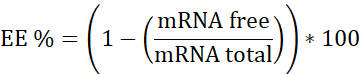
NOTE: First, Ribosomal RNA standard. Interpolate in this calibration curve the samples that do not contain Heparin. Second, mRNA:pBAE with Heparin. Interpolate in this calibration curve the sample that has Heparin. Third, Heparin. Used to assure the working concentration is in the linear range. - Calculate encapsulation efficiency (EE%) as follows:
- Prepare the three calibration curves
- Sample preparation
6. In vitro characterization
- Fluorescence microscopy for qualitative assessment
- Place the 96-well plate onto the microscope after 24 h of transfection. Start visualizing cells with the 10x objective.
NOTE: HeLa cells are used in this case. Cells are localized using the transmission mode (bright field), and the focus must be adjusted at this time.- First, apply a white balance to reference the software about the background of the samples. Then, acquire an image to overlay it with the fluorescence image during the analysis.
- Change the microscope mode to reflection mode (fluorescence) and move the filter wheel to the blue laser (488 nm) to visualize eGFP.
NOTE: At this point, the Exposure time and Gain must be adjusted. Gain must be adjusted in values between 3 and 5 to avoid artifacts and excess on background signal. The adjustment of the exposure time depends on the transfection efficiency (from ms to 1 s). - Acquire images for all the conditions or wells employing the same exposure time to compare all the samples.
- Place the 96-well plate onto the microscope after 24 h of transfection. Start visualizing cells with the 10x objective.
- Flow cytometry (FC) for quantitative assessment
- Use a multichannel pipette to prepare the 96-well plate for the flow cytometry experiment.
NOTE: When working with semi-adherent cells, such as in the JAWSII cell line, recover all the media volume and store it into another 96-well plate. Do not aspire to this media since it might contain a significant number of transfected cells. - Clean the cells (HeLa cells are used here) with 100 µL/well of 1x PBS and aspire it. Next, add 25 µL/well of trypsin and incubate it at 37 °C for 5 min.
- Once the cells are detached, add previously recovered media to stop the trypsin action and fix the cells, adding 31.25 µL/well of formalin 10% for 20 min (final concentration 2.5%).
NOTE: At this point, it is crucial to visualize the state of the cells by microscopy to ensure the detachment of the cells. It is highly recommended to pipette up and down several times to help the cells detach and avoid their agglomeration. This will produce a reliable result. - Turn on the flow cytometer and the software. In the software, set up the proper conditions for the experiment (type of plate, sample volume, and other parameters such as shaking, rinsing between samples.
NOTE: For step 6.2.5, the flow rate should be 120 µL/min. Mix one cycle every 1 min and rinse one cycle every one well. Mixing is essential to maintain the cells dispersed in the media and permit a better analysis of individual cells. In addition, rinsing is necessary to clean the microfluidics of the cytometer between samples. Finally, check whether there are enough buffers and whether all components are correctly connected. - Set up the appropriate parameters to quantify the percentage of positively transfected cells.
- First, view the flow data on (Forward scattered light) FSC versus SCC (Side scattered light) scatter plot to distinguish the cells from the debris. Then, another scatter plot comparing the amplitude (FSC-A) versus height (FSC-H) is plotted to gate and discriminate individual cells.
NOTE: The untreated population allows to gate the populations corresponding to positively transfected cells. Then, quantification of the positively transfected cells is performed on the proper channel on a histogram plot. Therefore, the positively transfected cells must be represented as a continuum of events on the histogram.
- First, view the flow data on (Forward scattered light) FSC versus SCC (Side scattered light) scatter plot to distinguish the cells from the debris. Then, another scatter plot comparing the amplitude (FSC-A) versus height (FSC-H) is plotted to gate and discriminate individual cells.
- Use a multichannel pipette to prepare the 96-well plate for the flow cytometry experiment.
7. In vitro functionality tests: capacity to activate model immune cells by using ovalbumin (OVA) as antigenic model mRNA
- Plate 10,000 cells/well (JAWSII cells are used here) in a 96-well plate the day before transfection.
NOTE: Plate as many wells as required to have triplicates for each condition. Let the cells attach to the plate at least for 12 h (an overnight incubation is recommended). - Prepare pBAEs NPs as described in the NOTE below to transfect 0.6 µg mRNA per well.
NOTE: As a positive control, transfect the cells using a transfection reagent. Here, we used 0.1 µg mRNA per well and 0.25 µL of the transfection reagent per well. mRNA codifying for OVA was purchased (see Table of Materials). It is polyadenylated, modified with 5-methoxyuridine, and optimized for mammalian systems, and is protected through an end-capping with Cap1 structure, a proprietary method from the supplier. - Incubate the 96-well plate for 24 h in a dry air incubator at 37 °C and 5% CO2.
NOTE: After this time, do not aspire the media since it might contain a certain number of cells. Recover the cells and save them in another well or plate. - Wash the cells remaining on the well with 25 µL of 1x PBS. Next, aspire it and add 25 µL of trypsin and incubate the plate for 5 min at 37 °C to detach the cells. Stop the trypsin reaction by adding the previously recovered media onto the correspondent well.
- Centrifuge the plate at 400 x g for 5 min at 4 °C. Aspire the media. Add 50 µL/well of a 1x PBS and 2.5% of formalin. Incubate it at 4 °C for 20 min to fix cells.
- Repeat steps 7.5 and 7.5.1. Then, add 50 µL/well of 1x PBS and 3% BSA (Blocking buffer) and incubate for 30 min at 4 °C. Again, repeat steps 7.5 and 7.5.1, and then add 50 µL/well of the primary antibody (mouse α-OVA) in 1x PBS and 3% BSA and incubate for 30 min at 4 °C.
- Repeat steps 7.5 and 7.5.1, and then wash the cells with 50 µL/well of 1x PBS. Aspire media, and then add secondary antibodies solution (α-mouse-AlexaFluor488/PerCP and Cy5.5-CD11b/APC-CD86) in 1x PBS and 3% BSA. Incubate it for 1 h at 4 °C.
- Centrifuge the plate at 400 x g for 5 min. Then, wash the cells with 50 µL/well of 1x PBS. Next, centrifuge (400 x g for 5 min), aspire, and then resuspend the cells in 100 µL/well of 1x PBS and 2.5% Formalin.
- Analyze it by flow cytometry, as described above (step 6.2).
Polymer synthesis and characterization
The OM-pBAE synthesis procedure is given in Figure 2. As Figure 2A shows, the first step to obtain the OM-pBAE is to synthesize the C6-pBAE by adding the amines (1-hexylamine and 5-amino-1-pentanol, ratio 1:1) to the diacrylate (1,4-butanediol diacrylate). This reaction is carried out at 90 °C for 20 h and with constant stirring. Afterward, a solution of oligopeptides is added to a solution of C6 polymer obtained from the previous reaction (Figure 2B). Finally, the synthesis is carried out using DMSO as a solvent at 25 °C with continuous stirring for 20 h. Figure 2C shows the structure of the OM-pBAE, which is the resulting product of polymerization. The resulting polymers are characterized by 1H-NMR (see Supplementary Figure S2). In the characterization, a positive result is the one with several monomer units (n + m in the figures) comprised between 7-10; distributed half one unit type and half the other; while any other number outside this range is a negative result.
Nanoparticle synthesis and physicochemical characterization
Table 3 shows the results of the physicochemical characterization of the GFP mRNA encapsulating nanoparticles as a proof of concept. It was previously demonstrated that size and surface charge do not show significant differences regardless of the mRNA type encapsulated14. Characterization has been performed both in freshly prepared and lyophilized nanoparticles to prove the stability of their physicochemical properties and to demonstrate that, as previously published, the freeze-drying process was adjusted to these formulations15. The hydrodynamic radius of the polyplexes is considered correct when it ranges around 150-220 nm. Besides, the PDI must be constant, approximately 0.2, but it is accepted up to 0.3. Therefore, neither size nor PDI showed significant differences before and after lyophilization, as shown here using representative results. Also, nanoparticle sizing was performed with NTA, to confirm the previously obtained results. Any value outside the ranges specified above is considered a negative result, and particles should be discarded for further use.
During the physicochemical characterization of this type of sample, it is convenient to compare the results obtained by DLS with the results obtained by NTA. Both techniques determine the hydrodynamic radius of the particles by measuring the diffusion coefficient. The number of nanoparticles counted per frame in NTA is between 80 and 100 to accurately track the particles. Surface charge was also characterized to ensure the positive charge of the nanoparticles, facilitating electrostatic interaction with the cell membrane. Finally, the morphology of the polyplexes was characterized by Transmission Electronic Microscopy (TEM). Figure 3 confirms the formation of spherical and monodisperse nanoparticles of an approximate size of 50 nm diameter. Smaller diameters were obtained by analyzing TEM images as both NTA and DLS measure the hydrodynamic diameters. Although a smaller size is always expected when performing electron microscopy measurements16,17, as compared to scattering measurements, it is essential to highlight here that the difference is almost 100 nm, which is usually not expected. This is attributed to the high hygroscopic character of these nanoparticles, given by both the encapsulated mRNA and the end terminal oligopeptides composing the polymer.
mRNA encapsulation efficiency determination
Another critical parameter to measure is the ability of the nanoparticles to encapsulate the mRNA, which is the active principle and needs to be encapsulated to achieve its protection from nucelases18,19,20. The protocol has been developed to analyze the encapsulation efficiency (EE; in %) of nucleic acid entrapment following the supplier's instructions. The obtained value gives an idea about the nucleic acid percentage that is successfully entrapped into the nanoparticles. RiboGreen dye, a fluorescent nucleic acid stain, is used for this purpose. The protocol consists of quantifying any RNA that remains accessible to the fluorescent dye. To quantify the total RNA, the nanoparticles need to be disassembled, and Heparin is used for this purpose.
Table 4 shows the encapsulation efficiency (EE%) of the OM-pBAE polyplexes encapsulating both GFP and OVA mRNA, as proof of concept. RiboGreen fluorescence assay allows calculating the amount of mRNA entrapped in the nanoparticles after disassembly by heparin addition to release their content. The efficiency values obtained must be higher than 80% and, as demonstrated here, do not show significant differences depending on the type of mRNA. Lower encapsulation efficiency values represent a negative result and constitute a signal for discarding these formulations. These results confirm the high encapsulation capacity of the mRNA of OM-pBAE, its versatility, and its promising application in gene delivery.
In vitro representative results
It is essential to determine the capacity of pBAEs polyplexes. First, to transfect and, second, to activate immune cells14. Transfection efficiency is assessed by two different techniques, using eGFP as the reporter mRNA: fluorescence microscopy to visually determine the reporter protein and flow cytometry expression to quantify the transfection efficiency. A fluorescent microscope equipped with a CCD camera, 5x, 10x, 20x, and 40x objectives and UV, blue and green filter lasers were employed to qualitatively determine the expression of eGFP protein in JAWSII cell line, a murine dendritic cell line. eGFP protein has an excitation maximum at 488 nm, corresponding to blue laser, and an emission maximum around 510 nm, corresponding to green color. To quantify the transfection efficiency expression of the target protein, flow cytometry analysis of the transfected cells is performed after 24 h of the transfection with pBAEs polyplexes with eGFP mRNA. A fixation step is helpful when cells have been transfected with fluorescent reporters such as GFP or RFP.
Figure 4 shows the qualitative analysis of the eGFP expression after 24 h of transfection in JAWSII cell line. Again, compared to the negative control, the eGFP reporter signal is significatively higher. Also, Table 5 shows the transfection efficiency displayed by OM-pBAE nanoparticles in JAWSII cell line. Again, the efficiency values are comparable to positive control performed with a classical transfection reagent.
The capacity of pBAEs NPs loaded with OVA mRNA to activate model immune cells has been assessed transfecting JAWSII cell line, a murine immature dendritic cell line. To determine the activation of the immune cells, two different markers: CD11b and CD86 were employed. First, select the proper fluorophores for the flow cytometer configuration. Here, PerCP-Cy5.5 (CD11b) and PE (CD86) are used. Immature and non-activated cell lines must show a CD11b-/CD86- profile, whereas activated cells must present a CD11b+/CD86+ profile. The blue laser at 488 nm is used to determine the target protein expression, and an anti-OVA antibody plus a secondary anti-mouse-Alexa488 antibody was employed. Table 6 shows the final panel employed in this experiment. Figure 5 shows the capacity of OVA mRNA-loaded OM-pBAE nanoparticles to activate DCs, which is indicative of the activation of the cellular immune response. Non-transfected cells show a low number of cells expressing the CD11b and CD86 membrane markers, whereas the mRNA OVA OM-pBAEs treated cells show a significatively increased expression of the CD11b and CD86 markers. This result suggests that OM-pBAE nanoparticles promote maturation. Altogether, these results confirm the capacity of OM-pBAEs nanoparticles to efficiently transfect dendritic cells and mediate the activation of the immune response promoting the maturation of immature DCs.
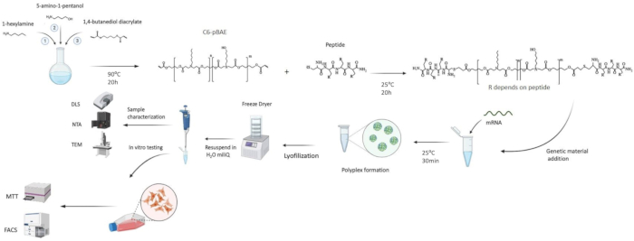
Figure 1: Schematic representation of the whole synthetic protocol. In this figure, the mRNA vaccine synthesis steps are detailed, including the critical characterization parameters. Please click here to view a larger version of this figure.
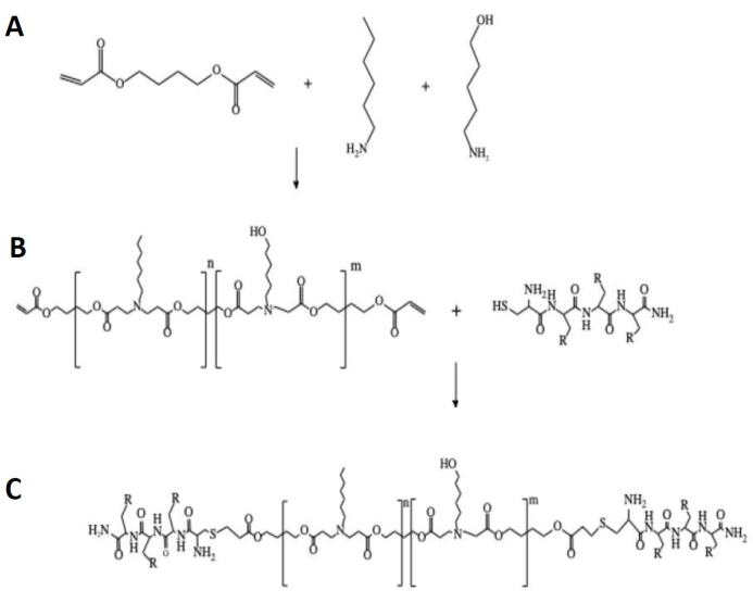
Figure 2: Synthesis of C6-pBAE oligopeptide end-modified (OM-pBAE). (A) Raw chemicals to perform the Michael addition. (B) pBAE + oligopeptide. (C) OM-pBAE. R can be Histidine (H) or Lysine (K). Please click here to view a larger version of this figure.
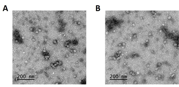
Figure 3: TEM images of fresh GFP mRNA encapsulating OM-pBAE nanoparticles. Polyplexes were negatively stained for electronic microscopy characterization. Monodisperse particles of approximately 60 nm diameter are shown. (A) and (B) images represent two different microscopies of the same sample. Please click here to view a larger version of this figure.
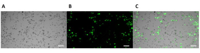
Figure 4: Fluorescence microscopy imaging of JAWSII transfected cells with OM-pBAE/eGFP mRNA nanoparticles. (A) Bright field image. (B) GFP fluorescence (in green). (C) Composite of the previous two images. Scale bars = 100 mm. Please click here to view a larger version of this figure.
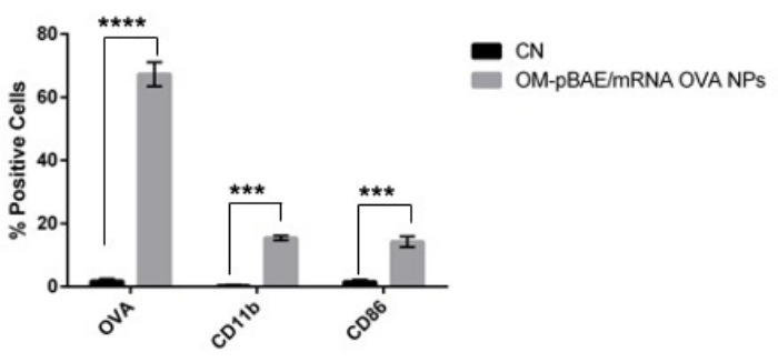
Figure 5: Activation of JAWSII after transfection with OM-pBAE/OVA mRNA nanoparticles. Negative control (CN) values correspond to the black label. Transfected cells correspond to the gray label. Bars correspond to the standard deviation (SD) of three replicates. Please click here to view a larger version of this figure.
| Material | Material | Microvesicles |
| Refractive Index | 1.4 | |
| Absorption | 0.01 | |
| Dispersant | Dispersant | Water |
| Temperature (°C) | 25 | |
| Viscosity (cP) | 0.8872 | |
| Refractive Index | 1.33 | |
| Temperature | Temperature (°C) | 25 |
| Equilibration time (s) | 10 | |
| Cell | Cell type | ZEN0040 |
Table 1: Parameters to create an SOP for size measurements.
| Material | Material | Microvesicles |
| Refractive index | 1.4 | |
| Absortion | 0.01 | |
| Dispersant | Dispersant | Water |
| Temperature (°C) | 25 | |
| Viscosity (cP) | 0.8872 | |
| Refractive index | 1.33 | |
| Temperature | Temperature (°C) | 25 |
| Equilibration time | 10 | |
| Cell | Cell type | DTS1070 |
Table 2: Parameters to create an SOP for zeta potential measurements.
| GFP mRNA/OM-pBAE | T (oC) | Hydrodinamic diameter (nm) | PDI | Surface charge (mV) | |
| NTA | DLS | ||||
| Fresh | 25 | 124 ± 32 | 134 ± 8 | 0.2 ± 0.04 | 32 ± 0.7 |
| Lyophilized | 25 | 135 ± 35 | 138 ± 2 | 0.17 ± 0.02 | 34 ± 0.7 |
Table 3: Physicochemical characterization of mRNA encapsulating OM-pBAE nanoparticles. GFP mRNA encapsulating OM-pBAE polyplexes were characterized immediately after preparation – fresh- and after lyophilization process to ensure the stability of the particles. DLS and NTA sizing, and surface charge data are shown.
| EE% | |
| GFP mRNA/OM-pBAE | 82.3 ± 1.5 |
| OVA mRNA/OM-pBAE | 83.1 ± 1.5 |
Table 4: Encapsulation efficiency (EE%) data of mRNA encapsulating OM-pBAE nanoparticles. Encapsulation efficiency percentages of GFP and OVA mRNA encapsulating OM-pBAE nanoparticles. RiboGreenfluorescence assay was performed on lyophilized samples to assess the amount of mRNA entrapped in the polyplexes.
| Sample | % GFP positive cells | SD |
| Negative Control | 1.85% | 0.21% |
| OM-pBAE/eGFP mRNA transfected cells | 57.79% | 5.28% |
Table 5: Transfection efficiency of OM-pBAE/eGFP mRNA nanoparticles in JAWSII cell line. Transfection efficiency was tracked by flow cytometry.
| Antibody | Cells stained | Fluorescent label |
| CD11b | Activated DCs | PerCP-Cy 5.5 |
| CD86 | Activated DCs | PE |
| α-mouse AlexaFluor488 | OVA positive cells | AlexaFluor 488 |
Table 6: Flow cytometry panel. This panel was used for flow cytometry studies to determine the in vitro functionality of the mRNA vaccine.
Supplementary information: See the Supplemental File with all Supplementary information included. Please click here to download below Files.
Table S1A: Ribosomal RNA standard.
Table S1B: RNA:pBAE with heparin standard.
Table S1C: Heparin standard.
Figure S2: 1H-NMR proton spectra of C6-pBAE and OM-pBAE. (A) 1H-NMR from the C6-pBAE. Chloroform-d was used as solvent (δ = 7.25 ppm). (B) 1H-NMR from the C6CH3. D2O was used as solvent (δ = 4.64 ppm).
