活斑马鱼幼虫的眼睛去除以检查视觉系统的神经支配依赖性生长和发育
Summary
本文解释了如何手术从活斑马鱼幼虫身上摘除眼睛,作为研究视网膜输入如何影响视神经结构生长和发育的第一步。此外,本文还提供了有关幼虫麻醉、固定和脑解剖的信息,然后是免疫组化和共聚焦成像。
Abstract
斑马鱼表现出非凡的终身生长和再生能力。例如,在胚胎发生期间建立的专用干细胞生态位支持整个视觉系统在眼睛和大脑中的连续生长。视网膜和视神经结构之间的协调生长确保了在眼睛和大脑中添加新神经元时准确的视网膜图谱图。为了解决视网膜轴突是否为调节颅骨干和祖细胞行为(如存活、增殖和/或分化)提供关键信息,有必要能够比较同一动物内和动物之间的神经支配和去神经支配的构造叶。
手术从活的幼虫斑马鱼中切除一只眼睛,然后观察视网膜,实现了这一目标。随附的视频演示了如何麻醉幼虫,电解磨钨针,并用它们去除一只眼睛。接下来,它展示了如何从固定的斑马鱼幼虫中解剖大脑。最后,该视频概述了免疫组织化学方案,并演示了如何在低熔点琼脂糖中安装染色胚胎以进行显微镜检查。
Introduction
该方法的目的是研究视网膜输入如何影响斑马鱼大脑视觉处理中心视神经技术的生长和发育。通过移除一只眼睛,然后比较视网膜的两侧,可以观察和归一化同一标本内的构造变化,从而能够比较多个标本。现代分子方法与该技术相结合,将深入了解视觉系统生长和发育以及轴突变性和再生的潜在机制。
感觉系统 – 视觉,听觉和躯体感觉 – 从外部器官收集信息并将该信息传递给中枢神经系统,生成中脑外部世界的“地图”1,2。视觉是几乎所有脊椎动物(包括许多鱼类)的主要感觉方式。视网膜是眼睛中的神经组织,它通过主要由光感受器,双极细胞和视网膜神经节细胞(RGC)组成的神经元回路收集信息,RGC是视网膜的投射神经元。RGC具有长轴突,它们穿过视网膜的内表面到达视神经头,在那里它们束曲并一起穿过大脑,最终终止于背中脑的视觉处理中心。这种结构在鱼类和其他非哺乳动物脊椎动物中被称为视技术,并且与哺乳动物的上丘脑同源3。
视神经结构是背中脑中双侧对称的多层结构。在斑马鱼和大多数其他鱼类中,视神经的每个叶仅接收来自对侧眼的视觉输入,使得左视神经终止于右构造叶,右视神经终止于左构造叶4 (图1)。像哺乳动物的对应物,上丘脑一样,视神经构造将视觉信息与其他感官输入(包括试听和躯体感觉)集成在一起,控制视觉注意力和眼球运动(如扫视1,5,6)的变化。然而,与哺乳动物上丘脑不同,视神经膜不断从称为构造增殖区7的内侧和尾部边缘附近的特化干细胞位产生新的神经元和神经胶质细胞。在视神经结构和中枢神经系统其他区域维持增殖祖细胞在一定程度上有助于斑马鱼8中记录的显着再生能力。
先前对盲人或独眼鱼大脑的研究表明,视神经结构的大小与其接受的视网膜神经支配量成正比9,10,11。在成年洞穴鱼中,其眼睛在早期胚胎发生时退化,视神经构造明显小于密切相关的目击面鱼9。洞穴鱼眼变性可以通过在胚胎发生过程中用表层鱼的晶状体替换内源性晶状体来阻止。当这些独眼洞穴鱼被饲养到成年期时,神经支配的构造叶比未支配的构造叶9含有约10%的细胞。同样,在用化学处理孵育以在同一个体内产生不同大小的眼睛的幼虫基利鱼中,具有更多神经支配的构造体的一侧更大并且包含更多的神经元10。来自成年金鱼视神经压碎实验的证据表明,神经支配促进增殖,当神经支配被破坏时,构造细胞增殖减少11。
证实并扩展了这些经典研究,最近的几份报告提供了数据,表明响应神经支配的增殖至少部分受到BDNF-TrkB途径12,13的调节。关于视神经构造细胞生长和发育的许多悬而未决的问题仍然存在,包括发育中的感觉系统如何应对损伤和轴突变性,哪些细胞和分子信号使视网膜输入能够调节视神经构造的生长,当这些机制变得活跃时,以及神经支配相关的增殖和分化是否使视网膜及其靶组织能够协调生长速率并确保准确的视网膜学映射。此外,关于活动依赖性发育还有更大的问题,可以通过用外科手术方法询问斑马鱼视觉系统来解决,如下所述。
为了研究神经活动(特别是来自视觉输入)改变细胞存活和增殖的细胞和分子机制,所述方法直接比较单个斑马鱼幼虫内神经支配和去神经支配的构造叶(图1)。该方法允许记录视神经组织中RGC轴突变性,并确认有丝分裂细胞的数量与神经支配相关。
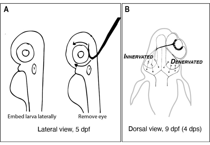
图1:单侧取眼前后斑马鱼幼虫的草图。 (A)在解剖显微镜下观察的5 dpf幼虫的图纸。每个幼虫都嵌入低熔点琼脂糖中,并横向定向,然后使用带有锋利钩状尖端的钨针将眼睛朝上舀出(在本例中为左眼)。(B) A中描述的手术产生的9 dpf幼虫的背视图图。只有来自右眼的三个高度模式化的RGC轴突显示脱发并与左脑叶中的神经元连接。缩写:dpf = 受精后的天数;dps = 手术后天数;RGC = 视网膜神经节细胞。 请点击此处查看此图的大图。
Protocol
Representative Results
Discussion
本文中描述的技术说明了研究斑马鱼脊椎动物视觉系统发育的众多方法之一。其他研究人员已经发表了解剖胚胎视网膜并进行基因表达分析的方法19 或可视化视神经结构30中的神经元活动。本文提供了一种探索视网膜差异输入如何影响视神经组织中细胞行为的方法。
为了确保手术后成功消灭眼部和幼虫存活,重要的是幼虫健康,充分麻醉,…
Declarações
The authors have nothing to disclose.
Acknowledgements
这项工作的资金主要由里德学院到KLC的启动资金,海伦斯塔福德研究奖学金基金到OLH,以及里德学院科学研究奖学金到YK。该项目始于史蒂夫威尔逊的实验室,与人力资源部门合作,人力资源部门得到了惠康信托学生奖学金(2009-2014)的支持。我们感谢Máté Varga,Steve Wilson和Wilson实验室的其他成员对这个项目的初步讨论,我们特别感谢Florencia Cavodeassi和Kate Edwards,他们是第一个教KLC如何在琼脂糖中安装胚胎并进行斑马鱼大脑解剖的人。我们还感谢Greta Glover和Jay Ewing帮助组装我们的钨针磨刀设备。
Materials
| Equipment and supplies: | |||
| Breeding boxes | Aquaneering | ZHCT100 | |
| Dow Corning high vacuum grease | Sigma or equivalent supplier | Z273554 | |
| Erlenmeyer flasks (125 mL) | For making Marc's Modified Ringers (MMR) with antibiotics for post-surgery incubation | ||
| Fine forceps – Dumont #5 | Fine Science Tools (FST) | 11252-20 | |
| Glass Pasteur pipettes | DWK Lifescience | 63A53 & 63A53WT | For pipetting embryos and larvae |
| Glass slides for microscopy | VWR or equivalent supplier | 48311-703 | Standard glass microscope slides can be ordered from many different laboratory suppliers. |
| Glassware including graduated bottles and graduated cylinders | For making and storing solutions | ||
| 2-part epoxy resin | ACE Hardware or other equivalent supplier of Gorilla Glue or equivalent | 0.85 oz syringe | https://www.acehardware.com/departments/paint-and-supplies/tape-glues-and-adhesives/glues-and-epoxy/1590793 |
| Microcentrifuge tube (1.7 mL) | VWR or equivalent supplier | 22234-046 | |
| Nickel plated pin holder (17 cm length) | Fine Science Tools (FST) | 26018-17 | To hold tungsten wire while sharpening and performing surgeries/dissections. |
| Nylon mesh tea strainer or equivalent | Ali Express or equivalent | For harvesting zebrafish eggs after spawning; https://www.aliexpress.com/item/1005002219569756.html | |
| Paper clip | For Tungsten needle sharpening device. | ||
| Petri dishes 100 mm | Fischer Scientific or equivalent supplier | 50-190-0267 | |
| Petri dishes 35 mm | Fischer Scientific or equivalent supplier | 08-757-100A | |
| Pipette pump | SP Bel-Art or equivalent | F37898-0000 | |
| Potassium hydroxide (KOH) | Sigma | 909122 | For Tungsten needle sharpening device. Make a 10% w/v solution of KOH in the hood by adding pellets to deionized water. |
| Power supply (variable voltage) | For Tungsten needle sharpening device. Any power supply with variable voltage will work (even one used for gel electrophoresis). | ||
| Sylgard 184 Elastomer kit | Dow Corning | 3097358 | |
| Tungsten wire (0.125 mm diameter) | World Precision Instruments (WPI) | TGW0515 | Sharpen to remove eye and dissect larvae. |
| Variable temperature heat block | The Lab Depot or equivalent supplier | BSH1001 or BSH1002 | Set to 40-42 °C ahead of experiments. |
| Wide-mouth glass jar with lid (e.g., clean jam or salsa jar) | For Tungsten needle sharpening device. | ||
| Wires with alligator clip leads | For Tungsten needle sharpening device. | ||
| Microscopes: | |||
| Dissecting microscope | Any type will work but having adjustable transmitted light on a mirrored base is preferred. | ||
| Laser scanning confocal microscope | High NA, 20-25x water dipping objective lens is recommended. Microscope control and image capture software (Elements) is used here but any confocal microscope will work. |
||
| Reagents for surgeries and dissections: | |||
| Calcium chloride dihydrate | Sigma | C7902 | For Marc's Modified Ringers (MMR) and embryo medium (E3). |
| HEPES | Sigma | H7006 | For Marc's Modified Ringers (MMR). |
| Low melting point agarose | Invitrogen | 16520-050 | Make 1% in embryo medium (E3) or Marc's Modified Ringers (MMR). |
| Magnesium chloride hexahydrate | Sigma | 1374248 | For embryo medium (E3). |
| Magnesium sulfate | Sigma | M7506 | For Marc's Modified Ringers (MMR). |
| Paraformaldehyde | Electron Microscopy Sciences | 19210 | Dilute 8% (w/v) stock with 2x concentrated PBS (diluted from 10x PBS stock). |
| Penicillin/Streptomycin | Sigma | P4333-20ML | Dilute 1:100 in Marc's Modified Ringers. |
| Phosphate buffered saline (PBS) tablets | Diagnostic BioSystems | DMR E404-01 | Make 10x stock in deionized water, autoclave and store at room temperature. Dilute to 1x working concentration. |
| Potassium chloride | Sigma | P3911 | For Marc's Modified Ringers (MMR) and embryo medium (E3). |
| Sodium chloride | Sigma | S9888 | For Marc's Modified Ringers (MMR) and embryo medium (E3). |
| Sodium hydroxide | Sigma | S5881 | Make 10 M and use to adjust pH of MMR to 7.4. |
| Sucrose | Sigma | S9378 | |
| Tricaine-S | Pentair | 100G #TRS1 | Recipe: https://zfin.atlassian.net/wiki/spaces/prot/pages/362220023/TRICAINE |
| Reagents for immunohistochemistry: | |||
| Alexafluor 568 tagged Secondary antibody to detect rabbit IgG | Invitrogen | A-11011 | Use at 1:500 dilution for wholemount immunohistochemistry. |
| DAPI or ToPro3 | Invitrogen | 1306 or T3605 | Make up 1 mg/mL solutions in DMSO; 1:5,000 dilution for counterstaining. |
| Dimethyl sulfoxide (DMSO) | Sigma | D8418 | A component of immunoblock buffer. |
| Methanol (MeOH) | Sigma | 34860 | Mixing MeOH with aqueous solutions like PBST is exothermic. Make the MeOH/PBST solutions at least several hours ahead of time or cool them on ice before using. |
| Normal goat serum | ThermoFisher Scientific | 50-062Z | A component of immunoblock buffer. Can be aliquoted in 1-10 mL volumes and stored at -20 °C. |
| Primary antibody to detect phosphohistone H3 | Millipore | 06-570 | Use at 1:300 dilution for wholemount immunohistochemistry. |
| Primary antibody to detect Red Fluorescent Protein (RFP; detects dsRed derivatives) | MBL International | PM005 | Use at 1:500 dilution for wholemount immunohistochemistry. |
| Proteinase K (PK) | Sigma | P2308-10MG | Make up 10 mg/mL stock solutions in PBS and use at 10 µg/mL. |
| Triton X-100 | Sigma | T8787 | Useful to make a 20% (v/v) stock solution in PBS. |
| Software for data analysis | |||
| ImageJ (Fiji) | freeware for image analysis; https://imagej.net/software/fiji/ | ||
| Rstudio | freeware for statistical analysis and data visualization; https://www.rstudio.com/products/rstudio/download/ | ||
| Adobe Photoshop or GIMP | Proprietary image processing software (Adobe Photoshop and Illustrator) are often used to compose figures). A freeware alternative is Gnu Image Manipulation Program (GIMP; https://www.gimp.org/) | ||
| Zebrafish strains | available from the Zebrafish International Resource Centers in the US (https://zebrafish.org/home/guide.php) or in Europe (https://www.ezrc.kit.edu/). Specialized transgenic strains that have not yet been deposited in either resource center can be requested from individual labs after publication. |
Referências
- Butler, A. B., Hodos, W. Optic tectum. Comparative Vertebrate Neuroanatomy: Evolution and Adaptation. , 311-340 (2005).
- Cang, J., Feldheim, D. A. Developmental mechanisms of topographic map formation and alignment. Annual Review of Neuroscience. 36 (1), 51-77 (2013).
- Basso, M. A., Bickford, M. E., Cang, J. Unraveling circuits of visual perception and cognition through the superior colliculus. Neuron. 109 (6), 918-937 (2021).
- Burrill, J. D., Easter, S. S. Development of the retinofugal projections in the embryonic and larval zebrafish (Brachydanio rerio). The Journal of Comparative Neurology. 346 (4), 583-600 (1994).
- Regeneration in the goldfish visual system. Webvision: The Organization of the Retina and Visual System Available from: https://webvision.med.utah.edu/book/part-x-repair-and-regeneration-in-the-visual-system/regeneration-in-the-goldfish-visual-system/ (2021)
- Regeneration in the visual system of adult mammals. Webvision: The Organization of the Retina and Visual System Available from: https://webvision.med.utah.edu/book/part-x-repair-and-regeneration-in-the-visual-system/regeneration-in-the-visual-system-of-adult-mammals/ (2021)
- Cerveny, K. L., Varga, M., Wilson, S. W. Continued growth and circuit building in the anamniote visual system. Developmental Neurobiology. 72 (3), 328-345 (2012).
- Lindsey, B. W., et al. Midbrain tectal stem cells display diverse regenerative capacities in zebrafish. Scientific Reports. 9 (1), 4420 (2019).
- Soares, D., Yamamoto, Y., Strickler, A. G., Jeffery, W. R. The lens has a specific influence on optic nerve and tectum development in the blind cavefish Astyanax. Developmental Neuroscience. 26 (5-6), 308-317 (2004).
- White, E. L. An experimental study of the relationship between the size of the eye and the size of the optic tectum in the brain of the developing teleost, Fundulus heteroclitus. Journal of Experimental Zoology. 108 (3), 439-469 (1948).
- Raymond, P., Easter, S., Burnham, J., Powers, M. Postembryonic growth of the optic tectum in goldfish. II. Modulation of cell proliferation by retinal fiber input. The Journal of Neuroscience. 3 (5), 1092-1099 (1983).
- Sato, Y., Yano, H., Shimizu, Y., Tanaka, H., Ohshima, T. Optic nerve input-dependent regulation of neural stem cell proliferation in the optic tectum of adult zebrafish. Developmental Neurobiology. 77 (4), 474-482 (2017).
- Hall, Z. J., Tropepe, V. Visual experience facilitates BDNF-dependent adaptive recruitment of new neurons in the postembryonic optic tectum. The Journal of Neuroscience. 38 (8), 2000-2014 (2018).
- Nusslein-Volhard, C., Dahm, R. . Zebrafish. , (2002).
- Cold Spring Harbor Protocols. Marc’s modified Ringer’s (MMR) (10X, pH 7.4). Cold Spring Harbor Protocols. , (2009).
- Turner, K. J., Bracewell, T. G., Hawkins, T. A. Anatomical dissection of zebrafish brain development. Brain Development. 1082, 197-214 (2014).
- Brady, J. A simple technique for making very fine, durable dissecting needles by sharpening tungsten wire electrolytically. Bulletin of the World Health Organization. 32 (1), 143-144 (1965).
- . ZFIN Tricaine recipe Available from: https://zfin.atlassian.net/wiki/spaces/prot/pages/362220023/TRICAINE (2018)
- Zhang, L., Leung, Y. F. Microdissection of zebrafish embryonic eye tissues. Journal of Visualized Experiments: JoVE. (40), e2028 (2010).
- . ZFIN protocols Available from: https://zfin.atlassian.net/wiki/spaces/prot/overview (2021)
- Engerer, P., Plucinska, G., Thong, R., Trovò, L., Paquet, D., Godinho, L. Imaging subcellular structures in the living zebrafish embryo. Journal of Visualized Experiments: JoVE. (110), e53456 (2016).
- ImageJ with batteries included. Fiji Available from: https://figi.sc/ (2021)
- O’Brien, J., Hayder, H., Peng, C. Automated quantification and analysis of cell counting procedures using ImageJ plugins. Journal of Visualized Experiments: JoVE. (117), e54719 (2016).
- Poggi, L., Vitorino, M., Masai, I., Harris, W. A. Influences on neural lineage and mode of division in the zebrafish retina in vivo. The Journal of Cell Biology. 171 (6), 991-999 (2005).
- Karlstrom, R. O., et al. Zebrafish mutations affecting retinotectal axon pathfinding. Development. 123 (1), 427-438 (1996).
- Harvey, B. M., Baxter, M., Granato, M. Optic nerve regeneration in larval zebrafish exhibits spontaneous capacity for retinotopic but not tectum specific axon targeting. PLOS ONE. 14 (6), 0218667 (2019).
- Robles, E., Filosa, A., Baier, H. Precise lamination of retinal axons generates multiple parallel input pathways in the tectum. Journal of Neuroscience. 33 (11), 5027-5039 (2013).
- Vargas, M. E., Barres, B. A. Why Is Wallerian degeneration in the CNS so slow. Annual Review of Neuroscience. 30 (1), 153-179 (2007).
- Hughes, A. N., Appel, B. Microglia phagocytose myelin sheaths to modify developmental myelination. Nature Neuroscience. 23 (9), 1055-1066 (2020).
- de Calbiac, H., Dabacan, A., Muresan, R., Kabashi, E., Ciura, S. Behavioral and physiological analysis in a zebrafish model of epilepsy. Journal of Visualized Experiments: JoVE. (176), e58837 (2021).
- Adams, S. L., Zhang, T., Rawson, D. M. The effect of external medium composition on membrane water permeability of zebrafish (Danio rerio) embryos. Theriogenology. 64 (7), 1591-1602 (2005).
- Fredj, N. B., et al. Synaptic activity and activity-dependent competition regulates axon arbor maturation, growth arrest, and territory in the retinotectal projection. Journal of Neuroscience. 30 (32), 10939-10951 (2010).
- Alberio, L., et al. A light-gated potassium channel for sustained neuronal inhibition. Nature Methods. 15 (11), 969-976 (2018).
- Kay, J. N., Finger-Baier, K. C., Roeser, T., Staub, W., Baier, H. Retinal ganglion cell genesis requires lakritz, a zebrafish atonal homolog. Neuron. 30 (3), 725-736 (2001).
- Gnuegge, L., Schmid, S., Neuhauss, S. C. F. Analysis of the activity-deprived zebrafish mutant macho reveals an essential requirement of neuronal activity for the development of a fine-grained visuotopic map. The Journal of Neuroscience. 21 (10), 3542-3548 (2001).
- Jeffery, W. R. Astyanax surface and cave fish morphs. EvoDevo. 11 (1), 14 (2020).
- Sieger, D., Peri, F. Animal models for studying microglia: The first, the popular, and the new. Glia. 61 (1), 3-9 (2013).
- Svahn, A. J., et al. Development of ramified microglia from early macrophages in the zebrafish optic tectum. Developmental Neurobiology. 73 (1), 60-71 (2013).
- Herzog, C., et al. Rapid clearance of cellular debris by microglia limits secondary neuronal cell death after brain injury in vivo. Development. 146 (9), (2019).
- Chen, J., Poskanzer, K. E., Freeman, M. R., Monk, K. R. Live-imaging of astrocyte morphogenesis and function in zebrafish neural circuits. Nature Neuroscience. 23 (10), 1297-1306 (2020).
