Summary

在啮齿动物中进行体内和离体电阻抗肌图

Published: June 08, 2022
doi:

Summary

本文详细介绍了如何对啮齿动物腓肠肌进行 体内 (使用表面和针状电极阵列)和 离体 (使用介电电池)电阻抗肌图。它将在小鼠和大鼠中演示该技术,并详细说明可用的修改(即肥胖动物,幼崽)。

Abstract

电阻抗肌图(EIM)是一种方便的技术,可用于临床前和临床研究,以评估肌肉组织的健康和疾病。EIM是通过在一系列频率(即从1 kHz到10 MHz)上向感兴趣的肌肉施加低强度,定向聚焦的电流并记录产生的电压来获得的。由此,可以获得几个标准阻抗分量,包括电抗、电阻和相位。当对切除的肌肉进行 离体 测量时,还可以计算组织的固有被动电特性,即电导率和相对介电常数。EIM已被广泛用于动物和人类,以诊断和跟踪各种疾病的肌肉改变,与简单的废用性萎缩有关,或作为治疗干预的措施。在临床上,EIM提供了跟踪疾病随时间发展并评估治疗干预影响的潜力,从而提供了缩短临床试验持续时间和减少样本量要求的机会。由于EIM可以在活体动物模型和人类中进行无创或微创,因此具有作为新型转化工具的潜力,可实现临床前和临床开发。本文提供了有关如何在小鼠和大鼠中进行 体内离体 EIM测量的分步说明,包括使该技术适应特定条件的方法,例如用于幼崽或肥胖动物。

Introduction

电阻抗肌图(EIM)提供了一种评估肌肉状况的强大方法,有可能诊断神经肌肉疾病,跟踪疾病进展和评估对治疗的反应123。它可以类似地应用于动物疾病模型和人类,允许从临床前到临床研究的相对无缝转换。使用四个线性放置的电极可以轻松获得EIM测量,其中两个外部电极在频率范围内(通常在1 kHz和大约2 MHz之间)施加无痛的弱电流,两个内部电极记录产生的电压1。从这些电压中,可以获得组织的阻抗特性,包括电阻(R),这是电流通过组织的难度的量度,以及组织的电抗(X)或“可充电性”,与组织的电荷存储能力(电容)有关。根据电抗和电阻,相位角(θ)通过以下公式Equation 1
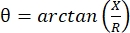
计算:,提供单个求和阻抗测量。这种测量可以使用任何多频生物阻抗设备获得。由于肌纤维本质上是长圆柱体,肌肉组织也是高度各向异性的,电流更容易沿着纤维流动而不是穿过它们45.因此,EIM通常在两个方向上进行:阵列沿着纤维放置,使电流平行于它们,并穿过肌肉,使电流垂直于它们流动。此外,在离体测量中,在阻抗测量池中测量已知体积的组织时,可以得出肌肉的固有电特性(即电导率和相对介电常数)6。

术语“神经肌肉疾病”定义了导致结构肌肉改变和功能障碍的各种原发性和继发性疾病。这包括肌萎缩侧索硬化症和各种形式的肌肉萎缩症,以及与衰老(例如肌肉减少症)、废用性萎缩(例如由于长时间卧床或微重力)甚至损伤相关的简单变化7。虽然病因很多,可能起源于运动神经元、神经、神经肌肉接头或肌肉本身,但EIM可用于检测由于许多这些过程引起的肌肉早期变化,并跟踪治疗的进展或反应。例如,在杜氏肌营养不良症 (DMD) 患者中,EIM 已被证明可以检测疾病进展和对皮质类固醇的反应8。最近的工作还表明,EIM对不同的废弃状态很敏感,包括部分重力9,就像在月球或火星上经历的那样,以及老化的影响1011。最后,通过将预测和机器学习算法应用于每次测量获得的数据集(多频率和方向依赖性数据),可以推断组织的组织学方面,包括肌纤维大小1213、炎症变化和水肿 14,以及结缔组织和脂肪含量1516

其他几种非侵入性或微创方法也用于评估人类和动物的肌肉健康,包括针状肌电图17 和成像技术,如磁共振成像,计算机断层扫描和超声1819。然而,与这些技术相比,EIM显示出明显的优势。例如,肌电图仅记录肌纤维膜的主动电特性,而不记录被动特性,因此无法提供对肌肉组成或结构的真实评估。在某种程度上,成像方法与EIM的关系更密切,因为它们也提供了有关组织结构和组成的信息。但从某种意义上说,它们提供了太多的数据,需要详细的图像分割和专家分析,而不仅仅是提供定量输出。此外,鉴于其复杂性,成像技术也受到所用硬件和软件细节的极大影响,理想情况下需要使用相同的系统,以便可以比较数据集。相比之下,EIM更简单的事实意味着它受这些技术问题的影响较小,并且不需要任何形式的图像处理或专家分析。

以下协议演示了如何使用非侵入性(表面阵列)和微创(皮下针阵列)技术以及在新鲜切除的肌肉上进行体外 EIM,在大鼠和小鼠中进行体内 EIM

Protocol

此处描述的所有方法均已获得贝斯以色列女执事医疗中心机构动物护理和使用委员会的批准,协议编号为(031-2019; 025-2019)。穿戴适当的个人防护装备来处理动物,并遵守IACUC关于所有动物工作的指导方针。 1. 体内 表面EIM 将动物放入麻醉盒中以诱导麻醉。注意:对于大鼠,使用1.5%-3.5%异氟烷和2O 2 L·min-1,对于小鼠,使用2%异氟烷和1 O<sub…

Representative Results

EIM可以在许多条件下获得,包括表面体内阵列(图1),针状体内阵列(图2A-F)和离体介电电池(图2G,H)。 EIM根据测量的阻抗值提供肌肉状况的近乎即时的快照。测量结果可快速采集,并生成简单的输出数据文件,无需任何特殊软件(<strong class="xfi…

Discussion

本文提供了在 齿动物体内和 体外进行EIM的基本方法。为了获得可靠的测量结果,执行一系列步骤至关重要。首先,需要正确识别感兴趣的肌肉,因为每块肌肉对疾病、治疗和病理都有不同的反应。必须注意,在一块肌肉(例如腓肠肌)上获得的数据不会提供与另一块肌肉(例如胫骨前肌)相同的信息。其次,需要仔细选择最佳的电极阵列来进行阻抗测量。虽然每种阵列类型都有?…

Declarações

The authors have nothing to disclose.

Acknowledgements

这项工作得到了查理基金和NIH R01NS055099的支持。

Materials

3D Printer Formlabs Inc. Form 2 Desktop 3D printer
3D Printer Shenzhen Creality 3D Technology Co. LTD Creality Ender 3 V2 3D printer
3M Micropore surgical tape Fisher 19-027761 and 19-061655 models 1530-0 and 1530-1
3M TRANSPORE surgical tape Fisher 18-999-380 and 18-999-381 models 1527-0 and 1527-1
Connector header vertical 10 POS 1 mm spacing Digi-Key (Sullins connector solution) S9214-ND (SMH100-LPSE-S10-ST-BK) Plastic spacer 1 mm holes for the rat in vivo array displayed in Figure 2A
Cotton-tipped applicators Fisher 22-363-172
Dental Wax Fisher NC9377103
Depilatory agent NAIR NA hair remover lotion with softening baby oil
Dumont #7b Forceps Fine Science Tools No. 11270-20 Used for dissection, Style: #7b, Tip Shape: Curved, Tips: Standard, Tip Dimensions: 0.17 mm x 0.1 mm, Alloy/Material: Inox, Length: 11 cm
Electronic Digital Caliper Fisher 14-648-17 Used to measure out the dimensions of the Gastrocnemius muscle
Epoxy adhesive dual cartridge 4 min work life Devcon series 14265, model 2217 Glue used in the rat in vivo array displayed in Figure 2A
Ex vivo dielectric impedance cell Custom NA Dielectric cells were 3D printed in the Rutkove laboratory
Graefe Forceps Fine Science Tools No. 11051-10 Used for muscle to place and adjust, Length: 10 cm, Tip Shape: Curved, Tips: Serrated, Tip Width: 0.8 mm, Tip Dimensions: 0.8 mm x 0.7 mm, Alloy/Material
Hair clipper Amazon NA Wahl professional animal BravMini+
Impedance Animal Device Myolex EIM1103 mView system – investigational electrical impedance myography device for use in animal research
In vivo needle arrays Custom NA Custom arrays using 27 G subdermal needles from Ambu. The construction was finalized using a 3D printer in the Rutkove laboratory
In vivo surface array Custom NA The in vivo surface array was printed and assembled in the Rutkove laboratory
Isoflurane Patterson Veterinary Supplies 07-893-8441 (NDC: 46066-755-04) Pivetal – 250 mL bottle
Non-woven gauze Fisher 22-028-559 2 x 2 inch
Polystyrene Weighing Dishes Fisher S67090A Dimensions (L x W x H): 88.9 mm x 88.9 mm x 25.4 mm
Razor Blades Fisher 12-640 Used to cut muscle to right dimensions, Single-edge carbon steel blades
Student Fine Scissors Fine Science Tools No. 91460-11 Used for dissection, Tips: Sharp-Sharp, Alloy/Material: Student Stainless Steel, Serrated: No, Tip Shape: Straight, Cutting Edge: 20 mm, Length: 11.5 cm, Feature: Student Quality
Subdermal needles 27 G Neuroline Ambu 745 12-50/24 Needles used in the rat in vivo array displayed in Figure 2A
Surgical Scissors – Sharp Fine Science Tools No. 14002-13 Used to cut skin, Tips: Sharp-Sharp, Alloy/Material: Stainless Steel, Serrated: No, Tip Shape: Straight, Cutting Edge: 42 mm, Length: 13 cm
TECA ELITE monopolar needle electrodes Natus 902-DMG50-S 0.46 mm diameter (26 G). Blue hub
Teknova 0.9% saline solution Fisher S5815 1000 mL sterile

Referências

  1. Rutkove, S. B., Sanchez, B. Electrical impedance methods in neuromuscular assessment: An overview. Cold Spring Harbor Perspectives in Medicine. 9 (10), 034405 (2019).
  2. Rutkove, S. B. Electrical impedance myography: Background, Current State, and Future Directions. Muscle & Nerve. 40, 936-946 (2009).
  3. Sanchez, B., Rutkove, S. B. Present uses, future applications, and technical underpinnings of electrical impedance myography. Current Neurology and Neuroscience Reports. 17 (11), 86 (2017).
  4. Garmirian, L. P., Chin, A. B., Rutkove, S. B. Discriminating neurogenic from myopathic disease via measurement of muscle anisotropy. Muscle & Nerve. 39 (1), 16-24 (2009).
  5. Rutkove, S. B., et al. Loss of electrical anisotropy is an unrecognized feature of dystrophic muscle that may serve as a convenient index of disease status. Clinical Neurophysiology. 127 (12), 3546-3551 (2016).
  6. Wang, L. L., et al. Assessment of alterations in the electrical impedance of muscle after experimental nerve injury via finite-element analysis. IEEE Transactions on Biomedical Engineering. 58 (6), 1585-1591 (2011).
  7. Katirji, B., Ruff, R. L., Kaminski, H. J. . Neuromuscular Disorders in Clinical Practice. , (2014).
  8. Rutkove, S. B., et al. Electrical impedance myography for assessment of Duchenne muscular dystrophy. Annals of Neurology. 81 (5), 622-632 (2017).
  9. Semple, C., et al. Using electrical impedance myography as a biomarker of muscle deconditioning in rats exposed to micro- and partial-gravity analogs. Frontiers in Physiology. 11, 557796 (2020).
  10. Kortman, H. G. J., Wilder, S. C., Geisbush, T. R., Narayanaswami, P., Rutkove, S. B. Age- and gender-associated differences in electrical impedance values of skeletal muscle. Physiological Measurement. 34 (12), 1611-1622 (2013).
  11. Clark, B. C., Rutkove, S., Lupton, E. C., Padilla, C. J., Arnold, W. D. Potential utility of electrical impedance myography in evaluating age-related skeletal muscle function deficits. Frontiers in Physiology. 12, 666964 (2021).
  12. Kapur, K., et al. Predicting myofiber size with electrical impedance myography: A study in immature mice. Muscle and Nerve. 58 (1), 106-113 (2018).
  13. Kapur, K., Nagy, J. A., Taylor, R. S., Sanchez, B., Rutkove, S. B. Estimating myofiber size with electrical impedance myography: a study in amyotrophic lateral sclerosis mice. Muscle and Nerve. 58 (5), 713-717 (2018).
  14. Mortreux, M., Semple, C., Riveros, D., Nagy, J. A., Rutkove, S. B. Electrical impedance myography for the detection of muscle inflammation induced by λ-carrageenan. PLoS ONE. 14 (10), 0223265 (2019).
  15. Pandeya, S. R., et al. Predicting myofiber cross-sectional area and triglyceride content with electrical impedance myography: A study in db/db mice. Muscle and Nerve. 63 (1), 127-140 (2021).
  16. Pandeya, S. R., et al. Estimating myofiber cross-sectional area and connective tissue deposition with electrical impedance myography: A study in D2-mdx mice. Muscle & Nerve. 63 (6), 941-950 (2021).
  17. Stålberg, E., et al. Standards for quantification of EMG and neurography. Clinical Neurophysiology. 130 (9), 1688-1729 (2019).
  18. Theodorou, D. J., Theodorou, S. J., Kakitsubata, Y. Skeletal muscle disease: Patterns of MRI appearances. British Journal of Radiology. 85 (1020), 1298-1308 (2012).
  19. Simon, N. G., Noto, Y., Zaidman, C. M. Skeletal muscle imaging in neuromuscular disease. Journal of Clinical Neuroscience. 33, 1-10 (2016).
  20. Kwon, H., Rutkove, S. B., Sanchez, B. Recording characteristics of electrical impedance myography needle electrodes. Physiological Measurement. 38 (9), 1748-1765 (2017).
  21. Kwon, H., Di Cristina, J. F., Rutkove, S. B., Sanchez, B. Recording characteristics of electrical impedance-electromyography needle electrodes. Physiological Measurement. 39 (5), 055005 (2018).
  22. Rutkove, S. B., et al. Characterizing spinal muscular atrophy with electrical impedance myography. Muscle and Nerve. 42 (6), 915-921 (2010).
  23. Schwartz, S., et al. Optimizing electrical impedance myography measurements by using a multifrequency ratio: A study in Duchenne muscular dystrophy. Clinical Neurophysiology. 126 (1), 202-208 (2015).
  24. Li, J., Pacheck, A., Sanchez, B., Rutkove, S. B. Single and modeled multifrequency electrical impedance myography parameters and their relationship to force production in the ALS SOD1G93A mouse. Amyotrophic Lateral Sclerosis and Frontotemporal Degeneration. 17 (5-6), 397-403 (2016).
  25. Hu, N., et al. Antisense oligonucleotide and adjuvant exercise therapy reverse fatigue in old mice with myotonic dystrophy. Molecular Therapy – Nucleic Acids. 23, 393-405 (2021).
  26. Sanchez, B., et al. Non-invasive assessment of muscle injury in healthy and dystrophic animals with electrical impedance myography. Muscle & Nerve. 56 (6), 85-94 (2017).
  27. Sanchez, B., Li, J., Bragos, R., Rutkove, S. B. Differentiation of the intracellular structure of slow- versus fast-twitch muscle fibers through evaluation of the dielectric properties of tissue. Physics in Medicine and Biology. 59 (10), 2369-2380 (2014).
  28. Shefner, J. M., et al. Assessing ALS progression with a dedicated electrical impedance myography system. Amyotrophic Lateral Sclerosis & Frontotemporal Degeneration. 19 (7-8), 555-561 (2018).
  29. Lungu, C., et al. Quantifying muscle asymmetries in cervical dystonia with electrical impedance: a preliminary assessment. Clinical Neurophysiology. 122 (5), 1027-1031 (2011).
  30. Wang, Y., et al. Electrical impedance myography for assessing paraspinal muscles of patients with low back pain. Journal of Electrical Bioimpedance. 10 (1), 103-109 (2019).
  31. Leitner, M. L., et al. Electrical impedance myography for reducing sample size in Duchenne muscular dystrophy trials. Annals of Clinical and Translational Neurology. 7 (1), 4-14 (2020).
  32. Rutkove, S. B., et al. Improved ALS clinical trials through frequent at-home self-assessment: a proof of concept study. Annals of Clinical and Translational Neurology. 7 (7), 1148-1157 (2020).
check_url/pt/63513?article_type=t

Play Video

Citar este artigo
Mortreux, M., Nagy, J. A., Zhong, H., Sung, D., Concepcion, H. A., Leitner, M., Dalle Pazze, L., Rutkove, S. B. Performing In Vivo and Ex Vivo Electrical Impedance Myography in Rodents. J. Vis. Exp. (184), e63513, doi:10.3791/63513 (2022).

View Video