Numerous novel virus-like sequences have been found in mosquitoes due to the extensive use of sequencing technologies. We provide an effective procedure for isolating and amplifying viruses using vertebrate and mosquito cell lines, which might serve as the basis for future studies on mosquito-associated viruses, including mosquito-borne and mosquito-specific viruses.
With the broad application of sequencing technologies, many novel virus-like sequences have been discovered in arthropods, including mosquitoes. The two main categories of these new mosquito-associated viruses are "mosquito-borne viruses (MBVs)" and "mosquito-specific viruses (MSVs)". These novel viruses might be pathogenic to both vertebrates and mosquitoes, or they could just be symbiotic with mosquitoes. Entity viruses are essential to confirm the biological characters of these viruses. Thus, a detailed protocol has been described here for virus isolation and amplification from field-collected mosquitoes. First, the mosquito samples were prepared as supernatants of mosquito homogenates. After centrifugation twice, the supernatants were then inoculated into either mosquito cell line C6/36 or vertebrate cell line BHK-21 for virus amplification. After 7 days, the supernatants were collected as the P1 supernatants and stored at -80 °C. Next, P1 supernatants were passaged twice more in C6/36 or BHK-21 cells while the cell status was being checked daily. When cytopathogenic effect (CPE) on the cells was discovered, these supernatants were collected and used to identify viruses. This protocol serves as the foundation for future research on mosquito-associated viruses, including MBVs and MSVs.
Mosquitoes are a group of important pathogenic arthropod vectors. There are approximately 3,500 species of mosquitoes in the family Culicidae1,2. The development of high-throughput sequencing technologies has led to the discovery of many novel, virus-like sequences in mosquitoes from different parts of the world3. Generally, these mosquito-associated viruses can be classified into two main groups: MBVs and MSVs.
MBVs are a group of diverse viruses that are causative agents of many human or animal illnesses, such as yellow fever virus (YFV), dengue virus (DENV), Japanese encephalitis virus (JEV), West Nile virus (WNV), and Rift Valley fever virus (RVFV)4. They have seriously threatened public health by causing severe morbidity and mortality in both humans and animals across the world. MBVs naturally sustain a life cycle between diverse hosts through transmission from an infected mosquito to a naïve host, as well as from a virus-infected host and to a feeding mosquito5. Therefore, these viruses can infect both mosquito cell lines and vertebrate cell lines in the lab1.
MSVs, which include Yichang virus (YCN), Culex flavivirus (CxFV), and Chaoyang virus (CHAOV), are a subgroup of insect-specific viruses1,6,7. In recent years, there has been a rise in the discovery of novel MSVs, and some of these MSVs have been found to have an impact on the transmission of MBVs. For example, CxFV, which can be a persistent infection in Culex pipiens, could suppress WNV replication at an early stage8. Another insect-specific flavivirus, cell-fusing agent virus (CFAV), has been found to inhibit the propagation of DENV and Zika virus (ZIKV) in Aedes aegypti mosquitoes9. Thus, this protocol is a useful approach for isolating mosquito-associated viruses and can help in further research into the distribution of pathogens related to mosquitoes and the control of mosquito-borne diseases.
1. Mosquito sampling and sorting
2. Mosquito grinding
3. Preparation of cells and cell culture maintenance medium
NOTE: Mosquito cell line C6/36 (Aedes albopictus RNAi-deficient) and vertebrate cell line BHK-21 (baby hamster kidney) were used for the virus amplification and isolation.
4. Virus isolation
NOTE: All steps were carried out in a biosafety level 2 (BSL-2) laboratory. The safety level requirement of the biosafety laboratory was determined by the biosafety risk assessment based on the regulations of different countries and regions. The process must be performed in a biosafety cabinet.
5. Detecting viral sequences by RT-PCR
After inoculation with the supernatants of the mosquito homogenates (P0), the C6/36 cells exhibited a wide intercellular space, and exfoliated cells were observed at 120 h (Figure 1A) compared to the uninoculated cells (control) at the same time (Figure 1B). After incubating the BHK-21 cells with the P3 supernatants, visible CPE was observed in the BHK-21 cells at 48 h (Figure 1C) in contrast to the control cells (Figure 1D). PCR was performed to determine viral species. The universal primers for the detection of flaviviruses, alphaviruses, and bunyaviruses were commercially synthesized (Table 1). A PCR product for the bunyavirus Ebinur Lake Virus was set as a positive control and added into Lane 1. The universal primers for bunyaviruses, flaviviruses, and alphaviruses were used to generate the PCR products for the viral supernatant, which were then added into Lane 2, 3, and 4, respectively. The estimated sizes of the PCR products for flaviviruses, alphaviruses, and bunyaviruses were 266 bp, 434 bp, and 251 bp, respectively. The bands were only visible in Lane 2 and the positive control Lane 1. As a result, the virus in the supernatants is likely to be a bunyavirus (Figure 2).
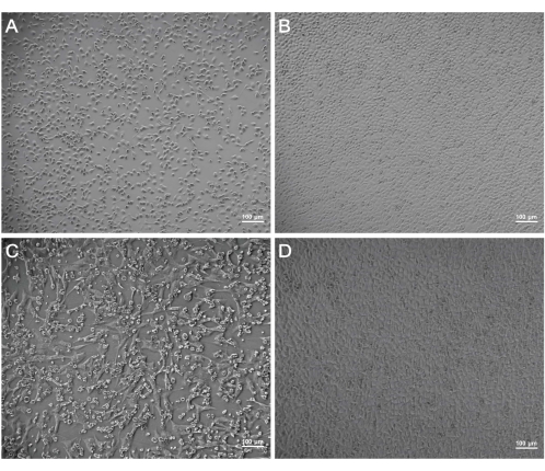
Figure 1: CPE observation of cells after the incubation with the viral supernatants. The status of C6/36 cells at 120 h post infection (CPE) (A) and that of the control cells at the same time (B). The status of BHK-21 cells at 48 hpi (C) and that of the control cells (D). Scale bars = 100 µm. Abbreviations: CPE = cytopathogenic effect; hpi = hours post infection. Please click here to view a larger version of this figure.
Figure 2: PCR results for viral supernatants. Lane 1 represent the positive control of bunyaviruses (Ebinur Lake Virus). Lanes 2-4 represented the PCR results of supernatants by using universal primers for bunyaviruses (251 bp), flaviviruses (266 bp), and alphaviruses (434 bp). Please click here to view a larger version of this figure.
Virus | Primer | Oligonucleotide (5'→3') | The size of PCR product (bp) | ||
Flaviviruses | F1 | TACAACATGATGGGAAAGAGAGAGAA | 266 | ||
F2 | GTGTCCCAGCCGGCGGTGTCATCAGC | ||||
Alphaviruses | M2w | YAGAGCDTTTTCGCAYSTRGCHW | 434 | ||
cMw3 | ACATRAANKGNGTNGTRTCRAANCCDAYCC | ||||
Bunyaviruses | BCS82C | ATGACTGAGTTGGAGTTTCATGATGTCGC | 251 | ||
BCS332V | TGTTCCTGTTGCCAGGAAAAT |
Table 1: Universal primers for the arbovirus detection.
The objective of this method was to offer a practical way for isolating mosquito-associated viruses using various cell lines. It is critical to add the Antibiotic-Antimycotic (Penicillin-Streptomycin-Amphotericin) to the supernatants of the mosquito homogenates to avoid contamination by bacteria or fungi. Mosquitoes and viral supernatants obtained in the field must be refrigerated at -80 °C to avoid repeated freeze-thaw cycles.
Another critical step in the protocol was grinding. Mosquito samples should be thoroughly powdered and stored on ice. Insufficiently ground mosquito tissues can induce cell death and may skew the findings of CPE analysis. Prior to virus isolation, it is necessary to employ normal mosquitoes to confirm the grinding parameters. Grinding must be done at low temperatures to prevent the virus from becoming inactive during the process.
This approach can effectively isolate and amplify mosquito-associated viruses, but is only acceptable for viruses that may display CPE in mammalian or insect cell lines. This approach is inappropriate for viruses that do not induce CPE. Using the protocol, we may obtain viral candidates that contain only one virus type or a mixture with different viruses. Further investigation — viral purification, morphological identification, and plaque analysis — is still needed to obtain the purified virus.
The authors have nothing to disclose.
This work was supported by the Wuhan Science and Technology Plan Project (2018201261638501).
0.22 µm membrane filter | Millipore | SLGP033RB | Polymer films with specific pore ratings.To remove cell debris and bacteria. |
24-well plates | CORNING | 3524 | Containers for cell |
75 cm2 flasks | CORNING | 430641 | Containers for cell |
a sterile 2 mL tube with 3 mm ceramic beads | |||
Antibiotic-Antimycotic | Gibco | 15240-062 | Antibiotic in the medium to prevent contamination from bacteria and fungi |
Automated nucleic acid extraction system | NanoMagBio | S-48 | |
BHK-21 cells | National Virus Resource Center, Wuhan Institute of Virology | ||
C6/36 cells | National Virus Resource Center, Wuhan Institute of Virology | ||
Centrifugal machine | Himac | CF16RN | Instrument for centrifugation of mosquito samples |
CO2 | |||
Dulbecco’s minimal essential medium (DMEM) | Gibco | C11995500BT | medium for vertebrate cell lines |
Ebinur Lake virus | Cu20-XJ isolation | ||
Feta Bovine Serum (FBS) | Gibco | 10099141C | Provide nutrition for cells |
high-speed low-temperature tissue homogenizer | servicebio | KZ-III-F | Instrument for grinding |
incubator (28 °C) | Panasonic | MCO-18AC | Instrument for cell culture |
incubator (37 °C) | Panasonic | MCO-18AC | Instrument for cell culture |
PCR tube | |||
penicillin-streptomycin | Gibco | 15410-122 | Antibiotic in the medium to prevent contamination from bacteria |
Penicillin-Streptomycin-Amphotericin B Solution | Gibco | 15240096 | |
Refrigerator (-80 °C) | sanyo | MDF-U54V | |
Roswell Park Memorial Institute medium (RPMI) | Gibco | C11875500BT | medium for mosiquto cell lines |
Screw cap storage tubes (2 mL) | biofil | FCT010005 | |
sterile pestles | Tiangen | OSE-Y004 | Consumables for grinding |
TGrinder OSE-Y30 electric tissue grinder | Tiangen | OSE-Y30 | Instrument for grinding |
The dissecting microscope | ZEISS | stemi508 | |
the light traps MXA-02 | Maxttrac | ||
The mosquito absorbing machine | Ningbo Bangning | ||
The pipette tips | Axygen | TF | |
The QIAamp viral RNA mini kit | QIAGEN | 52906 | |
Tweezers | Dumont | 0203-5-PO |