The present protocol describes the techniques for the systematic assessment of skull specimens to characterize anatomical and developmental variations and abnormalities of the teeth, periodontal disease, endodontal disease, and temporomandibular joint pathology.
Museum skull specimens represent a non-invasive, informative, and readily available means to study temporomandibular joint (TMJ) lesions, dental pathology, and anatomic variations in many mammalian species. Studying the teeth and jaws of an array of species can present a challenge requiring attention to detail and understanding of a species' normal anatomy. In the present article, a systematic and precise protocol for examining skull specimens is discussed that has been applied to a variety of mammals to define characteristic diseases in the oromaxillofacial region. The procedure outlined is simultaneously precise, repeatable, and adaptable to the highly differing skull and tooth shapes and anatomy across species. Specifically, specimens are examined for missing teeth, periodontal disease, endodontal disease, TMJ pathology, and anatomical variations. Results gleaned from research on museum specimens may reflect the natural history, health, and disease status of individuals and species. Furthermore, these data can inform ecological and conservation research efforts, as well as the care of captive individuals.
The development of jaws and teeth marks a critical time point in the evolution and development of vertebrates. While jaws initially developed as part of a mechanism of respiration in aquatic and marine species, teeth offered a new manner of apprehending and processing prey items1,2. Since the development of jaws and teeth, organisms have evolved innumerable variations in anatomy that correspond to their function and reflect the ecologic role to which they belong. Due to their mineralized nature, teeth and skulls represent a bounty of information that persists in the environment and fossil record and can offer myriad insights into the ecology, health status, and behavior of individuals and, by extension, species.
The acquisition of information pertaining to the teeth and jaws of animals and characterizing form and pathology has many benefits. Recognizing common disease processes can improve conservation efforts of wild species and optimize the care of captive animals3,4,5. For example, information gleaned from museum skull specimens has been used to make inferences on the exposure of the Baltic grey seal (Halichoerus grypus) and harbor seals (Phoca vitulina) to environmental pollutants such as organochlorines over time6,7, although a causative relationship between orofacial lesions and pollutants has not been confirmed. Furthermore, diseases of the oral cavity are some of the most prevalent diseases in domestic species, and understanding the oral health status of wild species may advance the clinical medicine and management of domestic species8,9.
As animals have developed such variation in normal craniofacial shape and dentition, it can be challenging to characterize and compare these aspects between species. Understanding the ecology and natural behavior of an organism, as well as its typical environment, is imperative before attempting to examine its skull. Doing so will drive the formation of questions and hypotheses about a particular species' dentition and inevitably enrich the conclusions from data analysis. For example, recognizing that the typical diet of the Southern sea otter (Enhydra lutris nereis) includes hard-shelled mollusks, crustaceans, and echinoderms is essential to contextualize the degree and effect of attrition and/or abrasion of the teeth10,11. Although one can assume the likelihood that an individual of a species will develop certain dental diseases, it is critical to have a systematic, precise, and reproducible protocol for evaluating dental pathology. This should include an appraisal of occlusion, anatomical and developmental findings, periodontal disease, endodontal findings, and temporomandibular joint (TMJ) pathology. Developing such a protocol with similar statistical analysis will allow for a detailed comparison of dental and TMJ disease from species to species. A systematic method has been utilized to characterize dental and temporomandibular joint pathology in many mammalian species and has proven to be translatable to organisms with diverse forms11,12,13,14,15,16,17,18,19,20,21,22,23,24.
To compare future data on additional species, it is important to have an accepted method for assessing diseases of the teeth and jaws that can be applied to a variety of species. This article aims to detail a standardized and organized approach for assessing the dental and TMJ pathology of skull specimens.
The present study was conducted using specimens from the Department of Ornithology and Mammalogy, California Academy of Sciences, San Francisco, the Museum of Vertebrate Zoology, University of California, Berkeley, and the Museum of the North, University of Alaska, Fairbanks. Permission to examine skull specimens and publish works from the data was obtained from the museums that own and manage each collection.
1. Specimen selection and documentation
2. Anatomical and developmental findings
3. Periodontal status
4. Fractured teeth and periapical lesions
5. Attrition/abrasion
6. Temporomandibular joint pathology
7. Trauma
8. Checking of other parameters
The current protocol results in a combination of objective and semi-subjective data, and the positive outcome depends on the accurate and repeatable assessment of specimens. Multiple observers with knowledge of the normal anatomy of the target species and an understanding of general dental and maxillofacial pathology ideally must be present to assess each specimen to minimize bias systematically. The assessment of each specimen must be discussed, and a consensus needs to be obtained. No threshold for the number of specimens has been suggested to ensure a representative sample, but published studies have included 76 to 1,205 individuals11,20. Additionally, a specimen free of any detectable pathology or minimal disease or anatomic variation must be found and documented to act as a standard for comparison (Figure 7). Furthermore, a successful outcome depends on the differentiation of pathology from artifactual damage. Often, specimens may incur artifactual damage during processing or storage. For this reason, for accurate data collection, the researcher must familiarize themselves with the general anatomy and pathophysiology of the maxillofacial region to understand the evidence of true chronic and acute disease processes. If there is evidence supporting that a finding is artifactual, such as evidence of post-mortem tampering with human tools or injuries to teeth or structures that would be biomechanically impossible in life, these findings must not be recorded as true pathology. If radiographs are utilized for specimen assessment, appropriate radiographic views need to be obtained for diagnostic quality38,39. Finally, pathology and anatomic anomalies must be documented with precise descriptions and/or high-resolution photographs.
Results may not be representative if normal anatomy is interpreted as pathology or if data are noted to have a high degree of variation between observers. Specimens must be examined carefully to preserve their quality as diligently as possible to allow for accurate repeat assessment in the future if necessary. Additionally, if meticulous record-keeping is not maintained, pathology or anatomical anomalies may be assigned to incorrect specimens, and the conclusions drawn from correlations from the data will be of questionable accuracy.
Data should be statistically analyzed to determine if there are differences in the frequency of abnormalities associated with the age of the individual at the time of death and sex. The frequency of dental abnormalities can be compared between different subspecies, geographic locations, and historic time points. Finally, data can be compared with previously published data on species of different genera.
The current protocol has proven to be an effective means of characterizing dental and TMJ abnormalities in various species11,12,13,14,15,16,17,18,19,20,21,22,23,24. The presentation of the results of future reports must follow a similar pattern to that of the previous manuscripts to maintain the completeness and to be able to compare results easily.
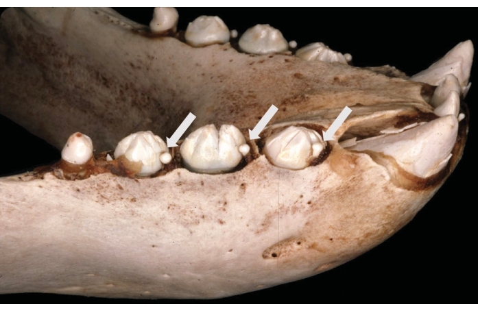
Figure 1: Persistent deciduous teeth in the Northern elephant seal (Mirounga angustirostris). Shown are persistent deciduous right mandibular first, second, and third premolar teeth (arrows) mesial to the corresponding permanent successor teeth. This figure has been modified from Abbott et al.12. Please click here to view a larger version of this figure.
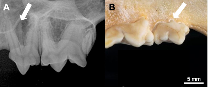
Figure 2: Supernumerary roots in the California bobcat (Lynx rufus californicus). (A) Radiographical and (B) gross appearance of a left maxillary third premolar tooth with an extra root (arrowed). This figure has been modified from Aghashani et al.19. Scale bar = 5 mm. Please click here to view a larger version of this figure.
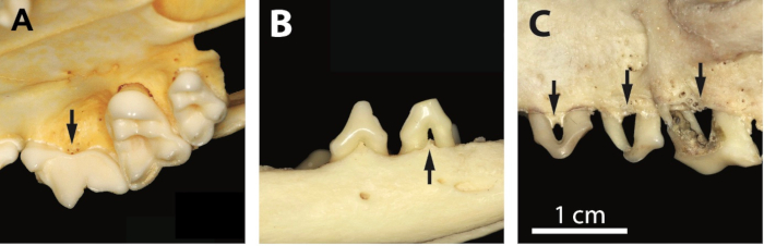
Figure 3: Stages of periodontitis in the grey fox (Urocyon cinereoargenteus). (A) Stage 2 associated with the right maxillary fourth premolar tooth, characterized by increased vascularization of the alveolar bone (arrow). (B) Stage 3 associated with the right mandibular second premolar tooth, characterized by moderate bone loss and furcation involvement (arrow). (C) Stage 4 associated with the left maxillary premolar teeth, characterized by severe alveolar bone loss (arrows). This figure has been modified from Evenhuis et al.22. Scale bar = 1 cm. Please click here to view a larger version of this figure.
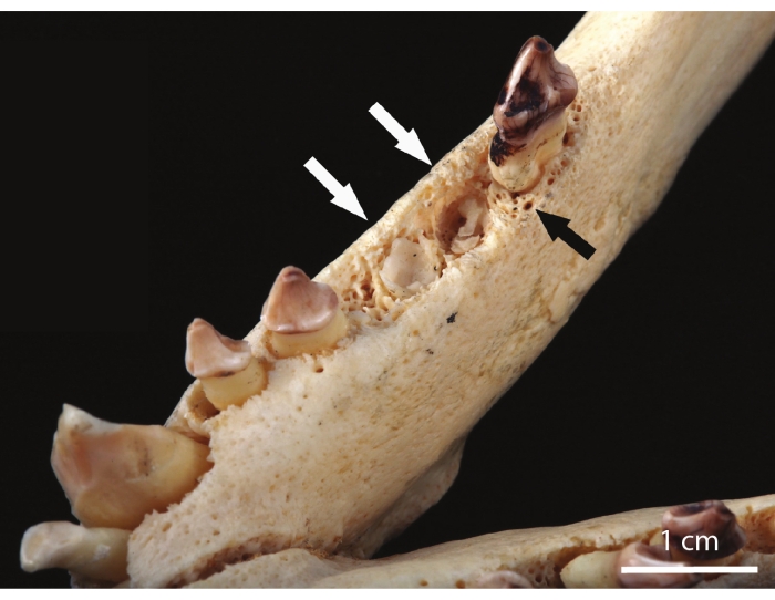
Figure 4: Fractured teeth in the Northern fur seal (Callorhinus ursinus). Root fractures of the right mandibular third and fourth premolar teeth (white arrows) and stage 2 periodontitis around the right mandibular first molar tooth (black arrow)13. Scale bar = 1 cm. Please click here to view a larger version of this figure.
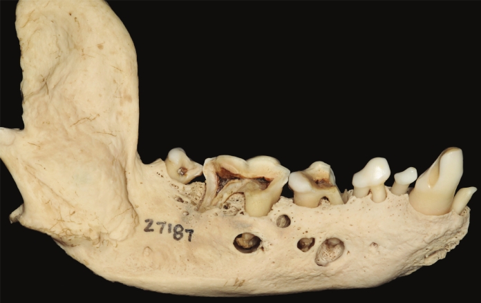
Figure 5: Periapical lesions in the Southern seal otter (Enhydra lutris nereis). Periapical disease associated with complicated crown root fractures of the right mandibular fourth premolar and first and second molar teeth in an adult female specimen. This figure has been modified from Winer et al.24. Please click here to view a larger version of this figure.
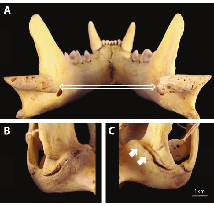
Figure 6: TMJ osteoarthritis in the California mountain lion (Puma concolor cougar). (A) Image of the irregular articular surface, subchondral bone exposure, and porosity of the mandibular condylar processes (open arrows). (B) The right TMJ exhibits no significant periarticular proliferation at the retroarticular process. (C) The left TMJ exhibits periarticular bony proliferation at the retroarticular process (closed arrows), partially encircling the mandibular head, resulting in partial ankylosis. This figure has been modified from Aghashani et al.21. Scale bar = 1 cm. Please click here to view a larger version of this figure.
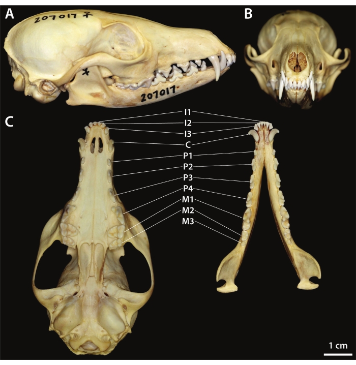
Figure 7: Normal dentition of the kit fox (Vulpes macrotis). (A) Right lateral view. (B) Rostral view. (C) Ventral view of the maxilla (left) and dorsal view of the mandibles (right). This figure has been modified from Yanagisawa et al.23. Scale bar = 1 cm. Please click here to view a larger version of this figure.
Degree of Periodontitis | Clinical Features | ||
Periodontitis stage 2 | Evidence of increased vascularity at the alveolar margin (more prominent vascular foramina in, and slightly rougher texture of, the bone of the alveolar margin) | ||
Periodontitis stage 3 | Rounding of the alveolar margin; moderate horizontal or vertical bone loss | ||
Periodontitis stage 4 | Widening of the periodontal space; severe horizontal or vertical bone loss; tooth mobile in the alveolus; furcation exposure |
Table 1: Summary of clinical features of progressive stages of periodontitis.
Fracture Type | Description | |
Enamel fracture | A chip fracture or crack of the enamel only. | |
Uncomplicated crown fracture | A fracture involving enamel and dentine, but not exposing the pulp. | |
Complicated crown fracture | A fracture involving enamel and dentine, with pulp exposure. | |
Uncomplicated crown–root fracture | A fracture involving enamel, dentine and cementum, but not exposing the pulp. | |
Complicated crown–root fracture | A fracture involving enamel, dentine and cementum, with pulp exposure. | |
Root fracture | A fracture affecting dentine, cementum and the pulp. |
Table 2: Summary of clinical features of tooth fracture types.
Stage of Attrition/abrasion | Description | |
Attrition/abrasion stage 1 | Mild wear of enamel, without dentine exposure | |
Attrition/abrasion stage 2 | Exposure of dentine on the cuspal tip, without tertiary dentine formation | |
Attrition/abrasion stage 3 | Exposure of dentine on the cuspal tip, with tertiary dentine formation | |
Attrition/abrasion stage 4 | Pulp cavity exposure secondary to attrition/abrasion |
Table 3: Summary of clinical features of tooth attrition and abrasion.
Severity of Osteoarthritis (OA) | Clinical Features | ||
Mild OA | There is any evidence of early lesions of periarticular new bone formation/osteophytes with minimal or no subchondral bone changes. | ||
Moderate OA | There is periarticular new bone formation and/or subchondral bone changes are more pronounced. | ||
Severe OA | All previously described signs are present and more pronounced, or if subchondral bone lysis is present. Partial or complete ankylosis may be observed. |
Table 4: Summary of clinical features of TMJ osteoarthritis.
The anatomy of the teeth and jaws is a quintessential example of divergent evolution and is a true reflection of a species' natural history, behavior, and health status. An individual's oral health may play directly into their survival and fitness. The current study outlines a systematic, reproducible, and detailed manner of assessing the dental health and TMJ abnormalities of museum specimens that may reflect pathology in live populations.
Despite dental diseases universally affecting mammals of a wide variety of genera, this topic remains underexplored. Understanding dental and craniofacial disorders in wild, captive, and domestic species has implications for conservation efforts, zoological husbandry, companion animal healthcare, and animal-related industry3,32,33,40,41. Therefore, studies on dental diseases could be key for improving and understanding animal health as a whole.
Critical steps in the current assessment algorithm include research into a species' natural behavior prior to data collection, understanding the target species' normal anatomy, and systematically and precisely assessing each tooth and anatomical landmark. It is also imperative to report results in a systematic and detailed manner so that comparisons between species can be made. Failure to complete these steps in an organized manner can lead to missing or inaccurate data or a misinterpretation of findings.
The system presented successfully characterizes many species, but the method has limitations. First, the examination is performed on museum specimens, and the quality of the specimens is subject to their preparation, care, and storage method. Therefore, artifacts may arise and could be inadvertently interpreted as true pathology present ante mortem. Additionally, the study population is inevitably biased toward individuals found in regions that humans could access easily. Furthermore, studies conducted in such a manner could overestimate the prevalence of dental diseases or craniofacial abnormalities that truly exist in a species. These abnormalities could reduce an individual's fitness. In contrast, the study design cannot evaluate diseases of the oral and facial soft tissues. It is also important to consider how individuals in a collection died and were collected, as this may impact the observed frequency of pathology. Individuals dispatched in large-scale hunting or trapping events may show increased tooth fractures or skull trauma prevalence compared with the general population22. In contrast, populations that experienced acute or subacute large-scale mortality events that do not have a known effect on the craniofacial region may provide a more accurate representation of dental diseases and TMJ abnormalities at a given time point42,43. Finally, while the current study method offers a comprehensive analysis of possible dental and TMJ abnormalities in a given species, one cannot conclude how the abnormalities discovered may impact the fitness of an individual or the evolutionary pressures to which a species is subject.
In conclusion, a systematic review of museum specimens has proven to be effective in characterizing dental anomalies, dental disease, and TMJ and craniofacial pathology in many species. The current methods offer a non-invasive means of data collection that can reflect the potential for disease in wild populations. The continuation of research into the dental pathology of wild and captive species is necessary to understand the health status and biological niche of these species and offer evidence-based means of optimizing management.
The authors have nothing to disclose.
The authors thank the Department of Ornithology and Mammalogy, California Academy of Sciences, San Francisco, the Museum of Vertebrate Zoology, University of California, Berkeley, and the Museum of the North, University of Alaska, Fairbanks, for making their collections available for this research.
Arctos Collaborative Collection Management Solution | https://arctosdb.org | ||
Disposible Nitrile Gloves | |||
Double-Ended Dental Explorer/Probe, #2 Handle | Hu Friedy | 541-5860 | |
High resolution digital camera | |||
Light source | |||
Magnifying glass (Optional) | |||
Surgical Magnification Loupes (Optional) | Surgitel | EVC00TTL |