Presented here is a new version of expansion microscopy (ExM), Magnify, that is modified for up to 11-fold expansion, conserving a comprehensive array of biomolecule classes, and is compatible with a broad range of tissue types. It enables the interrogation of the nanoscale configuration of biomolecules using conventional diffraction-limited microscopes.
The nanoscale imaging of biological specimens can improve the understanding of disease pathogenesis. In recent years, expansion microscopy (ExM) has been demonstrated to be an effective and low-cost alternative to optical super-resolution microscopy. However, it has been limited by the need for specific and often custom anchoring agents to retain different biomolecule classes within the gel and by difficulties with expanding standard clinical sample formats, such as formalin-fixed paraffin-embedded tissue, especially if larger expansion factors or preserved protein epitopes are desired. Here, we describe Magnify, a new ExM method for robust expansion up to 11-fold in a wide array of tissue types. By using methacrolein as the chemical anchor between the tissue and gel, Magnify retains multiple biomolecules, such as proteins, lipids, and nucleic acids, within the gel, thus allowing the broad nanoscale imaging of tissues on conventional optical microscopes. This protocol describes best practices to ensure robust and crack-free tissue expansion, as well as tips for handling and imaging highly expanded gels.
Biological systems exhibit structural heterogeneity, from the limbs and the organs down to the levels of proteins at the nanoscale. Therefore, a complete understanding of the operation of these systems requires visual examination across these size scales. However, the diffraction limit of light causes challenges in visualizing structures smaller than ~200-300 nm on a conventional fluorescence microscope. In addition, optical super-resolution methods1,2,3, such as stimulated emission depletion (STED), photo-activated localization microscopy (PALM), stochastic optical reconstruction microscopy (STORM), and structured illumination microscopy (SIM), though powerful, present their own challenges, as they require expensive hardware and reagents and often have slow acquisition times and a poor ability to image large volumes in 3D.
Expansion microscopy4 (ExM) provides an alternative means of circumventing the diffraction limit of light by covalently anchoring biomolecules into a water-swellable polymer gel and physically pulling them apart, thus rendering them resolvable on conventional optical microscopes. A multitude of ExM protocol variants have been developed since the original publication of ExM less than a decade ago, and these protocols allow the direct incorporation of proteins5,6,7, RNA8,9,10, or lipids11,12,13 into the gel network by altering the chemical anchor or expanding the sample further (thus improving the effective resolution) either in a single step14 or multiple iterative steps15,16. Until recently, no single ExM protocol could retain these three biomolecule classes with a single commercially available chemical anchor while providing a mechanically sturdy gel that could expand ~10-fold in a single expansion round.
Here, we present Magnify17, a recent addition to the ExM arsenal that uses methacrolein as the biomolecule anchor. Methacrolein forms covalent bonds with tissue like that of paraformaldehyde, ensuring that multiple classes of biomolecules can be retained within the gel network without requiring various specific or custom anchoring agents. Additionally, this technique can expand a broad spectrum of tissues up to 11-fold, including notoriously challenging samples such as formalin-fixed paraffin-embedded (FFPE) clinical samples. Previous methods for expanding such mechanically rigid samples required harsh protease digestion, rendering the antibody labeling of proteins of interest impossible after the sample had been expanded. In contrast, this technique achieves the expansion of FFPE clinical samples using a hot denaturing solution, thus preserving whole protein epitopes within the gel, which can be targeted for post-expansion imaging (Figure 1).
All the experimental procedures involving animals were conducted in accordance with the National Institutes of Health (NIH) guidelines and were approved by the Institutional Animal Care and Use Committee at Carnegie Mellon University. Human tissue samples were commercially obtained.
1. Preparation of the stock reagents and solutions
NOTE: Refer to the Table of Materials for a list of the reagents used.
2. Tissue preparation for archived and freshly prepared clinical tissue slides
NOTE: The tissue pre-processing steps differ based on how the specimens are prepared.
3. Tissue preparation for paraformaldehyde-fixed mouse brain
4. Gelling
NOTE: This protocol is suitable for all tissue types prepared for use with this technique.
5. Sample digestion and tissue expansion
NOTE: This protocol is suitable for all tissue types.
6. Post-expansion biomolecule profiling
If the protocol has been successfully completed (Figure 1), the sample will appear clear and flat after heat denaturation; any folding or wrinkling indicates incomplete homogenization. A successfully expanded sample will be 3-4.5-fold larger than before expansion in 1x PBS and 8-11-fold larger when fully expanded in ddH2O. Figure 3 shows example pre- and post-expansion images of 5 µm thick FFPE human kidney sample processed using this protocol and successfully expanded over 8-fold. The tissue was first stained with antibodies for ACTN4 along with DAPI to visualize the nuclear DNA. The specimen was then imaged using a spinning disk confocal microscope (Figure 3A). The tissue was treated following the above protocol, including the post-expansion staining of the same targets, and fully expanded in water (Figure 3B) before re-imaging. Following heat denaturation, biomolecules other than proteins may be stained for and imaged as well, as shown in Figure 4. Lipophilic dyes can be applied to reveal the cell and mitochondria membrane structures in the mouse brain (Figure 4A). Additionally, nucleic acids may be imaged through FISH, as in the FFPE normal human lymph node tissue shown in Figure 4B,C.
Figure 5 demonstrates a likely outcome if the sample is not properly homogenized. Following expansion, the tissue was stained with antibodies for ACTN4 and vimentin, along with DAPI to visualize the nuclear DNA and wheat germ agglutinin (WGA) to label the carbohydrates. The tissue was expanded 3.5-fold in PBS before imaging using a spinning disk confocal microscope. In one kidney section (Figure 5A,C), the crack-free expansion of a glomerulus can be seen. In a separate section (Figure 5B,D), cracking can be clearly seen in the tissue.
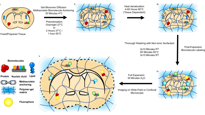
Figure 1: Schematic of the Magnify workflow. The pre-processing of clinically archived tissue slides is first performed based on the storage format. Free-floating paraformaldehyde-fixed sections need only be washed in PBS. The samples are then incubated in a gel monomer solution, including methacrolein to anchor the biomolecules to the hydrogel. In situ polymerization is performed prior to heat denaturation with urea, SDS, and EDTA. The samples are then thoroughly washed and stained using conventional immunostaining protocols, FISH protocols, or lipophilic dyes. The samples are then expanded in pure water before imaging. This figure has been modified from Klimas et al.17. Please click here to view a larger version of this figure.
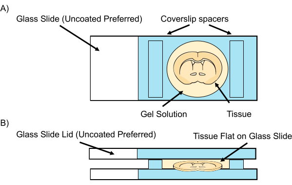
Figure 2: Tissue sample gelation chamber. (A) On each side of the tissue, two spacers, such as two pieces of #1.0 cover glass, are placed before the gel monomer solution is allowed to diffuse at 4 °C. To prevent compression, the spacers should be thicker than the tissue slices. (B) A lid, such as a second glass slide, is used to cover the sample before polymerization at 37 °C. Please click here to view a larger version of this figure.
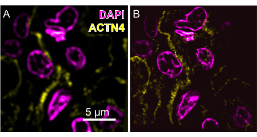
Figure 3: Example pre-expansion images of a human kidney tissue section. (A) An image taken at 60x magnification (1.4 NA) compared to (B) an image of the same field of view post-expansion with Magnify taken at 40x magnification (1.15 NA, expansion factor: 8.15x). Magenta, DAPI; Yellow, ACTN4. The maximum intensity of the post-expansion images is projected over 25 frames. Please click here to view a larger version of this figure.
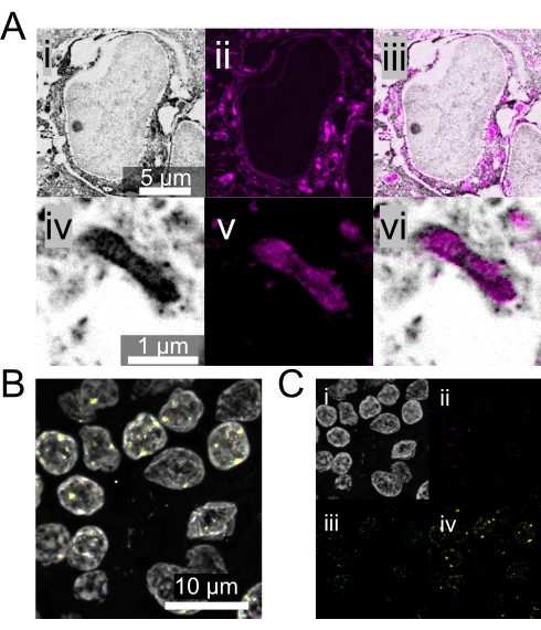
Figure 4: Alternative biomolecule staining strategies with Magnify. (A) (i and iv) NHS-ester pan-protein labeling of fully-expanded mouse brain tissue. (ii and v) Lipophilic dye (DiD) labeling of the same mouse brain tissue. (iii and vi) The channels merged. (B) DNA FISH with Magnify using FFPE normal human lymph node tissue. Expansion factor: 3.5x in 1x PBS. White, DAPI; Magenta, serine/threonine kinase 1 gene; Blue, APC/C activator protein CDH1 gene; Yellow, human satellite sequence S4. (C) Individual channels associated with B. All the images were obtained using a spinning disk confocal microscope at 40x magnification (1.15 NA). Please click here to view a larger version of this figure.
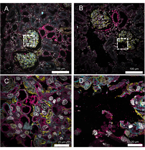
Figure 5: Representative results of 5 µm thick FFPE kidney samples. (A) Crack-free expansion. White, DAPI; Yellow, vimentin; Cyan, alpha-actinin 4; Magenta, wheat germ agglutinin. (B) Expanded kidney section exhibiting cracking. Cracking, distortions, and the loss of labeled targets can be the result of inadequate anchoring and/or homogenization. (C,D) Zoomed-in images of the boxed regions in A and B, respectively. All the images were obtained using a spinning disk confocal microscope at 10x (A,B; 0.45 NA) or 60x (C,D; 1.2 NA) magnification. Please click here to view a larger version of this figure.
Table 1: Gel monomer solution composition. Please click here to download this Table.
Table 2: Homogenization buffer composition. Please click here to download this Table.
Table 3: Summary of the methacrolein and homogenization conditions for validated tissues. Please click here to download this Table.
Here, we present the Magnify protocol17, an ExM variant that can retain multiple biomolecules with a single chemical anchor and expand challenging FFPE clinical specimens up to 11-fold with heat denaturation. The key changes in this protocol that distinguish it from other ExM protocols include the use of a reformulated gel that remains mechanically robust even when fully expanded, as well as the use of methacrolein as the biomolecule anchor. The most critical steps in this protocol are as follows: 1) the composition of the final gel solution; 2) the timing of the gelation steps; 3) the setup of the gelation chamber; 4) the parameters for sample homogenization; and 5) the sufficient washing of SDS from the sample before the post-expansion staining.
The most critical parameter for this protocol is the composition of the final gelling solution, particularly the concentration of the biomolecule anchor methacrolein. Different methacrolein concentrations are required to expand different tissue types (Table 3), and care must be taken to ensure this value is well-matched to the sample to be expanded. Over-anchoring with methacrolein can result in a reduced expansion factor and a loss of epitopes available for post-expansion staining, while under-anchoring, especially in FFPE clinical samples, can result in tissue cracks or distortion. Therefore, the methacrolein concentration must be optimized for unvalidated tissue types, although the optimal concentration will likely fall near or within the range presented here. While we expect that this protocol will work for most tissue types, even those we have not yet validated, it is known not to work for samples containing bone.
Beyond methacrolein, using an incorrect concentration of a critical gel ingredient (4HT or TEMED) can result in the complete failure of the experiment, either due to incomplete or no gelling (excessive 4HT) or premature gelation (excessive TEMED, reduced 4HT, or adding APS before the other components). To prevent premature gelation, it is also necessary to keep the sample and gel at 4°C and exposed to air for 30 min and to only cover the sample and place it at 37°C in a humidified chamber after the diffusion process is complete.
When constructing the gel chamber, including spacers between the two uncoated glass slides is critical, especially for thicker tissue sections such as PFA-fixed mouse brain. A lack of spacers can cause tissue compression, resulting in distorted images and inaccurate data.
The sample homogenization depends on the digestion time with the digestion buffer, as well as the digestion buffer’s temperature, and composition. Inadequate homogenization can cause distortions and reduced expansion factors compared to the reported value for a given tissue type. If incomplete homogenization is suspected, the digestion time can be increased, particularly in the case of thicker tissues.
A simple yet crucial step is the adequate washing-out of the SDS from the sample with a non-ionic surfactant (such as C12E10) after the homogenization step. Any leftover SDS can result in suboptimal or entirely hindered antibody binding. Fortunately, if this is suspected, further washing and the re-application of the antibodies will often be a satisfactory solution.
The protocol provides a cost-effective alternative to current super-resolution imaging and electron microscopy techniques to interrogate nanoscale structures in various tissue samples, including in FFPE clinical specimens. The use of methacrolein and heat denaturation allows the post-expansion profiling of any biomolecules that are preserved during tissue fixation (this precludes the imaging of lipids in FFPE samples, for instance). Our protocol, as an extension of the ExM framework, is modular and likely compatible with other techniques such as optical super-resolution methods (STED18, STORM19) or iterative expansion microscopy (iExM)15. However, these have yet to be tested with the presented protocol, and the large expansion factors may present challenges, especially with fluorophore dilution. Additionally, the large size of the samples after full expansion in water necessitates care when handling (although the gel used here is more resilient than previous high-expansion factor ExM gel formulations, such as ten-fold robust expansion microscopy (TREx), to handling miscues17), and creative solutions are occasionally required to transfer and image these fully expanded gels. For instance, using wide-based implements such as a thin sheet of plastic to transfer the fully expanded gels rather than a paintbrush, or using custom-made large imaging plates (in our hands, this means laser-cut or 3D-printed plates with large pieces of #1.5 coverglass adhered to the bottom; these can be seen in the accompanying video). Most importantly, this method broadens the applicability of nanoscale imaging by allowing the nanoscale imaging of common biological and pathological sample preparations on conventional wide-field or confocal microscopes.
The authors have nothing to disclose.
This work was supported by Carnegie Mellon University and the D.S.F. Charitable Foundation (Y.Z. and X.R.), the National Institutes of Health (N.I.H.) Director's New Innovator Award DP2 OD025926-01, and the Kauffman Foundation.
4-hydroxy-TEMPO (4HT) | Sigma Aldrich | 176141 | Inhibitor |
6-well glass-bottom plate (#1.5 coverglass) | Cellvis | P06-1.5H-N | |
Acrylamide | Sigma Aldrich | A8887 | Gel Monomer component |
Ammonium persulfate (APS) | Sigma Aldrich | A3678 | Initiatior |
DAPI (1 mg/mL) | Thermo Scientific | 62248 | |
Decaethylene glycol mono dodecyl ether (C12E10) | Sigma Aldrich | P9769 | Non-ionic surfactant |
Diamond knife No. 88 CM | General Tools | 31116 | |
Ethanol | Pharmco | 111000200 | |
Ethylenediaminetetraacetic acid (EDTA) 0.5 M |
VWR | BDH7830-1 | Homogenization Buffer Component |
Forceps | |||
Glycine | Sigma Aldrich | G8898 | Homogenization Buffer Component |
Heparin | Sigma Aldrich | H3393 | |
Methacrolein | Sigma Aldrich | 133035 | Anchoring Agent |
Micro cover Glass #1 (24x60mm) | VWR | 48393 106 | |
Micro cover Glass #1.5 (24x60mm) | VWR | 48393 251 | |
N,N,N′,N′- Tetramethylethylenediamine (TEMED) |
Sigma Aldrich | T9281 | Accelerator |
N,N′-Methylenebisacrylamide (Bis) | Sigma Aldrich | M7279 | Gel Monomer component |
N,N-dimethylacrylamide (DMAA) | Sigma Aldrich | 274135 | Gel Monomer component |
Nunclon 4-Well x 5 mL MultiDish Cell Culture Dish | Thermo Fisher | 167063 | |
Nunclon 6-Well Cell Culture Dish | Thermo Fisher | 140675 | |
Nunc™ 15mL Conical | Thermo Fisher | 339651 | |
Nunc™ 50mL Conical | Thermo Fisher | 339653 | |
Orbital Shaker | |||
Paint brush | |||
pH Meter | |||
Phosphate Buffered Saline (PBS), 10x Solution | Fischer Scientific | BP399-1 | |
Polyethylene glycol 200 | Sigma Aldrich | P-3015 | |
Proteinase K (Molecular Biology Grade) | Thermo Scientific | EO0491 | |
Razor blade | Fischer Scientifc | 12640 | |
Safelock Microcentrifuge Tubes 1.5 mL | Thermo Fisher | 3457 | |
Safelock Microcentrifuge Tubes 2.0 mL | Thermo Fisher | 3459 | |
Sodium acrylate (SA) | AK Scientific | R624 | Gel Monomer component |
Sodium azide | Sigma Aldrich | S2002 | |
Sodium chloride | Sigma Aldrich | S6191 | |
Sodium citrate tribasic dihydrate | Sigma Aldrich | C8532-1KG | |
Sodium dodecyl sulfate (SDS) | Sigma Aldrich | L3771 | Homogenization Buffer Component |
Tris Base | Fischer Scientific | BP152-1 | Homogenization Buffer Component |
Triton X-100 | Sigma Aldrich | T8787 | |
Urea | Sigma Aldrich | U5378 | Homogenization Buffer Component |
Xylenes | Sigma Aldrich | 214736 | |
20x SSC | Thermo Scientific | AM9763 | |
Tween20 | Sigma Aldrich | P1379 | |
poly-L-lysine | Sigma Aldrich | P8920 |