Materialography is a method for microscopic structure imaging and analysis of structural components of solid materials. Quantitative image analysis methods, such as X-ray tomography, are helpful to characterize varied microstructures.
However, these often involve costly instrumentation. Optical microscope-based materialography is an affordable alternative to study solid materials. In a previous video on materialography, we covered the topic of sample preparation for optical materialography.
This video will now illustrate how to analyze the images of the prepared sample, using the principles of statistical methods and quantification of three-dimensional structure of a solid material.
From optical materialography, images are analyzed according to three main characteristics: porosity, grain density, and effective density.
Let us first look at the porosity. It is defined as the fraction of the volume of a material that is unoccupied by atoms. This void portion in a material determines its mechanical, electrical, and optical properties. It also affects its permeability. Statistically, the porosity is estimated on a representative two-dimensional slice of a sample, by the void area normalized by the total imaged area. By analyzing multiple images of the same sample, one obtains the mean void area of a sample. Similarly, by rasterizing the images, the average number of points, or pixels, line in void, normalized by the total probe points, gives the mean void points of a sample.
The second characteristic of polycrystalline materials is the grain density. Taking a rasterized image, it is estimated by quantifying the number of intersections of a grain with test lines. The mean number of intersections for all images is indicative of the average lateral dimension of a crystal grain. For high-porosity materials, the mean grain density can also be found through the mean porosity.
The third characteristic is the effective density. This takes into account the volume of pores in a material, and the global density of the material. Here the porosity can either be defined by the parameters A or P. We will now see how to analyze these three characteristics on images obtained from optical materialography.
The quantitative analysis of optical materialography images requires the pre-requisite procedure of sample preparation. Please refer to the video materialography part one for appropriate sample preparation protocol in four steps: cutting, mounting, polishing, and etching.
Let us now consider the prepared sample of a toroidal inductor core sample. Multiple images of the same sample are needed to perform the optical materialography analysis.
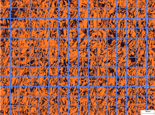
Use a digital analytical software where pixels can be categorized based on their brightness and counted accordingly. If not available, the analysis can be done by hand. Identify the void areas. Under Analyze in the menu, select Set Scale and choose the distance in pixels. Then select Image, Type, and 8-bit to change the image to grayscale. In the Process menu, select Binary and Make Binary to maximize the contrast of your image. Finally, choose Analyze Particles from the Analyze menu to measure the void area in micrometer units.
Take the sum of the void areas and normalize it by the total imaged area to obtain the parameter A. Repeat for all images to get the mean parameter A. Then, overlay a grid on the image. The intersection points are the test points. Count the number of test points. Identify porosity areas and count the total number of test points inside them. Normalize by the total number of test points to obtain the parameter P.
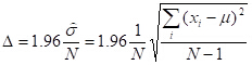
Repeat the calculation for all images to estimate the mean parameter P and the sampling error delta, where sigma is the standard deviation, n is the number of images, X-I is the sample I, and U is the sample average.
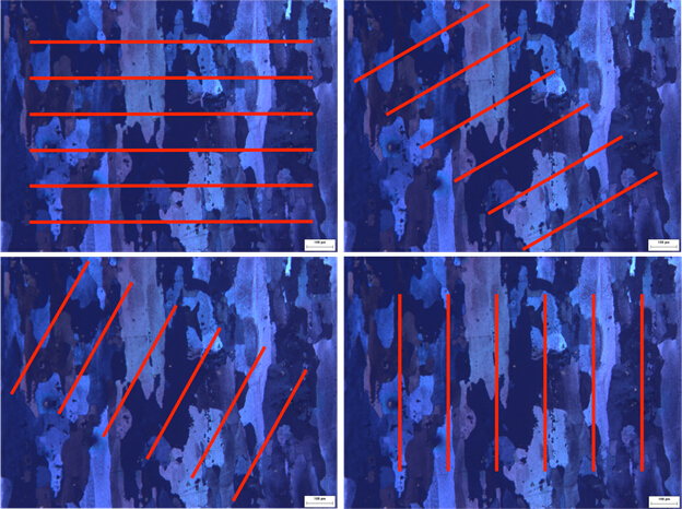
In the second step of the analysis, identify boundaries between neighboring grains, then superimpose a set of horizontal test lines on the image. Count the number of intersections between the test lines and the grain boundaries and evaluate the parameter I-L.
Repeat this step by rotating the lines by 90 degrees. Then repeat for all images. Calculate the mean intercept grain size in the horizontal direction and vertical direction. At last the grain size can be estimated.
Finally, rotate the lines at 30 degrees and 60 degrees and compare with previous vertical and horizontal cases. Observe the grain shape and the preferred angle of orientation. This is an indication of the anisotropy level of the sample.
Quantitative analysis of microscopic structure of solid materials with optical microscopy is useful for various applications. The study of grain size and shape in minerals contributes to the understanding of rock formation in extreme conditions.
For this reason materialographic analysis proves to be a useful method for planetary exploration. Polycrystalline samples may show various orientations of their grains. For example, in alloys used for oil pipelines, the orientational distribution function directly influences the axial and transverse mechanical strength of these alloys.
Materialography is routinely used to verify the quality of alloys that serve to build oil pipelines.
You’ve just watched Jove’s introduction to optical materialography. You should now understand the principles of image analysis used to investigate the microscopic structures of solids. You should also know how to determine porosity, grain size, and density for different materials.
Thanks for watching.