Institutional Animal Care and Use Committee at Drexel University approved all procedures (#20704).
1. Animal Arrival and Acclimation
2. Day of Experiment
3. Induction and Maintenance of Anesthesia
4. Monitoring and Care
5. Brachial Plexus Surgery
6. Biomechanical Testing
A representative load-time plot and strains from four segments of BP plexus (between four markers) are shown in Figure 5 and Figure 6, respectively. The obtained failure load of 8.3 N at 35% average failure strain reports the biomechanical responses of neonatal BP when subjected to stretch. Some regions of the nerve undergo higher strains than others, indicative of non-uniform injury along the length of the nerve. The camera data allows reporting the location of failure being proximal to the foramen.
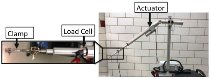
Figure 1: Details of in vivo mechanical testing device including the actuator, load cell, and clamp. Please click here to view a larger version of this figure.
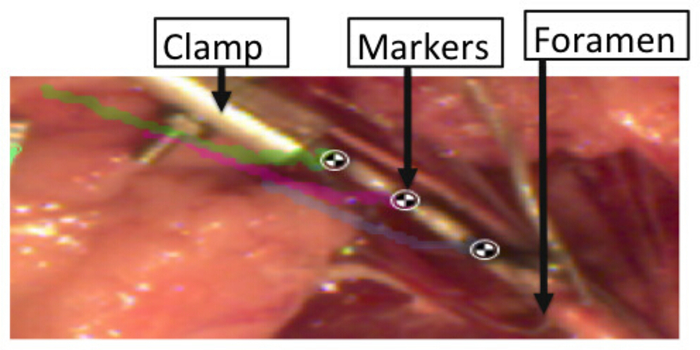
Figure 2: Markers placed over the BP segments to record strains sustained by the tissue during stretch. Please click here to view a larger version of this figure.
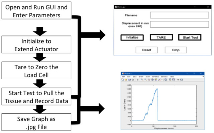
Figure 3: Steps for data acquisition using graphical user interface. Please click here to view a larger version of this figure.
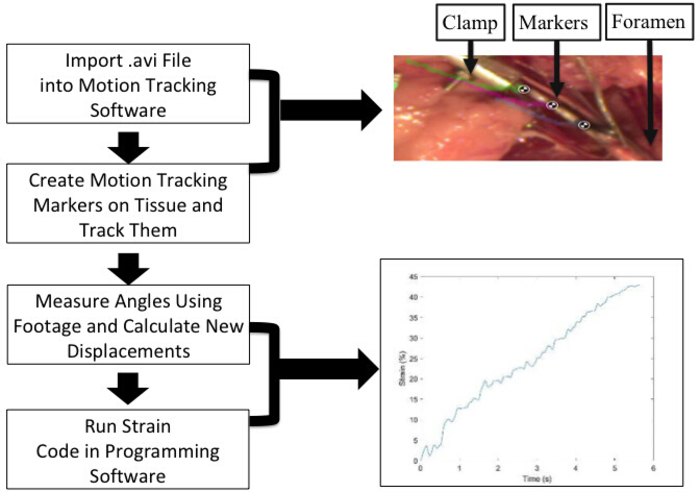
Figure 4: Marker tracking and strain analysis details. Test videos saved in AVI format are imported in the tracking software. Strain between each marker and the first and last markers are obtained as detailed. An average of between markers strains is used to report the failure strains. An example of nerve stretch with three markers and the calculated average strain-time plot are shown here, with reported failure strains of 43%. Please click here to view a larger version of this figure.
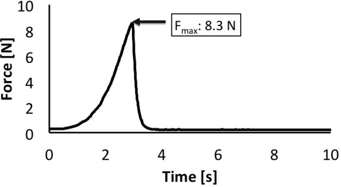
Figure 5: Maximum load reported during failure. Load cell attached to the actuator acquires the load data during stretch. The data are used to obtain a load-time plot as shown. Please click here to view a larger version of this figure.
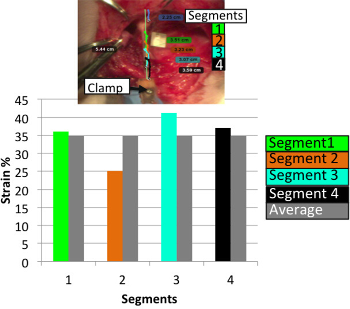
Figure 6: Strains reported in four different segments of the stretched plexus. Strains are calculated between each marker and compared against average strains obtained from all four segments (between each of the two adjacent markers). Some regions of the nerve undergo higher strains than others and the average strains indicative of non-uniform injury along the length of the nerve. Please click here to view a larger version of this figure.
Omega Subminature Tension & Compression Load Cell | Omega | LCM201-200N | 200N load cell |
Basler acA640-120uc camera | Basler | acA640-120uc | |
Feedback Linear Actuator | Progressive Automations | PA-14P | 10" stroke, 150lb force, 15mm/s speed |
Motion Tracking Software | Kinovea | N/A | Open Source |
Proramming Software – MATLAB | Mathworks | N/A | version 2018A |
Surgical instruments | |||
Forceps | Fine Science Tools Inc | 11006-12 and 11027-12 or 11506-12 | |
Hemostats | Fine Science Tools Inc | 13009-12 | |
Scissors | Fine Science Tools Inc | 14094-11 or 14060-09 |
Neonatal brachial plexus palsy (NBPP) is a stretch injury that occurs during the birthing process in nerve complexes located in the neck and shoulder regions, collectively referred to as the brachial plexus (BP). Despite recent advances in obstetrical care, the problem of NBPP continues to be a global health burden with an incidence of 1.5 cases per 1,000 live births. More severe types of this injury can cause permanent paralysis of the arm from the shoulder down. Prevention and treatment of NBPP warrants an understanding of the biomechanical and physiological responses of newborn BP nerves when subjected to stretch. Current knowledge of the newborn BP is extrapolated from adult animal or cadaveric BP tissue instead of in vivo neonatal BP tissue. This study describes an in vivo mechanical testing device and procedure to conduct in vivo biomechanical testing in neonatal piglets. The device consists of a clamp, actuator, load cell, and camera system that apply and monitor in vivo strains and loads until failure. The camera system also allows monitoring of the failure location during rupture. Overall, the presented method allows for a detailed biomechanical characterization of neonatal BP when subjected to stretch.