3D-printed fixtures were used to test 8-week old murine supraspinatus and Achilles tendons. All mechanically tested samples failed at the enthesis, as characterized by microCT scans, visual inspection, and video analysis after tensile tests. A one-to-one comparison of the previous and current methods for supraspinatus tendon testing in our laboratory is shown in Figure 3. In the previous method28,29,30, the humerus bone was embedded in epoxy and a paper clip was placed over the humeral head in an effort to prevent growth plate fracture. 4-6 hours were necessary to allow for the epoxy to fully cure (Figure 3), allowing for only 6-8 specimens to be tested in a typical day. A further limitation of the approach was the user-dependent effectiveness of the paper clip placement for preventing growth plate fracture. The testing results using these prior methods were highly variable, with coefficients of variation on the order of 30% for most parameters and growth plate failure rates of approximately 10%–20%. As summarized in Figure 3, specimen preparation time using the new methods was decreased to 5–10 minutes, making it practical to test 16–20 samples per day. Furthermore, growth plate failures were eliminated.
Compared to methodology reported by others for testing murine tendons14,15,17,25,28,29,30,31,32,33, the new methods were more efficient and reproducible. For supraspinatus tendons, structural properties such as maximum load (3.8 ± 0.6 N) and stiffness (12.7 ± 1.8 N/mm), as well as normalized material properties such as maximum stress (8.7 ± 3.0 MPa), and modulus (51.7 ± 13.5 MPa) had considerably lower coefficients of variations compared to results from the literature (Table 1). For the Achilles tendon, mechanical properties such as maximum load (7.8 ± 1.1 N) and stiffness (13.2 ± 1.9 N/mm) had lower coefficients of variations compared to results from the literature19,21,22,32,33,34,35,36,37,38, whereas maximum stress (24.2 ± 5.4 MPa) and modulus (73.2 ± 22.1 MPa) had coefficients of variations similar to those reported in the literature (Table 2).
Animal sex had a significant effect on the mechanical properties of the supraspinatus and Achilles tendons (Figure 4). When comparing male and female supraspinatus tendons, there were significant increases in maximum force (p = 0.002) and work to yield (p = 0.008). There were trends between the two groups for stiffness (p = 0.057), stress (p = 0.068), modulus (p = 0.061) and resilience (p = 0.078). When comparing male and female Achilles tendons, there were significant increases in maximum stress (p = 0.0006) and resilience (p = 0.0019). There were trends between the two groups for work to yield (p = 0.079), and modulus (p = 0.074) and no difference for maximum force (p = 0.1880) and stiffness (p = 0.6759).
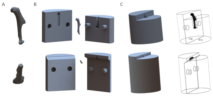
Figure 1: Representative 3D models of fixtures for the humerus (top row) and the calcaneus (bottom row). (A) 3D models of the bones. (B) Disassembled models of the fixtures. (C) Assembled models of the fixtures. Please click here to view a larger version of this figure.
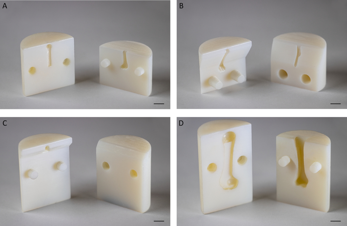
Figure 2: Representative 3D printed fixtures. (A) Fixture for biomechanical testing of supraspinatus tendons of 8-week old mice at an angle of 180° between the humerus and supraspinatus tendon. (B) Fixture for biomechanical testing of supraspinatus tendons of 8-week old mice at an angle of 135° between the humerus and supraspinatus tendon. (C) Fixture for biomechanical testing of murine Achilles tendons at an angle of 120° between the calcaneus and Achilles tendon. (D) Fixture for biomechanical testing of supraspinatus tendons of adult Sprague Dawley rats at an angle of 180° between the humerus and supraspinatus tendon. Scale bar: 5 mm. Please click here to view a larger version of this figure.
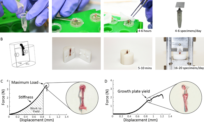
Figure 3: Comparison of previous and current methods for mechanical testing of murine supraspinatus tendons. (A) Previous specimen preparation methods used in our laboratory prior to mechanical testing: the humerus was potted in epoxy up to the humeral head to stabilize the bone, a paper clip was placed over the humeral head to prevent growth plate fracture, and, for the epoxy to cure, the specimens were left in room temperature for 4-6 hours prior to mechanical testing. (B) Specimen preparation methods used in the current study (Steps 1.2 and 2.1.4): Top left shows a 3D representation of the fixtures as produced by a solid modeling program. The 3D printed fixtures are reusable and easily assembled and disassembled. The bone end of the specimen is inserted into the fixtures, securing the growth plate and exposing the tendon for gripping and testing. The tendon end is glued between a folded thin tissue paper and inserted into the grips. Preparation time for each specimen is 10–15 minutes. (C) Representative load-deformation curves for tensile testing of supraspinatus tendon using current methods. (D) Representative load-deformation curve for tensile testing of supraspinatus tendon showing a growth plate failure. Please click here to view a larger version of this figure.
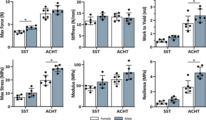
Figure 4: Sex effect on the mechanical properties of supraspinatus (SST) and Achilles (ACHT) tendons. There was a significant effect of sex on many of the mechanical properties based on unpaired t-tests (*sex effect, p < 0.05). Data shown as mean ± standard deviation. Please click here to view a larger version of this figure.
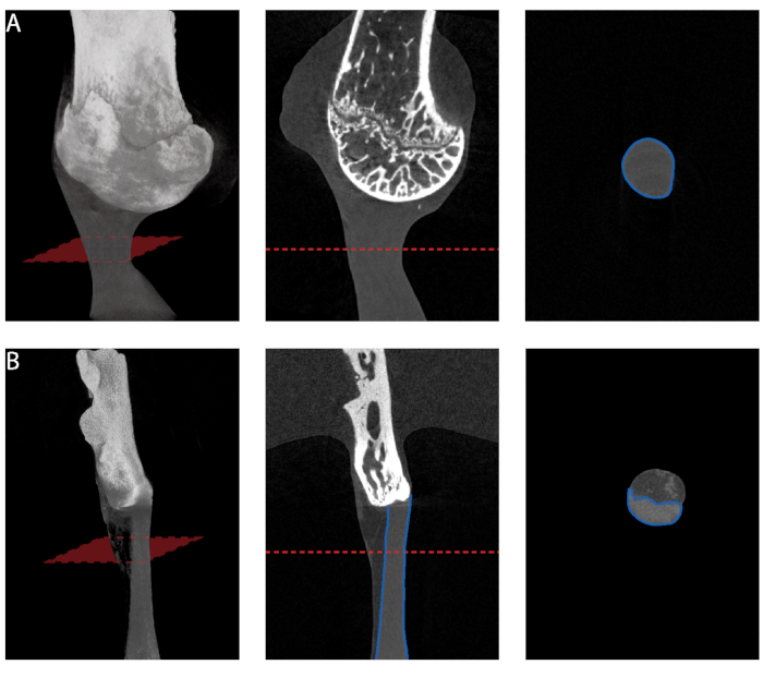
Figure 5: Cross-sectional area measurement from microCT. (A) Minimum cross-sectional area measurement along the length of supraspinatus tendon. (B) Minimum cross-sectional area measurement along the length of Achilles tendon. Only the tendon proper should be selected for measurement. Please click here to view a larger version of this figure.
| Structural Properties | Material Properties | |||||||||||
| Animals | Max Force (N) | Stiffness (N/mm) | Max Stress (Mpa) | Modulus (MPa) | ||||||||
| Autor | N | Background | Mean ± SD | COV(%) | Mean ± SD | COV (%) | Mean ± SD | COV (%) | Mean ± SD | COV (%) | ||
| Beason et al. Journal of Shoulder and Elbow Surgery (2013)15 | 10 | C57Bl/6 | 0.93±0.34 | 36.56 | 95.1±39.8† | 41.85 | 3.40±1.56 | 45.88 | 312.8±127.0 | 40.60 | ||
| Bell et al. Journal of Orthopaedic Research (2014)31 | 6 | C57Bl/6 | 1.22 ± 0.52 | 42.62 | 2.37 ± 1.6 | 67.51 | NR | NR | ||||
| Cong et al. Journal of Orthopaedic Research (2018)17 | 8 | C57Bl/6 | 5.38 ± 2.404# | 44.68 | 4.25 ± 1.67# | 39.29 | NR | NR | ||||
| Connizzo et al. Annals of Biomedical Engineering (2014)32 | 10 | NR (db/+) | NR | 84.44 ± 27.23*† | 32.25 | NR | 476 ± 186.27* | 39.13 | ||||
| Connizzo et al. Journal of Biomedical Engineering (2013)14 | NR | C57/BL6 | NR | NR | NR | 297 ± 148.90* | 50.13 | |||||
| Deymier et al. Acta Biomaterialia (2019)28 | 12 | CD-1 IGS Mouse (WT) | 5.0 ± 0.7 | 14 | 9.2 ± 2.9 | 31.52 | 33 ± 35 | 106.06 | NR | |||
| Eekhoff et al. Journal of Biomedical Engineering (2017)33 | 13 | Eln +/+ | NR | 8.50 ± 2.95 | 34.71 | 5.96 ± 3.23 | 54.19 | 101.2 ± 50.8 | 50.20 | |||
| Killian et al. FASEB Journal (2016)29 | 8 | C57BL/6 | NR | NR | 7.79 ± 2.61* | 33.50 | 58.32 ± 31.73* | 54.41 | ||||
| Schwartz et al. Bone (2014)25 | 20 | CD-1 IGS Mouse (WT) | 4.11 ± 0.79* | 19.22 | 8.58 ± 3.78* | 44.06 | 12.29 ± 5.95* | 48.41 | 133.80 ± 59.41* | 44.40 | ||
| Schwartz et al. Development (2015)30 | 12 | (Rosa-DTA (DTA) x Gli1-CreERT2 ) ScxCre;Smofl/fl (WT) | 4.16 ± 0.29* | 6.97 | 11.04 ± 1.98* | 17.93 | 26.24 ± 5.81 | 22.14 | 121.89 ± 44.18 | 36.25 | ||
| Average COV | 27.34 | Average COV | 38.64 | Average COV | 51.70 | Average COV | 45.02 | |||||
| New Method | 10 | C57BL/6J | 3.79 ± 0.62 | 16.41 | 12.73 ± 1.81 | 14.20 | 8.71 ± 3.04 | 34.91 | 51.67 ± 13.54 | 26.20 | ||
Table 1: Mechanical properties of supraspinatus tendons. Mean ± SD and coefficient of variation (COV) for structural and material properties estimated using new methods compared to ones reported in the literature. [NR: not reported, * estimated from figure(s), # standard deviation calculated from reported standard error, † measured deformation using optical stain lines].
| Structural Properties | Material Properties | |||||||||
| Animals | Max Force (N) | Stiffness (N/mm) | Max Stress (Mpa) | Young's modulus (MPa) | ||||||
| Autor | N | Background | Mean ± SD | COV(%) | Mean ± SD | COV (%) | Mean ± SD | COV (%) | Mean ± SD | COV (%) |
| Boivin et al. Muscles, Ligaments and Tendons Journal (2014)19 | 6 | Non-diabetic lean control mice | 8.1 ± 0.6 | 7.41 | 3.9 ± 0.7 | 17.95 | NR | 16 ± 3.7 | 23.13 | |
| Connizzo et al. Annals of Biomedical Engineering (2014)32 | 10 | db/+ | NR | 20.39 ± 2.43* | 11.92 | NR | 152.94 ± 44.12* | 28.85 | ||
| Eekhoff et al. Journal of Biomechanical Engineering (2017)33 | 8 | Eln +/+ | NR | 18.86 ± 3.37 | 17.87 | 10.55 ± 2.97 | 28.15 | 443.8 ± 131.7 | 29.68 | |
| Mikic et al. Journal of Orthopaedic Research (2006)34 | 20 | C57BL/6-J x 129SV/J | NR | NR | 18 ± 5 | 27.78 | 61 ± 20 | 32.79 | ||
| Probst et al. Journal of Investigative Surgery (2000)22 | 20 | BALB/c | 8.4 ± 1.1 | 13.10 | 6.3 ± 1.2 | 19.05 | NR | NR | ||
| Shu et al. Peer J (2018)21 | 9 | C57BL/6 | 9.6 ± 3.84 | 39.96 | 8.19 ± 3.63 | 44.32 | 27.55 ± 10.54 | 38.26 | NR | |
| Sikes et al. Journal of Orthopaedic Research (2018)35 | 7 | C57BL/6 | NR | NR | 19.53 ± 7.03 | 0.36 | 62.82 ± 20.20 | 32.16 | ||
| Wang et al. Journal of Orthopaedic Research (2006)36 | 9 | A/J | 8.4 ± 1.2 | 14.29 | 12.2 ± 2.8 | 22.95 | 78.2 ± 8.6 | 11.00 | 713.9 ± 203.7 | 28.53 |
| Wang et al. Journal of Orthopaedic Research (2006)36 | 8 | C57BL/6J | 10.2 ± 1.4 | 13.73 | 13.1 ± 2.5 | 19.08 | 97.4 ± 11.4 | 11.70 | 765.1 ± 179.6 | 23.47 |
| Wang et al. Journal of Orthopaedic Research (2006)36 | 7 | C3H/HeJ | 12.5 ± 1.7 | 13.60 | 14.1 ± 3.2 | 22.70 | 97.5 ± 10.9 | 11.18 | 708.6 ± 127.8 | 18.04 |
| Wang et al. Journal of Orthopaedic Research (2011)37 | 7 | C57BL/6 | 6.6 ± 1.7 | 25.76 | 8.2 ± 1.4 | 17.07 | 13.4 ± 3.7 | 27.61 | 86.8 ± 15.5 | 17.86 |
| Zhang et al. Matrix Biology (2016)38 | NR | CD-1 and C57BL/6J | 6.73 ± 3.74* | 55.57 | 12.03 ± 3.34* | 27.76 | 25.4 ± 15.14* | 59.61 | 632.31 ± 113.79* | 18.00 |
| Average COV | 22.93 | Average COV | 22.07 | Average COV | 23.96 | Average COV | 25.25 | |||
| New Method | 12 | C57BL/6J | 7.8 ± 1.08 | 13.91 | 13.19 ± 1.86 | 14.08 | 24.16 ± 5.42 | 22.45 | 73.17 ± 16.14 | 22.06 |
Table 2: Mechanical properties of Achilles tendons. Mean ± SD and COV for structural and material properties estimated using new methods compared to ones reported in the literature. [NR: not reported, * estimated from figure(s), # standard deviation calculated from reported standard error].
Supplemental Files. Please click here to download this file.