Part 1: Plant growth and maintenance
Nicotiana benthamiana plants used in the assay should be about 7 weeks old. They should be trimmed at least 4-5 days prior to the assay to remove all axillary branches and flowers. It is a good idea to remove axillary branches soon after they emerge in order to make the plants more manageable.
Part 2: Bacterial culture
DAY 1:
- Plates or cultures for all the strains used in the assay are initiated at the same time. For the Pseudomonas spp. which include 2 inducers and 3 challenge strains, streak bacteria onto KBM plates containing the appropriate antibiotics from frozen glycerol stocks (Table 2). A small amount of bacteria should be spread on the center of the plate. Incubate the plate at 30°C for about 18-24 hours. For Agrobacterium, start a liquid culture in 2 ml LB from a fresh plate and grow overnight at 250 r.p.m., 30°C.
DAY 2:
- For Pseudomonas, add 150-200 μl of liquid KBM to the bacteria and spread them over the entire plate using a sterile glass spreader. Put the plate back in the incubator and let it grow for another 20-24 hours. There should be a bacterial lawn on the plate the next day, indicating good growth. For Agrobacterium, start a secondary culture in about 5-10 ml LB with 20 μM acetosyringone and let it grow overnight to 20 hours at 250 r.p.m., 30°C.
Part 3: Preparing plants for the assay
DAY 2:
- One day before the assay the plants should be moved to a room at 22-24°C, ~35-40% RH and constant light.
- Mark circles on leaves that are at least 1.5 cm in diameter using a thick black marker. Two to four circles can be marked per leaf. The circles should be well spaced and preferably not cross a large vein. Choose well-expanded leaves. Avoid older leaves or leaves that are tough or thick to the touch and avoid marking circles on the lower portions of the leaf.
Marking circles a day prior to the experiment is not essential to the protocol. However, it saves time if there are large numbers of plants being assayed, and makes it easier to achieve precise timing between the induction of PTI and challenge inoculations.
Part 4: Induction of PTI
DAY 3:
- Harvest the cells from the plate in 10 mM MgCl2 for P. fluorescens and P. putida. For Agrobacterium, centrifuge the culture to collect cells and resuspend in 10mM MgCl2 + 10 mM MES pH 5.6. Repeat the centrifugation and resuspend in the MgCl2 -MES solution. Measure the optical density (O.D.) of the cells at 600 nM. Adjust the O.D.s to the final required values as described in Table 2. Pseudomonas should be finally resuspended in 10 mM MgCl2 and Agrobacterium in the MgCl2 –MES solution. A total volume of 25 ml is sufficient to inoculate about 40-50 spots.
- Infiltrate the PTI inducers into the pre-marked circles on the leaves using a 1 ml needleless syringe. Write down the order in which the plants were infiltrated and the exact time when the infiltration was begun.
Part 5: Challenge inoculation
- Prepare P. syringae pv. tomato DC3000, P.s.t. DC3000 ΔhopQ1-1 and P.s. pv. tabaci for the challenge as described in part 4.1 and Table 2.
- Seven hours after the inducer infiltration, perform the challenge infiltration in the same order of plants as the induction was done. Generally, a point on the periphery of the first inoculation circle can be used as the center of the second inoculation circle. If there are multiple spots on a leaf then make sure the infiltrations do not overlap.
- Plate serial dilutions of all the cultures used in the assay on KBM or LB plates containing the appropriate antibiotics to determine the exact C.F.U. number.
Part 6: Scoring for breakdown of PTI
Day 5:
- Look for the appearance of cell death due to ETI in the spots that were challenged with P.s.t. DC3000. Cell death inside the overlapping area of infiltration indicates a breakdown of PTI and should be scored as a positive phenotype.
Day 7:
- Look for the appearance of cell death due to disease in the spots that were challenged with P.s.t. DC3000 ΔhopQ1-1 or P.s. pv. tabaci. Score for a positive phenotype in the same manner as described in part 6.1.
When the control plants (that should not be compromised for PTI) begin to show cell death in the overlapping area of infiltration, stop making any further observations.
Part 7: Representative results
Figure 1 shows the outcome of an assay when P. fluorescens was used as the inducer and P.s.t. DC3000 as the challenge.
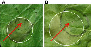
Figure 1. P. fluorescens was infiltrated onto N. benthamiana leaves (black circle) to induce PTI and 7 hours later, the spot was challenged with P.s.t DC3000 (white circle). Plants silenced for FLS2 showed a breakdown of PTI in the region where P. fluorescens was infiltrated (A), as seen by cell death. Control plants that were not silenced showed no cell death in the overlapping area due to induction of PTI (B). Red arrows indicate lack of or presence of cell death in the overlapping area of infiltration. Photographs were taken 2 days after the infiltrations. Please click here to see a larger version of figure 1.
| PTI inducer | Cell death-eliciting challenge | Nature of cell death | Code |
| Pseudomonas fluorescens 55 | Pseudomonas syringae pv tomato (P.s.t.) DC30006 | ETI | Pf/DC |
| P. putida KT2440 | P.s.t. DC3000 | ETI | Pp/DC |
| P. fluorescens 55 | P.s.t. DC3000 ΔhopQ1-16 | Disease | Pf/Q1-1 |
| Agrobacterium tumefaciens GV2260 | P. syringae pv. tabaci 11528R | Disease | Agro/Ptab |
Table 1: Combinations of PTI inducing and cell death-eliciting microbes used in the cell death assay for PTI.
| Bacterial strain | Selection medium | Final O.D. used in experiment | Corresponding C.F.U./ml |
| P. fluorescens 55 | KBM Ampicillin (100 ug/ml) | 0.5 | 1 x 10⁹ |
| P. putida KT2440 | KBM Ampicillin (100 ug/ml) | 0.5 | 1 x 10⁸ |
| A. tumefaciens GV2260 | LB Rifampicin (100 ug/ml) | 0.5 | 5 x 10⁸ |
| P.s.t. DC3000 | KBM Rifampicin (100 ug/ml) | 0.02 | 2 x 10⁷ |
| P.s.t. DC3000 ΔhopQ1-1 | KBM Rifampicin (100 ug/ml) | 100 fold dilution of 0.1 | 1 x 10⁶ |
| P. syringae pv. tabaci 11528R | KBM Rifampicin (100 ug/ml) | 100 fold dilution of 0.1 | 1 x 10⁶ |
Table 2:Culture conditions and inoculum levels used in the assay. The O.D. and corresponding C.F.U. levels may vary between different brand of spectrophotometers.