Cell Preparation
- Human keratinocytes (FEP-1811) were grown in Keratinocyte-Serum Free Medium (K-SFM; Invitrogen) supplemented with epidermal growth factor, bovine pituitary extract and 20 μg/ml gentamicin, at 37°C and 5% CO2.
- A single cell suspension was prepared by detaching with trypsin-EDTA (0.05% v/v)
- Cells were seeded in 8-well Lab Tek II microchamber slides (10,000 cells/well) and slides were incubated for 3 days at 37°C and 5% CO2.
Irradiation
- Cells were irradiated on ice with 2 Gy using a 137Cs source (Gammacell 1000 Elite irradiator; Nordion International, ON, Canada; 20.6 seconds/Gy)
- Unirradiated control and 2 Gy irradiated cells were incubated for 1 hour at 37°C and 5% CO2.
Immunofluorescence staining
- Media was tipped off and cells were washed with 300μl of PBS (w/o Ca2+ or Mg2+) per well and were rotated on an orbital mixer for 5 minutes.
- The buffer was tipped off and 100μl of freshly prepared 4% (v/v) paraformaldehyde was added to each well and slides were incubated at room temperature for 10 minutes.
All incubations were performed in a humidified staining trough - Cells were then washed with PBS (w/o Ca2+ or Mg2+). Slides were placed in a Coplin jar and rotated on an orbital mixer for 5 minutes. This wash step was repeated a further two times.
- The buffer was tipped off and excess PBS was gently blotted.
- Cells were permeabilized using 100ml Triton X-100 (0.1% v/v) per well and a 10 minute incubation at room temperature.
- Cells were washed with PBS (w/o Ca2+ or Mg2+) as described in steps 3 and 4 above.
- Non-specific protein binding was blocked with 100ml of BSA (1% v/v) per well and 20 minute incubation at room temperature.
- Excess BSA was tipped off and 100μl of primary mouse monoclonal anti-phospho histone-H2AX antibody (diluted 1:500, in 1% BSA; Millipore), was added to each well for a 1 hour incubation at room temperature.
- Cells were washed with PBS (w/o Ca2+ or Mg2+) as described in steps 3 and 4 above and incubated with 100μl of secondary antibody (Alexa Fluor 488 goat anti-mouse IgG diluted 1:500, in 1% BSA; Invitrogen) per well for 45 minutes at room temperature in the dark. (Diluted antibody was kept in the dark throughout the procedure)
- Cells were washed with PBS (w/o Ca2+ or Mg2+) as described in steps 3 and 4 above. However, exposure to light was minimized using foil.
- Nuclear counterstaining was performed with 100ml TOPRO3 (diluted 1:500; Invitrogen) per well and a 10 minute incubation at room temperature
- Cells were washed with PBS (w/o Ca2+ or Mg2+) as described in step 10 above.
- The chambers were carefully removed from the slides, excess moisture was blotted and slides were allowed to air dry.
- One drop of ProLong GOLD anti-fade solution (Invitrogen) was added per well and slides were mounted (22×50 mm coverslip) and any excess liquid around the edges of slide was blotted.
- The slides were kept in the dark for a further 30 minutes at room temperature before sealing with nail polish.
- The slides were stored overnight at 4°C in the dark before analysis.
Microscopy / Analysis
- Zeiss LSM510 Meta Confocal Microscpe used to acquire images using the standard GFP (for γH2AX – Alexa Fluor 488 goat anti-mouse IgG) and far red lasers (for TOPRO-3). Typically, a 63 x oil immersion objective lens is used.
Images are acquired in a Z-series pattern with a step size of 0.5 μm. A step size of 0.5 μm was chosen to minimize loss of foci present in different planes in the nuclei. During analysis, individual planes are deconvoluted and stacked to produce a maximum projected image to minimize the overlap of foci (Top-hat filter applied). - Metamorph (Molecular Devices, USA) was used to analyse number of foci.
The program quantitates the number of foci in each cell after the threshold has been applied to exclude background. The information is logged in a Microsoft Excel spreadsheet for further analysis.
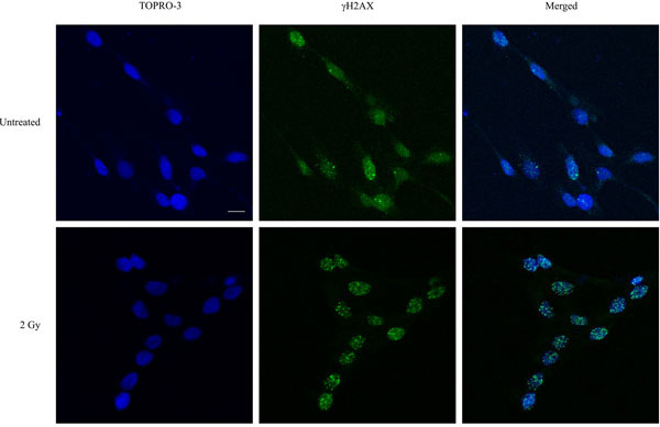
Figure 1. Immunofluorescence visualization of γH2AX foci (green) in untreated human keratinocytes and in cells irradiated with 2 Gy and incubated for a further 1 hour at 37°C, 5% CO2. DNA was stained with TOPRO-3 (blue). Images were acquired using a Zeiss LSM 510 Meta Confocal microscope. Bar = 10 μm.
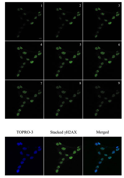
Figure 2. Immunofluorescence visualization of γH2AX foci (green) in human keratinocytes and in cells irradiated with 2 Gy and incubated for a further 1 hour at 37°C, 5% CO2. Images were acquired using a Zeiss LSM 510 Meta Confocal microscope using 0.5 μm Z-sectioning (1-9) to ensure all foci were acquired. The images was then stacked for quantitation using Metamorph. DNA was stained with TOPRO-3 (blue). The stacked γH2AX and blue images were stacked for visualization. Bar = 10 μm.
