The protocol provides a reliable and optimized approach to the isolation of nuclei from solid tumor specimens for multiome sequencing using the 10x Genomics platform, including recommendations for tissue dissociation conditions, cryopreservation of single-cell suspensions, and assessment of isolated nuclei.
Multiome sequencing, which provides same-cell/paired single-cell RNA- and the assay for transposase-accessible chromatin with sequencing (ATAC-sequencing) data, represents a breakthrough in our ability to discern tumor cell heterogeneity-a primary focus of translational cancer research at this time. However, the quality of sequencing data acquired using this advanced modality is highly dependent on the quality of the input material.
Digestion conditions need to be optimized to maximize cell yield without sacrificing quality. This is particularly challenging in the context of solid tumors with dense desmoplastic matrices that must be gently broken down for cell release. Freshly isolated cells from solid tumor tissue are more fragile than those isolated from cell lines. Additionally, as the cell types isolated are heterogeneous, conditions should be selected to support the total cell population.
Finally, nuclear isolation conditions must be optimized based on these qualities in terms of lysis times and reagent types/ratios. In this article, we describe our experience with nuclear isolation for the 10x Genomics multiome sequencing platform from solid tumor specimens. We provide recommendations for tissue digestion, storage of single-cell suspensions (if desired), and nuclear isolation and assessment.
As our knowledge of tumor biology grows, the importance of analyzing heterogeneous cells across the tumor microenvironment has also increased1,2. The ability to acquire single-cell RNA and the assay for transposase-accessible chromatin with sequencing (ATAC-sequencing) data from the same cell in a paired-cell fashion (multiome sequencing) provides a significant advance towards this end3,4. These experiments are expensive and time-consuming, however, and the quality and impact of the data acquired are highly dependent on the quality of the experimental conditions and materials. Standardized protocols for nuclei isolation have been published5,6. Fresh and heterogeneous tissues require protocol optimization since freshly isolated cells from solid tumor specimens are more fragile than those isolated from cell lines.
Another consideration is that for solid tumors, surgical specimens are often not available from the operating room until late in the day. As such, it is generally not feasible to proceed directly from sample acquisition to nuclei capture without a cryopreservation step. In our experience, freezing a single-cell suspension yields the highest-quality nuclei (rather than flash-frozen whole tissue or other modalities of preservation). This is particularly true for enzymatic tissue types with high RNase content such as the pancreas.
Tissue digestion conditions also need to be designed to maximize cell yield without sacrificing quality7. In the context of solid tumor types with dense desmoplastic matrices8, the extracellular matrix must be gently broken down for cell release. Additionally, because the cell types isolated are heterogeneous, conditions should be adjusted to support the total cell population. Human pancreatic cancer (pancreatic ductal adenocarcinoma) samples are used in the described protocol. Pancreatic cancer represents a highly desmoplastic tumor type, which portends relatively sticky tissue and cells. Moreover, as pancreatic tumor specimens available for research also tend to be relatively small, efforts are made to maximize the quantity of cells captured.
Isolation of nuclei requires the most optimization in terms of cell lysis conditions and timing, as well as reagent types and ratios. Handling the nuclei over the course of isolation also requires great care. In this article, we describe our experience optimizing nuclear isolation for the 10x Genomics multiome sequencing platform from solid tumor tissue (Figure 1). We provide recommendations for tissue digestion, cryopreservation of single-cell suspensions (if desired), and nuclear isolation.
Human pancreatic cancer (pancreatic ductal adenocarcinoma) samples were acquired according to an IRB-approved protocol in our laboratory. Informed consent was obtained from patients for tissue collection. Tissue was transported from the operating room to the laboratory and then processed as follows.
1. Tissue dissociation (digestion)
2. Cryopreservation
3. Nuclei isolation
To isolate high-quality nuclei from patient solid tumor specimens for multiome sequencing (Figure 1), the tumor tissue was dissociated and a single-cell suspension was cryopreserved (Figure 2A–D). The cell suspension was then thawed at the time of planned multiome capture. Nuclei capture was conducted with optimized lysis buffer reagents and timing to maximize both quality and yield (Figure 3A–D). Representative nuclei images show appropriate size and shape (Figure 4A–C). The nuclei circled in pale grey show mild stippling of the envelope.
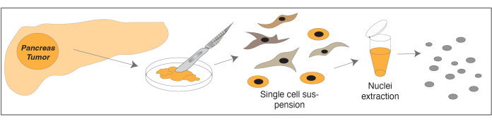
Figure 1: Schematic of nuclei isolation workflow. Schematic showing the basic workflow from a whole tumor tissue specimen to a single-nucleus suspension ready for submission for nuclei capture, multiome library preparation, and ultimately scRNA- and ATAC-sequencing. Abbreviations: scRNA = single-cell RNA; ATAC-seq = assay for transposase-accessible chromatin with sequencing. Please click here to view a larger version of this figure.
Figure 2: Tissue digestion, cryopreservation, cell assessment, and centrifugation steps. (A) Equipment setup for mincing tumor tissue specimens; (B) cell suspension cryopreservation; (C) microscopic evaluation of cell suspension; (D) approach to centrifuging for this protocol. Please click here to view a larger version of this figure.
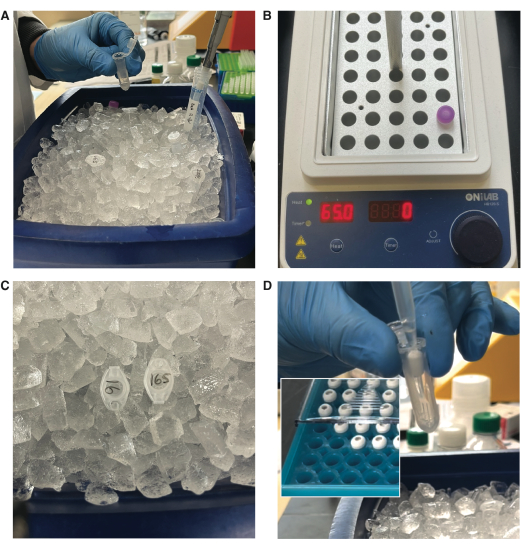
Figure 3: Preparation of reagents, nuclei extraction, and filtering. (A) Preparation of nuclei extraction reagents; (B) dissolution of digitonin at 65 °C prior to use; (C) incubation of samples for cell lysis on ice; (D) filtering of extracted nuclei. Please click here to view a larger version of this figure.
Figure 4: Representative microscopic images of nuclei harvested from complex tumor tissue specimens. (A–C) Representative images of nuclei harvested with and without Trypan Blue staining. (B,C) The nuclei circled in pale grey show mild stippling of the nuclear envelope. 10x and 40x objectives used. Scale bars = 50 µm. Please click here to view a larger version of this figure.
Solution Name | Components | ||
Digest Buffer | 0.5-1 mg/mL collagenase type IV | ||
100 U/mL DNase I | |||
0.1% Poloxamer 188 | |||
20 mM HEPES | |||
1 mM CaCl2 | |||
3-5% fetal bovine serum (FBS) in Medium 199 | |||
1X Cell Lysis Buffer | 10 mM Tris-HCl (pH 7.4) | ||
10 mM NaCl | |||
3 mM MgCl2 | |||
2% BSA (rather than 1%) | |||
0.10% Tween-20 | |||
0.1% Nonident P40 Substitute | |||
0.01% Digitonin | |||
1 mM DTT | |||
1 U/µL RNAse inhibitor in Nuclease-free water | |||
Cell Lysis Dilution Buffer | 10 mM Tris-HCl (pH 7.4) | ||
10 mM NaCl | |||
3 mM MgCl2 | |||
2% BSA | |||
1 mM DTT | |||
1 U/µL RNAse inhibitor in Nuclease-free water | |||
Wash Buffer | 10 mM Tris-HCl (pH 7.4) | ||
10 mM NaCl | |||
3 mM MgCl2 | |||
2% BSA | |||
0.10% Tween-20 | |||
1 mM DTT | |||
1 U/µL RNAse inhibitor in Nuclease-free water |
Table 1: Solutions used in this protocol.
Untangling the heterogeneous cell populations present in the tumor microenvironment is an active area of focus in cancer biology. Similarly, complex tissues exist in benign pathologies such as wound healing and fibrosis. Multiome sequencing has emerged as a powerful tool permitting the acquisition of same-cell paired scRNA- and ATAC-seq data. This protocol describes the isolation of nuclei, which demands optimization in the setting of processing fresh, fragile, small tumor specimens. Here we provide a protocol for nuclei isolation from desmoplastic solid tumor tissue specimens. We have found this approach to yield reliable and high-quality data even when cell suspensions are frozen prior to nuclei isolation and capture.
Increasing the albumin concentration throughout the assay is helpful in limiting cell and downstream nuclei clumping, which can be a problem when working with tumor samples. In terms of tissue digestion, digestion kit reagents can also be used with this protocol if desired (see Table of Materials). If used, we would still recommend following the every-30-min digest/quench and buffer exchanges to maximize the yield of viable cells. Following tissue dissociation, red blood cell lysis can be performed per the manufacturer's protocol. A gradient cell separation protocol can also be performed at the discretion of the researcher (see Table of Materials). We do not perform these protocols routinely, but we have applied such protocols to similar samples without notable differences in results. If significant, small debris is noted after nuclei filtering, Fluorescence-Activated Nuclei Sorting (FANS) can be considered for further purification. A detailed discussion of FANS, including protocol recommendations, is available but beyond the scope of this protocol. Of note, there are protocols in the literature that consider a fixation step prior to cell lysis. However, we prefer to proceed with non-fixed tissue and nuclei as scATAC-seq works best on unfixed tissue9.
For cell lysis, as the RNase inhibitor is quite costly, it is reasonable to scale down the prepared volume of the Cell Lysis Buffer (and the Cell Lysis Dilution Buffer) compared with the volumes provided in the published protocol, if desired, as long as the ratios of reagents stay proportional. When assessing and counting the harvested nuclei, a LIVE/DEAD Viability/Cytotoxicity Kit can be applied at the discretion of the researcher. Trypan blue dye can also be used to provide contrast for viewing the nuclei and to help discern nuclei from unlysed cells if needed. Finally, it is worth noting that we have applied the same cell lysis conditions with adjusted reagents per protocol for single-cell ATAC-seq with excellent results10.
Critical steps in the protocol are precise timing of lysis and gentle but expedient handling of the nuclei. Undermixing will lead to incomplete or inconsistent cell lysis; however, overmixing will shear the chromatin. As noted in other nuclei isolation protocols, nuclei mixing should be accomplished by pipetting rather than vortexing of the sample as the shearing forces can damage the fragile nuclei11. In this regard, it is important that the Cell Lysis Buffer be prepared fresh just prior to use on each occasion. Pipette Tip cell straining (filtering) after the final centrifugation step is key in our experience to prevent nuclei clumping prior to capture. This filtering can be repeated prior to loading if clumps are still detected on the final count or develop during transport of the specimen prior to loading, keeping in mind the additional loss in volume and concentration with each filtering step added. Of note, there should be no significant time pause between nuclei isolation and capture.
Limitations of this protocol are that tissue dissociation and particularly, the cell lysis incubation timing and conditions may need to be optimized for different solid tumor types12. This protocol has been optimized for desmoplastic solid tumors such as breast carcinoma and pancreatic adenocarcinoma and has also been successfully applied to non-tumor desmoplastic tissues such as parenchymal fibrosis, but other tumor types may require additional adjustments. It is optimized for both fresh cell suspensions as well as frozen cell suspensions with excellent nuclei yield from both of these. We recommend pursuing such optimization sequentially starting with tissue dissociation, and once an optimal single-cell suspension has been achieved, moving on to the optimization of the cell lysis conditions to yield an optimal single-nucleus suspension.
Single-cell ATAC-sequencing and more recently, multiome sequencing present a tremendous breakthrough tool for untangling solid tumor cellular heterogeneity. Cell subtypes can be delineated based on both their chromatin accessibility and gene expression characteristics, and the influence of transcription factor accessibility and expression on tumor behavior can be examined directly. These methodological advances are being broadly applied given their tremendous potential to uncover novel therapeutic targets. As such, the development of optimized protocols to obtain these data is of utmost importance. Here we share this protocol for nuclei isolation in the context of pancreatic cancer tissue-a cancer type for which few therapies exist and all with limited efficacy to date.
The authors have nothing to disclose.
We would like to acknowledge the Stanford Functional Genomics Facility (SFGF), particularly Dhananjay Wagh and John Coller, and 10x Genomics for their assistance with optimizing our experiments. We would also like to thank Drs. George Poultsides, Monica Dua, Brendan Visser, and Byrne Lee for their assistance in acquiring patient specimens. We would like to acknowledge Art and Elaine Taylor, the Rantz Foundation, and Warren and Judy Kaplan for their generous support of our research efforts. Funding sources include NIH grants 1F32CA23931201A1 (D.S.F.), 1R01GM116892 (M.T.L.), 1R01GM136659 (M.T.L), Goldman Sachs Foundation (J.A.N., D.S.F., M.T.L.), the Damon Runyon Cancer Research Foundation (D.D., M.T.L.), the Gunn/Olivier Fund, the California Institute for Regenerative Medicine, Stinehart/Reed Foundation, and the Hagey Laboratory for Pediatric Regenerative Medicine. Sequencing was obtained using machines purchased with NIH funds (S10OD025212, S10OD018220, and 1S10OD01058001A1).
100, 70, and 40 μm Falcon cell strainers | ThermoFisher | ||
10x Genomics Nuclei Buffer (20x) | 10x Genomics | 2000153/2000207 | |
Bambanker | Wako, Fisher Scientitic | NC9582225 | |
BSA | Miltenyi Biotec | 130-091-376 | |
Calcium Chloride | Sigma Aldrich | 499609 | |
Collagenase (Collagenase Type IV) | ThermoFisher | 17104019 | |
Digitonin | Thermo Fisher | BN2006 | |
DNase I | Worthington | LS006330 | |
DTT | Sigma Aldrich | 646563 | |
Dulbecco’s Modified Eagle Medium F-12 | Thermo Fisher | 11320082 | |
Fetal Bovine Serum | Thermo Fisher | 10438026 | |
Flowmi 40 μm Pipette Tip Cell Strainer | Sigma Aldrich | BAH136800040 | |
HEPES | Sigma Aldrich | H3375 | |
Histopaque-1119 Gradient Cell Separation solution | Sigma Aldrich | 11191 | |
Medium 199 | Sigma Aldrich | M2520 | |
MgCl2 | Sigma Aldrich | M1028 | |
Miltenyi GentleMACSTM digest kit | |||
NaCl | Sigma Aldrich | 59222C | |
Nalgene Cryo "Mr. Frosty" Freezing Container | ThermoFisher | 5100-0001 | |
Nonident P40 Substitute | Sigma Aldrich | 74385 | |
Poloxamer 188 | Sigma | P5556 | |
Rnase inhibitor | Sigma Aldrich | 3335399001 | |
Tris-HCl | Sigma Aldrich | T2194 | |
Tween-20 | Thermo Fisher | 85113 |