The severe acute respiratory syndrome coronavirus 2 (SARS-CoV-2) causes coronavirus disease 2019 (COVID-19). The World Health Organization declared a public health emergency of international concern on 30 January 2020 and a pandemic on 11 March 2020. The pandemic resulted in over 760 million cases and 6.87 million deaths as of the date this article was written1.
The impact of this virus has highlighted the need for better, more accurate, faster, and more widely available surveillance tools to improve infectious disease detection and control2,3. During the pandemic, SARS-CoV-2 diagnostic tests were based on detecting nucleic acid, antibodies, and proteins, but RT-PCR detection of nucleic acid is the gold standard4. However, RT-PCR has some limitations; it requires specialized equipment, infrastructure, and personnel trained in molecular biology, limiting its application to specialized laboratories. Further, it is time-consuming (4-6 h), not including the time to transport the specimens to the laboratory, which can take days5. These constraints prevent efficient sample processing and obtaining the information required for contingency planning and epidemiological management.
Reverse transcription-loop-mediated isothermal amplification (RT-LAMP) has several advantages over RT-PCR, making it an appealing strategy for designing future point-of-care diagnostic tests (POCT), particularly in resource-constrained settings6. First, it is greatly specific because it uses between four and six primers that recognize six to eight areas in the target sequence, be it DNA or RNA7,8. Second, because it operates at a constant temperature, it does not require sophisticated equipment such as real-time thermal cyclers to generate the amplification, nor does it necessitate highly trained personnel to operate it. Third, the reaction time is very short (~60 min), and reagents that are not very specialized are employed, which makes it a cost-effective tool6. Given the foregoing and the health emergency caused by the COVID-19 pandemic, this technique can be viewed as an alternative diagnostic method that is quick, inexpensive, and simple to implement in any research laboratory9.
The protocol for standardizing and implementing an RT-LAMP to detect SARS-CoV-2 by colorimetric methods using a thermocycler and a water bath is described in this article (Figure 1). Critical points, their limitations, and alternatives to advance them are discussed.
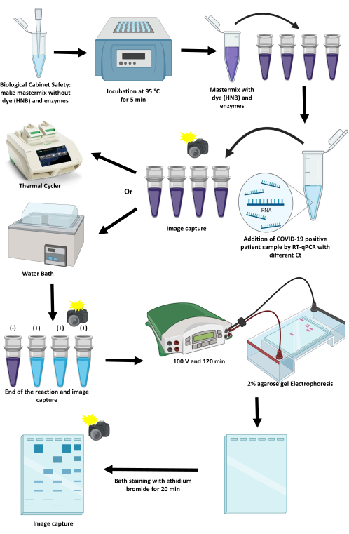
Figure 1: Scheme of the protocol for amplifying SARS-CoV-2 using the RT-LAMP technique. Please click here to view a larger version of this figure.
The implementation of the protocol starts by designing the set of primers for each target gene following the protocol described above. In June 2020, 5,000 SARS-CoV-2 genomes were obtained from the NextStrain database, with a 10% representativeness of Colombian genomes. These sequences were aligned to obtain the consensus sequence that was used in the primer design process. Table 1 shows the primers set chosen for primers RdRp/Hel and RdRp. The primer set for gene N amplification was obtained from a previously published report14.
The first step in the standardization of the protocol was to avoid NTC amplification. One of the most important parameters that must be verified in this regard is determining the optimal melting temperature (Tm) and Bst 3.0 concentration for the set of primers. A temperature gradient was used to determine the best Tm for the amplification (Figure 4A). For the set of primers used in this protocol, the optimal Tm was 66.3 °C (Figure 4A). Furthermore, different concentrations of the Bst 3.0 enzyme were evaluated, with 3.2 lU/µL being the optimal concentration for that reaction (Figure 4B). The concentrations provided by Lu et al.15 were used for the remaining reagents (Table 3, Table 4, and Table 5).
Once the Tm and Bst 3.0 optimal concentration was determined, the amplification process was carried out. The patient samples were provided by University Hospital Fundación Valle del Lili. The positive and negative samples were previously amplified using a conventional RT-PCR protocol, and the viral RNA was then used for RT-LAMP amplification using the protocol described here and the conditions listed. The amplification of N, RdRp/Hel, and RdRp genes in the patient samples but not in NTC is shown in Figure 5A,B. Given that the RT-LAMP is an appealing strategy for designing POCT in the future, the amplifications in this protocol were implemented in a conventional thermal cycler and a water bath (Figure 6).
The second step in standardizing the protocol was to define the colorimetric strategy, so phenol red and neutral red were the first dyes evaluated; for its evaluation, different dyes concentration was probed (Figure 7), but no color change was observed after amplification with any of the concentrations tested. This result could be explained by the fact that both dyes are pH indicators, meaning they are sensitive to the pH of the sample, especially if the viral RNA was eluted in Buffer TE, which was the case for the patient samples evaluated with this protocol. Hydroxy Naphthol Blue (HNB) was investigated because its color changes with changes in Mg2+ concentration, which would be reduced during the amplification reaction as a cofactor of the polymerase enzyme. In this case, different concentrations of HNB were tested to determine which allowed for the best color change after amplification, which occurs in the reaction with 125 µM of HNB (Figure 8). A complete description of reagents employed in the preparation of the amplification mix for N and RdRp/Hel genes by colorimetric LAMP is included in Table 5.
After determining the best conditions for colorimetric detection, patient samples with varying numbers of viral genomes were amplified. Figure 9 shows the amplification of the samples, in which a change of color occurs in the samples according to the concentration of the viral genome. The amplification results were also visualized using agarose electrophoresis, confirming the amplification of patient samples but not NTC.
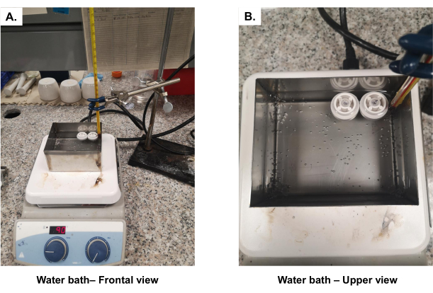
Figure 2: Diagram of the water bath used to amplify SARS-CoV-2 genes using the LAMP technique. (A) Front view and (B) top view. Please click here to view a larger version of this figure.
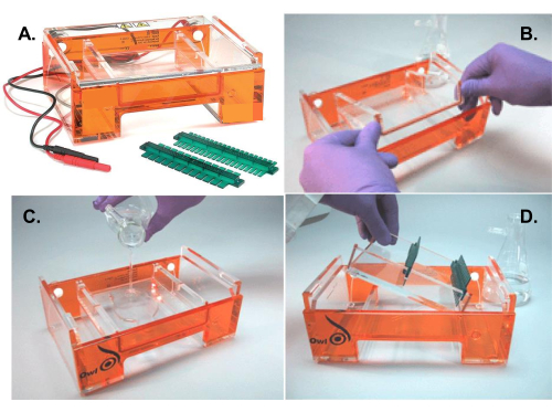
Figure 3: Electrophoresis chamber assembly. (A) Diagram of the electrophoresis chamber used to separate the PCR products. (B) Formation of the internal chamber for the preparation of the agarose gel. (C) Addition of the molten agarose in the internal chamber. (D) Disassembly of the inner chamber to accommodate the gel in the running position. Please click here to view a larger version of this figure.
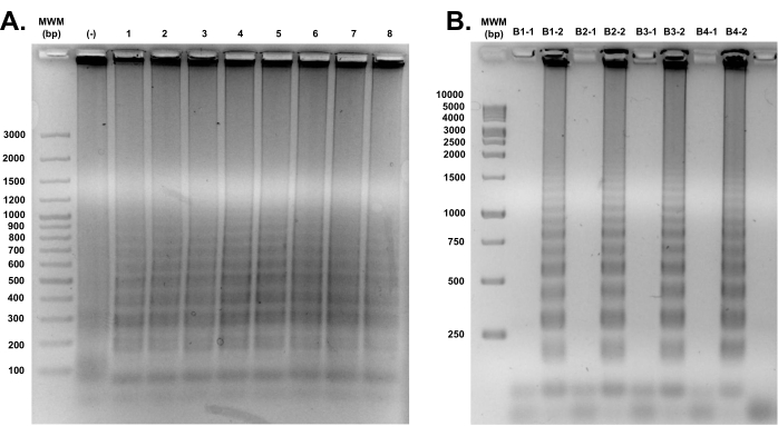
Figure 4: Standardization of the amplification conditions of the primers and the enzyme Bst 3.0. (A) Each lane shows a different gradient temperature, ranging from 63 °C to 67 °C. From left to right, molecular weight marker (MWM), No Template Control (-), lane 1: 67 °C; lane 2: 66.8 °C; lane 3: 66.3 °C; lane 4: 65.5 °C; lane 5: 64.6 °C; lane 6: 63.9 °C; lane 7: 63.4 °C; lane 8: 63 °C. (B) B1: 8.0 U/µL of Bst 3.0; B2: 6.4 U/µL of Bst 3.0; B3: 4.8 U/µL of Bst 3.0; B4: 3.2 U/µL of Bst 3.0; 1: No Template Control; and 2: patient sample EEDD8 (Ct = 23.39). Please click here to view a larger version of this figure.
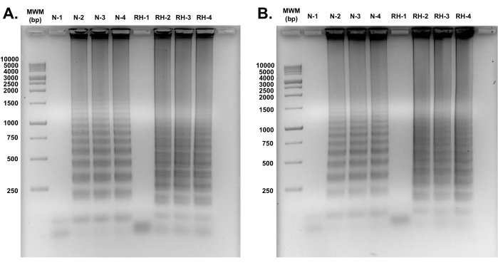
Figure 5: Agarose gel electrophoresis of the amplification products of the N and RdRp/Hel genes present in the SARS-CoV-2 virus using the LAMP technique. (A) Replicate 1 and (B) Replicate 2, where MWM: molecular weight marker; 1: No Template Control; 2: patient sample E1123 (Ct = 19.95); 3: patient sample E1324 (Ct = 26.01); 4: patient sample EEDD10 (Ct = 30.09); RH: RdRp/Hel gene and N: N gene. Please click here to view a larger version of this figure.
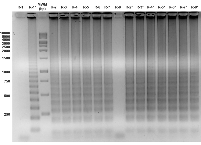
Figure 6: Agarose gel electrophoresis of the amplification products of the RdRp gene present in the SARS-CoV-2 virus using the LAMP technique. The reaction was carried out in (A) a thermal cycler and (B) a water bath system. MPM: molecular weight marker; 1: No Template Control; 2: patient sample E1123 (Ct = 19.95); 3: patient sample E1757 (Ct = 23.67); 4: patient sample E1604 (Ct = 23.98); 5: patient sample E1245 (Ct = 25.99); 6: patient sample E1324 (Ct = 26.01); 7: patient sample EEDD7 (Ct = 26.56); 8: patient sample 24 (Ct = 37.99) and R: RdRp gene. *Refers to the amplifications that were subjected to 60 min of reaction. Please click here to view a larger version of this figure.
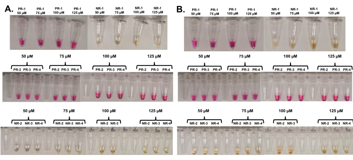
Figure 7: Amplification of the RdRp gene present in the SARS-CoV-2 virus using the LAMP technique with colorimetric detection. Tubes (A) before and (B) after the reaction, where 1: No Template Control; 2: patient sample E1123 (Ct=19.95); 3: patient sample E1757 (Ct=23.67); 4: patient sample E1324 (Ct=26.01); PR: Phenol Red; and NR: Neutral Red. For each dye, four concentrations were evaluated in the final reaction (50 µM, 75 µM, 100 µM, and 120 µM). Please click here to view a larger version of this figure.
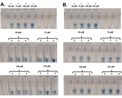
Figure 8: Amplification of the RdRp gene present in the SARS-CoV-2 virus by the colorimetric LAMP technique, using the blue hydroxynaphthol indicator. Tubes (A) before and (B) after the reaction, where 1: No Template Control; 2: patient sample E1594 (Ct=20.75); 3: patient sample E990 (Ct=22.67); 4: patient sample E1245 (Ct=25.99). For this dye, four concentrations were evaluated in the final reaction (50 µM, 75 µM, 100 µM, and 125 µM). Please click here to view a larger version of this figure.
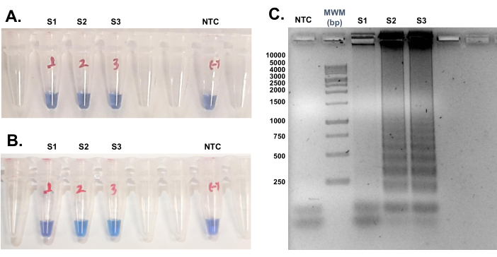
Figure 9: Amplification of the samples. Tubes (A) before and (B) after reaction by LAMP using the blue hydroxynaphthol indicator. (C) Agarose gel electrophoresis of the amplification products of the N gene present in the SARS-CoV-2 virus using the colorimetric LAMP technique. MPM: molecular weight marker; NTC: No Template Control; S1: patient sample E1594 (Ct = 20.75); S2: patient sample E990 (Ct = 22.67); S3: patient sample E1245 (Ct = 25.99). Please click here to view a larger version of this figure.
| Target Gene | Primer | Primer Sequence (5’ → 3’) | |||
| N (Zhang et al., 2020) |
N-F3 | TGGCTACTACCGAAGAGCT | |||
| N-B3 | TGCAGCATTGTTAGCAGGAT | ||||
| N-FIP | TCTGGCCCAGTTCCTAGGTAGTCCAGACGAATTCGTGGTGG | ||||
| N-BIP | AGACGGCATCATATGGGTTGCACGGGTGCCAATGTGATCT | ||||
| N-LF | GGACTGAGATCTTTCATTTTACCGT | ||||
| N-LB | ACTGAGGGAGCCTTGAATACA | ||||
| RdRp | R-F3 | CTATGGTGGTTGGCACAA | |||
| R-B3 | TTGAGCACACTCATTAGCT | ||||
| R-FIP | GCATGGCTCTATCACATTTAGGATA-GTTTATAGTGATGTAGAAAACCCTC | ||||
| R-BIP | ACATGCTTAGAATTATGGCCTCAC-TCTATAGAAACGGTGTGACAAG | ||||
| R-LB | TGTTCTTGCTCGCAAACATACAACG | ||||
| RdRp/Hel | RH-F3 | GGTATTGGGAACCTGAGTT | |||
| RH-B3 | GACAAGACTAATTTATGTGATGTTG | ||||
| RH-FIP | GCAAAGAACACAAGCCCCAACTTATGAGGCTATGTACACACC | ||||
| RH-BIP | TTCACAGACTTCATTAAGATGTGGTACATGGTCGTAACAGCAT | ||||
| RH-LB | GCTTGCATACGTAGACCATTCTT | ||||
Table 1: Primer sequences for SARS-CoV-2 detection by RT-LAMP.
| Component | Stock (µM) | Primer Mix 10x (µM) | Volume (µL) |
| Forward Outer Primer (F3) | 100 | 2 | 12.5 |
| Backward Outer Primer (B3) | 100 | 2 | 12.5 |
| Forward Inner Primer (FIP) | 100 | 8 | 50 |
| Backward Inner Primer (BIP) | 100 | 8 | 50 |
| Loop Forward (LF) | 100 | 4 | 25 |
| Loop Backward (LB) | 100 | 4 | 25 |
| Nuclease-free Water* | — | — | 450 |
| Total Volume | 625 | ||
Table 2: Preparation of 10x RT-LAMP primer mix. The RdRp gene primer mix does not contain the LF primer; therefore, replace this volume with Nuclease-free water. *Instead of Nuclease-free water, 10 mM Tris pH 8.0 prepared in DEPC 0.1% water can be used.
| Item | Reagents | Final concentration for 25 μL | 1 sample (μL) |
| 1 | 10x Buffer | 1x | 2.5 |
| 2 | 100 mM MgSO4 | 4 mM + 2 mM in buffer = 6 mM | 1.0 |
| 3 | 10 mM dNTPs | 1.4 mM | 3.5 |
| 4 | 10x Mix Primers | 1x [ 0.2 μM F3/B3; 0.8 μM FIP/BIP; 0.4 μM LB] | 2.5 |
| 5 | Bst 3.0 DNA pol (8000 IU/mL) | 3.2 IU | 0.4 |
| 6 | RTx (15000 IU/mL) | 1.5 IU | 0.1 |
| 7 | Q5 DNA pol (2000 IU/mL) | 0.15 IU | 0.1 |
| 8 | Nuclease-free Water | N/A | 11.9 |
| 9 | RNA sample | N/A | 3.0 |
| 10 | Total Reaction Volume | 25 | |
Table 3: Preparation of the RdRp gene amplification mix by LAMP.
| Item | Reagents | Final concentration for 25 μL | 1 sample (μL) |
| 1 | 10x Buffer | 1x | 2.5 |
| 2 | 100 mM MgSO4 | 6 mM + 2 mM in buffer = 8 mM | 1.5 |
| 3 | 10 mM dNTPs | 1.4 mM | 3.5 |
| 4 | 10x Mix Primers | 1x [ 0.2 μM F3/B3; 0.8 μM FIP/BIP; 0.4 μM LF/LB] | 2.5 |
| 5 | Bst 3.0 DNA pol (8000 IU/mL) | 3.2 IU | 0.4 |
| 6 | RTx (15000 IU/mL) | 1.5 IU | 0.1 |
| 7 | Q5 DNA pol (2000 IU/mL) | 0.15 IU | 0.1 |
| 8 | Nuclease-free Water | N/A | 11.4 |
| 9 | RNA sample | N/A | 3.0 |
| 10 | Total Reaction Volume | 25 | |
Table 4: Preparation of the amplification mix of the N-A and RdRp/Hel genes by LAMP.
| Item | Reagents | Final concentration for 25 μL | 1 sample (μL) |
| 1 | 10x Buffer | 1x | 2.5 |
| 2 | 100 mM MgSO4 | 6.5 mM + 2 mM in buffer = 8.5 mM | 1.6 |
| 3 | 10 mM dNTPs | 1.4 mM | 3.5 |
| 4 | 10x Mix Primers | 1x [ 0.2 μM F3/B3; 0.8 μM FIP/BIP; 0.4 μM LF/LB] | 2.5 |
| 5 | 1 mM Hydroxy naphthol blue | 125 μM | 3.1 |
| 6 | Bst 3.0 DNA pol (8000 IU/mL) | 3.2 IU | 0.4 |
| 7 | RTx (15000 IU/mL) | 1.5 IU | 0.1 |
| 8 | Q5 DNA pol (2000 IU/mL) | 0.15 IU | 0.1 |
| 9 | Nuclease-free Water | N/A | 8.2 |
| 10 | RNA sample | N/A | 3.0 |
| 11 | Total Reaction Volume | 25 | |
Table 5: Preparation of the amplification mix of the N-A and RdRp/Hel genes by colorimetric LAMP.
| Temperature | Time |
| 66.3 °C | 60 min |
| 80 °C | 5 min |
| 4 °C | ∞ |
Table 6: Thermal conditions used for the amplification of the RdRp, N-A, and RdRp/Hel genes by LAMP.
| Dye | pH Dependent | Color Before Reaction | Color After Reaction | Comments | ||
| Hydroxy naphthol blue | No | Violet | Sky Blue | With this dye the magnesium concentration is critical and must be between 8 mM and 8.5 mM in the final reaction. In this way, the color transition from violet to sky blue is guaranteed. | ||
| Cresol red | Yes | Fuscia | Yellow | The presence of buffers in the RNA eluates or in the reagents can prevent the reduction of pH and affect the color change when the reaction is finished. Therefore, in the case of RNA and primers, it is recommended not to use TE buffer for elution and resuspension/dilution, respectively. | ||
| Neutral Red | Yes | Faint yellow or Faint orange | Fuscia | |||
| Phenol Red | Yes | Fuscia | Yellow | |||
Table 7: Comparison of the dyes used in the visual detection of colorimetric LAMP.
