A subscription to JoVE is required to view this content. Sign in or start your free trial.

Method Article

An Assay to Detect Autoantibodies in Human Serum using a Hippocampal Neuronal Culture

719 views

⸱

July 8th, 2025

In This Article

Abstract

Source: Cunquero, M., et al. Hippocampal Neuronal Cultures to Detect and Study New Pathogenic Antibodies Involved in Autoimmune Encephalitis. J. Vis. Exp. (2022)

This video demonstrates a technique for identifying antibodies against specific antigens in a sample using a rat hippocampal neuronal culture. By exposing cultured neurons to patient samples containing autoantibodies targeting N-Methyl-D-aspartate receptors (NMDARs), the presence of autoantibodies bound to the neuronal surface is revealed through immunostaining.
Protocol

All procedures involving sample collection have been performed in accordance with the institute's IRB guidelines.

PART 1: Establishment of dissociated, fetal, rodent hippocampal neuronal cultures

1. Preparative steps

  1. Poly-L-Lysine (PLL) coating of the plating surfaces (3 days before dissection)
    1. Prepare the borate buffer by adding 2.38 g of boric acid and 1.27 g of borax to 500 mL of distilled water and stirring for 15 min until dissolved. Under a hood, sterilize the buffer solution by filtering (0.2 µm pore size), label, and store at room temperature ....

Access restricted. Please log in or start a trial to view this content.

Results

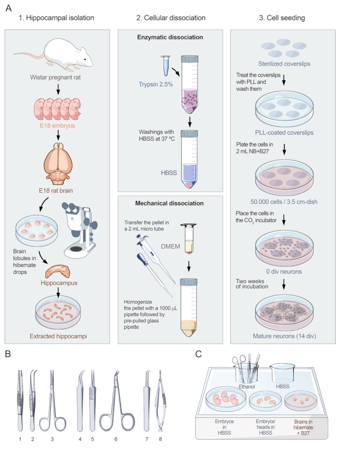
Hippocampal cell isolation and seeding diagram; shows enzymatic, mechanical dissociation steps.
Figure 1: Visual protocol for primary cultures of hippocampal neurons. (A) Flowchart showing the three parts of the protocol for preparing dissociated-cell cultures of hippocampal neurons from embryonic rats at E18. The protocol is divided i.......

Access restricted. Please log in or start a trial to view this content.

Disclosures

No conflicts of interest declared.

Materials

List of materials used in this article
NameCompanyCatalog NumberComments
10 cm Cell culture dishNunc12-565-020
12 mm round coverslipsFisherNC9708845
20x NA 0.75 S Fluor air objectiveNikonCFI Super Fluor 20X
3.5 cm Cell culture dishNunc12-565-90
6 cm Cell culture dishNunc12-565-94
B27 supplementGibco17504-044
Beaker 100 mLPirex-
BoraxSigma-AldrichB9876
Boric AcidSigma-AldrichB0252
Curved forcepsFST11009-13
D-GlucoseSigma-AldrichD9434
DMEM High Glucose (4.5 g/L), without L-Glutamine, without Phenol RedCapricornDMEM-HXRXA
Female Wistar rat (18-days pregnant)Janvier-
Fetal Bovine Serum (FBS)BiowestS181B-500
Fine-angled forcepsFST11251-35
Fine-curved forcepsFST11272-30
Fine-straight forcepsFST11251-23
FITC filter cubeNikonStandard Series
ForcepsFST11000-12
Goat anti-Human AF488InvitrogenA11013
HBSSCapricornHBSS-1A
HEPESSigma-AldrichH3375
Hibernate-E mediumGibcoA12476-01
Horse Serum (HS)Thermofisher26050088
Human anti-NMDAR antibody (CSF)Patient Sample-
Human anti-NMDAR antibody (Serum)Patient Sample-
ImageJ/FijiNIHv1.50i
Inverted fluorescence microscopeNikonEclipse TE2000-U
Phosphate-Buffered SalineGibco10010023
Poly-L-Lysine (PLL)Peptide internationalOKK-35056
Polystyrene ice tray--
Precision spring-scissorsFST15000-08
ProLong Gold with DAPI (antifading mounting media)Molecular ProbesP36941
ScissorsFST14068-12
Sodium pyruvateBiowestL0642-100
Stereo microscopeZeissStemi 2000
Surgery scissorsFST14081-09
Trypsin 2.5%Gibco15090046
Water, sterile endotoxine freeSigma-AldrichW3500

Tags

Autoantibody DetectionNMDAR AutoantibodiesTissue DissociationCell SeedingImmunostaining ProtocolFluorescent LabelingSecondary AntibodyDAPI Nuclei StainingFluorescence Microscopy