Parkinson’s disease is a neurodegenerative disorder that results from the degeneration of dopaminergic neurons in the central nervous system, causing locomotion defects. Rotenone models Parkinson’s disease in Drosophila. This paper outlines two assays that characterize both spontaneous and startle-induced locomotion deficiencies caused by rotenone.
Parkinson’s disease is a neurodegenerative disorder that results from the degeneration of dopaminergic neurons in the central nervous system, primarily in the substantia nigra. The disease causes motor deficiencies, which present as rigidity, tremors and dementia in humans. Rotenone is an insecticide that causes oxidative damage by inhibiting the function of the electron transport chain in mitochondria. It is also used to model Parkinson’s disease in the Drosophila. Flies have an inherent negative geotactic response, which compels them to climb upwards upon being startled. It has been established that rotenone causes early mortality and locomotion defects that disrupt the flies’ ability to climb after they have been tapped downwards. However, the effect of rotenone on spontaneous movement is not well documented. This study outlines two sensitive, reproducible, and high throughput assays to characterize rotenone-induced deficiencies in short-term startle-induced locomotion and long-term spontaneous locomotion in Drosophila. These assays can be conveniently adapted to characterize other Drosophila models of locomotion defects and efficacy of therapeutic agents.
Locomotion deficiencies are a major symptom of Parkinson’s disease and are largely caused by deterioration of dopaminergic neurons of the substantia nigra1. Rotenone is a ketonic insecticide that has been studied extensively to model Parkinson’s motor deficits in Drosophila2-6. Rotenone causes oxidative damage by blocking the oxidative phosphorylation pathway, which ultimately causes cell death7. Dopaminergic neurons are more prone to rotenone toxicity, making the effects of the chemical primarily motor based2,7. By inducing Parkinson’s disease symptoms in flies, we can better understand the disease and remedy its symptoms6,8-11. Drosophila provides a good model for studying this effect because they are genetically tractable, easy to maintain, and have a rapid life cycle.
Several studies have shown that rotenone causes short-term startle-induced locomotion defects in Drosophila—when flies are maintained on rotenone-supplemented food, they show a slower negative geotactic response after startle2-6. Their failure to climb upwards in a vial apparatus as quickly as control trials is indicative of startle-induced locomotion defects.
The effect of rotenone on long-term, spontaneous movement is not well described. Drosophila activity monitors (DAMs) have been successfully used to monitor movement in Drosophila circadian rhythm studies12,13. Flies are placed in individual tubes, which are loaded into the DAM. This apparatus is equipped with an infrared sensor, which counts the number of times a fly breaks the infrared beam. These counts can be used as a measure of undisturbed locomotion and activity12,13. By placing flies in a DAM, the effect of rotenone on their long-term locomotion can be characterized. This study describes methods to measure short-term startle-induced locomotion and long-term spontaneous locomotion in order to better understand the effects of rotenone mediated motor deficiencies. Characterization of locomotion deficiencies mimicking Parkinson’s disease are important because they allow for the study of other compounds which may reverse these locomotion defects.
1. Drosophila Startle-induced Locomotion Assay
2. Drosophila Spontaneous Locomotion Assay
Drosophila Startle-induced Locomotion Assay
Wildtype, canton-S, flies showed a robust negative geotactic response with only approximately 88% and 5% of flies in the top and bottom sections respectively, of the double-vial apparatus after 30 sec (Figure 1). Flies exposed to 125 μM and 250 μM rotenone for 3 days showed a slight decrease in the number of flies in the top section and slight increase in the number of flies in the bottom section. Flies exposed to 500 μM rotenone showed significant defect in the negative geotactic response (p < 0.05 ANOVA, Bonferroni pair wise comparison) as evidenced by fewer flies in the top section and more flies in bottom section as compared to control flies (Figure 1). This defect in negative geotactic response due to inability to swiftly climb in the apparatus is indicative of a defect in startle-induced locomotion.
Drosophila Spontaneous Locomotion Assay
Wildtype, canton-S, flies showed 0.57 counts per minute as a measure of spontaneous mobility on the fourth day in the DAM (Figure 2). Flies exposed to 125 μM rotenone showed a similar level of spontaneous locomotion. By contrast, flies exposed to 250 μM and 500 μM rotenone showed approximately 50% lower measures (p < 0.05 ANOVA, Bonferroni pair wise comparison) of spontaneous locomotion (Figure 2). These flies moved at about 0.20 counts per minute, which is indicative of a rotenone-induced defect in spontaneous locomotion.
To account for initial discrepancies (if any) in locomotion not caused by rotenone exposure, we subtracted locomotion data between day 4 and day 3. Control flies showed an increase of about 0.1 counts per minute in spontaneous locomotion between days, while flies exposed to 125 μM rotenone exhibited a slight decrease of about 0.15 counts per minute (Figure 3). Flies exposed to 250 μM and 500 μM rotenone displayed more severe decreases in locomotion between days, with differences being approximately 0.3 and 0.5 (p < 0.05 ANOVA, Bonferroni pair wise comparison) counts per minute respectively. This data suggests a deficiency in spontaneous locomotion over time with exposure to rotenone and confirms single day analysis mentioned above–flies exposed to higher dosages of rotenone showed a decrease in spontaneous locomotion. Taken together, these methods reliably measure rotenone-induced deficiencies in spontaneous and startle-induced locomotion.
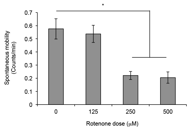
Figure 1. Startle-induced locomotion plot of flies exposed to increasing doses of rotenone. Wild type, canton-S, male flies were exposed to different dosages of rotenone for 3 days and surviving flies (8-12) were then tapped into the bottom of the double vial apparatus. Flies exposed to 500 μM rotenone show a significant decrease in the percent of flies in the top (A) and increase in the percent of flies in the bottom section (B) of the apparatus after 30 sec. This is indicative of a deficiency in startle response in flies exposed to rotenone. Columns represent the average percentage of 6 independent experiments. Error bars represent the standard error of mean; * p < 0.05 ANOVA, Bonferroni pair wise comparison.
Figure 2. Spontaneous locomotion plot of flies exposed to increasing doses of rotenone. Wild type, canton-S, male flies were exposed to different dosages of rotenone and counts per min on the fourth day after exposure were plotted. Counts were measured in a DAM. Flies exposed to 250 μM and 500 μM rotenone show a reduction in counts per min. This is indicative of a deficiency in spontaneous locomotion in flies exposed to rotenone. Columns represent the average counts per min on fourth day of 5 independent trials. Error bars represent the standard error of mean; * p < 0.05 ANOVA, Bonferroni pair wise comparison.
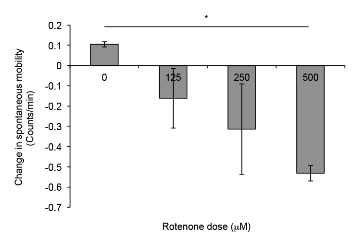
Figure 3. Change in spontaneous locomotion plot of flies exposed to increasing dosages of rotenone. Wild type, canton-S, male flies were exposed to different dosages of rotenone and the difference in counts per min on the third and fourth day after exposure were plotted. Counts were measured in a DAM. There is a dose dependent trend for decline in spontaneous locomotion with flies exposed to higher doses having a more negative change in locomotion. This is indicative of a decrease in movement. Columns represent the average change in locomotion per minute of 5 independent trials. Error bars represent the standard error of mean; * p < 0.05 ANOVA, Bonferroni pair wise comparison.
In this study, we describe two procedures for measuring both long-term spontaneous locomotion and short-term startle-induced locomotion in a rotenone-induced Drosophila model of Parkinson’s disease. One can also measure these locomotion characteristics in flies exposed to other pharmacological agents known to model Parkinson’s disease e.g., paraquat14, genetic models of Parkinson’s disease e.g., alpha-synuclein mutants15, and other fly models of diseases affecting locomotion. For both methods, alternative methods and modifications can be considered. Flies can be anesthetized using ice, which could alleviate limitations of CO2 anesthetization for example lag in data collection to let CO2 effect wear off.
In the startle-induced locomotion assay, since flies show circadian variation in mobility, it is important to collect data at the same time of the day between experiments. It is also important to introduce flies into the testing apparatus without anesthesia. Contrary to most startle response locomotion assays, which rely on a snapshot of passing a threshold in a predetermined amount of time10,11,15,16, our approach, similar to rapid iterative negative geotaxis (RING) assay17,18, monitors the movement at successive multiple instances over a period of time. This approach of continuous monitoring of the distribution of flies in different zones may resolve subtle differences between treatment groups. Additionally, our approach of calculating the percentage of flies in multiple zones of the arena may help minimize the contribution of outliers in the data.
We also systematically decided at which time point to take data to compare the treatment groups. After taking data every 5 sec for 1 min, we plotted the data and found that the most notable differences between treatments could be seen at 30 sec. After this time point, flies exposed to rotenone are able to compensate for their locomotor defects. Therefore we recommend users to optimize the timing of data acquisition to best resolve the differences between their control and experimental sets. This approach also has the advantage of determining relative locomotor deficiencies between pharmacological agents and/or genetic models. For example, a more toxic chemical than rotenone may show most notable differences earlier than 30 sec time point.
For the long-term spontaneous locomotion assay, since flies are anesthetized to introduce them in the tubes, do not consider data from first 24-48 hr to allow for the anesthesia to wear-off and acclimation of flies in the monitor tubes. Another consideration for this assay is the relative position of the tube and the motion sensor in DAM, which we think can impact the spontaneous locomotion data. We placed the tubes containing flies in the monitor so that the sensor was monitoring the one-third span of the tube farthest from the food and not the middle of the tube as is usually done in traditional use of DAM in circadian studies. This allowed us to examine the ability of the flies to traverse at least two-thirds of the tube’s length and led to more consistent data. It is likely that the activity counts can be impacted in rotenone-fed flies due to movement bursts and/or twitching phenotype. Other possible confounding factors for the activity counts could be a gustatory and/or an olfactory aversion/attraction towards rotenone and other chemicals of interest to the user. Therefore additional video tracking17,19 can be employed to complement the DAM data for a more thorough analysis of the locomotion phenotype.
In a scenario where experimental flies have similar activity counts as compared to control flies, it is possible that they differ in the circadian distribution of movement since flies are more active around light on/off and off/on transition in a 12 hr light-12 hr dark cycle. Therefore, it would be helpful to determine the counts per min at multiple time points and bin lengths in a 24-hour period to determine the exact distribution of locomotion. In conclusion, this assay, due to its ability to assess movement characteristics not limited to movements in response to startle, will provide new insights into locomotion defects and characterization of remedial strategies.
The authors have nothing to disclose.
The authors would like to thank Qiuli Wang, Language Resource Center, Colby College, for technical assistance with video processing and Eric Thomas, department of music, Colby College, for providing the background music. This project was supported by grants from the National Center for Research Resources, INBRE (P20RR016463-12), the National Institute for General Medical Sciences (P20 GM103423-12), Nationals Institutes of Health and Science Division Grant, Colby College (STA). JL and LWM were supported by grants from Summer Scholar Fund, Colby College.
Standard narrow vials | Genesee Scientific | 32-120 | |
Rotenone | Sigma | R8875 | Store in freezer, make fresh for each experiment |
Dimethyl Sulfoxide (DMSO) | Sigma | D8418 | Solvent for rotenone |
Instant Drosophila medium | Carolina Biological | Formula 4-24 | |
Drosophila activity monitor (DAM) | Trikinetics | DAM2 | trikinetics.com |
DAM tubes | Trikinetics | Tubes 5X65 mm | |
Recipe for Rotenone +food (125 mM dose) | Make 62.5 mM rotenone stock solution in DMSO by dissolving 25 mg rotenone in 1 ml DMSO | ||
For 125 mM dose, add 10 mM rotenone stock in DMSO to 5 ml water. |