Experimental animal research plays a pivotal role in the development of clinical transplantation practice. The porcine orthotopic liver transplantation model (OLTx) closely resembles human conditions and is frequently used in clinically oriented research. The following protocol contains all information for a reliable porcine OLTx model using an active porto-caval-jugular shunt.
Method Article
Technique of Porcine Liver Procurement and Orthotopic Transplantation using an Active Porto-Caval Shunt
In This Article
Summary
Abstract
The success of liver transplantation has resulted in a dramatic organ shortage. Each year, a considerable number of patients on the liver transplantation waiting list die without receiving an organ transplant or are delisted due to disease progression. Even after a successful transplantation, rejection and side effects of immunosuppression remain major concerns for graft survival and patient morbidity.
Experimental animal research has been essential to the success of liver transplantation and still plays a pivotal role in the development of clinical transplantation practice. In particular, the porcine orthotopic liver transplantation model (OLTx) is optimal for clinically oriented research for its close resemblance to human size, anatomy, and physiology.
Decompression of intestinal congestion during the anhepatic phase of porcine OLTx is important to guarantee reliable animal survival. The use of an active porto-caval-jugular shunt achieves excellent intestinal decompression. The system can be used for short-term as well as long-term survival experiments. The following protocol contains all technical information for a stable and reproducible liver transplantation model in pigs including post-operative animal care.
Introduction
Orthotopic liver transplantation (OLTx) is the only treatment option for patients with end-stage liver disease or advanced hepatocellular carcinoma. For the last 25 years, the number of candidates on the waiting list has gradually increased and now far exceeds the number of available grafts. In most transplant regions, 20 to 30% of patients on the waiting list for liver transplantation die without receiving an organ transplant or are delisted due to progression of disease. Strategies to increase the donor pool and, thus, the number of available grafts, are desperately required. Extended criteria organ allocation, prolonged graft preservation, and induction of immunological tolerance still represent major clinical challenges1-3. Hence, experimental OLTx research is pivotal in order to optimize clinical OLTx practice.
Porcine OLTx is a well-established experimental model that resembles human OLTx in many ways including liver size, anatomy, and physiology4-6. Thus, it has become a standard experimental method in research fields such as surgical techniques, physiology, immunology, preservation, and ischemia-reperfusion injury. Numerous techniques of graft procurement, recipient hepatectomy, and, particularly, vascular reconstruction, have been described in literature5. The choice of the appropriate technique varies according to the researcher’s preference and technical capability.
In contrast to the human scenario, splanchnic congestion during the anhepatic phase represents an important problem in porcine OLTx. Subsequent intestinal ischemia and congestive vascular damage can cause severe hemodynamic instability, jeopardizing the pig survival and, thus, the success of the experiment7-9. Therefore, sufficient intestinal decompression is mandatory, especially in less technically refined experimental settings.
Using an active porto-caval-jugular shunt for the duration of the anhepatic phase is a reliable option to avoid intestinal congestion. The system can be used for early reperfusion experiments as well as long-term survival scenarios. The following protocol contains all information for a stable and reproducible liver transplantation model in pigs, including donor liver procurement, recipient operation including hepatectomy and end-to-end vessel reconstruction techniques, and post-operative care.
Protocol
All animals received humane care in compliance with the ‘‘Principles of Laboratory Animal Care’’ formulated by the National Society for Medical Research and the ‘‘Guide for the Care of Laboratory Animals’’ published by the National Institutes of Health, Ontario, Canada. The Animal Care Committee of the Toronto General Research Institute approved all studies.
1. Organ Retrieval
- House male Yorkshire pigs between 30 and 35 kg in the research facility for 1 week before transplantation to prevent a stress-induced physical reaction (which can alter the perfusion’s outcome10,11) and to acclimatize the animals to the housing conditions. Fast the pig for a minimum of 6 hrs prior to induction of anesthesia.
- Anesthetize the donor pig by an intramuscular (i.m.) injection of a mixture of ketamine (25 mg/kg), atropine (0.04 mg/kg), and midazolam (0.15 mg/kg).
- Prior to intubation, ensure the pig spontaneously breathes 2 L of oxygen dosed with 5% of isoflurane. In supine position, spray the vocal chords with 2% lidocaine 2 min before intubation to avoid vocal cord spasms. For a 35 kg pig, use a 6.5 Fr tracheal tube. Block the tracheal tube with 3 - 5 ml of room air.
- After intubation, use capnometry to confirm correct intubation. Monitor the heart rate and oxygen saturation by pulse oximetry at the pig’s tail. Lower the isoflurane vaporizer to 2%.
- Adjust the depth of anesthesia by minimal alveolar concentration (MAC) values; aim for 2 - 2.5 MAC. Set the ventilator to 14 - 16 breaths/min and a tidal volume of 10 - 15 ml/kg bodyweight.
- Place an 18 G intravenous (i.v.) catheter in one of the ear veins to allow infusion of Ringer’s Lactate solution (200 ml/hr). Scrub the pig and cover it with sterile drapes.
- After ensuring sterile conditions, make a midline laparotomy followed by a left lateral extension. Use a towel to cover large and small bowels before moving them to the left side.
- Divide the falciform ligament and the triangular ligament using a cautery.
- Release the liver from the diaphragm on the right side using an electro-cautery; use scissors for the upper part between the cava and diaphragm. Dissect the infrahepatic cava down to the branch of the adrenal vein on the right side and the renal vein on the left side.
- Separate the infrahepatic cava and distal aorta from each other; ligate aortic branches to the spine; isolate and free renal arteries from adherent tissue. Surround each renal artery with a 2-0 tie.
- Cranial to the left renal vein, dissect the aorta and mesenteric artery. Surround the mesenteric artery with a 2-0 tie.
- After opening the peritoneum cranial to the mesenteric artery, carefully follow the aorta towards the coeliac trunk. Dissect the coeliac trunk caudally to the portal vein; surround the splenic and left gastric arteries, which branch posteriorly off the coeliac trunk. Dissect the coeliac trunk off the portal vein.
- Release the portal vein by a peritoneal incision between the pancreas and portal vein. Tie off veins draining from the pancreas to the portal vein.
- Separate the bile duct from the hepatoduodenal ligament and divide it distally after ligation.
- Ligate the lymph vessels within the hepatoduodenal ligament to prevent lymphatic leakage. Divide the gastroduodenal artery and right gastric arteries between ties. Ligate smaller veins.
- Dissect the aorta behind the diaphragm between the heart and coeliac trunk. Place a 2-0 tie around the aorta cranial to the coeliac trunk.
- Remove the gallbladder and cauterize any bleeding from the gallbladder bed.
- Open up the diaphragm. Administer 1,000 iU/kg donor weight of heparin intracardially or i.v. Set isoflurane to 5% (> 2.5 MAC) in order to achieve a deeper anesthetic level. For a donor after circulatory death (DCD) model, induce cardiac arrest by intracardial injection of 40 mval KCl 3 min after heparin administration. Set cardiac arrest as the starting point of warm ischemia.
- Tie off the previously set ties around the renal, splenic, mesenteric, and left gastric arteries. Tie off the aorta distally between renal and iliac arteries and cannulate the aorta with an organ flush line.
- Tie off the portal vein as proximally as possible and cannulate it with another organ flush line.
- After closing the pre-set tie around the proximal aorta, flush the liver with 2 L of cold University of Wisconsin (UW) solution using dual perfusion via aorta (pressure bag) and portal vein (gravity-driven).
- Excise the liver, leaving all remaining vessels long. Leave a generous diaphragmatic rim around the suprahepatic cava. Place the liver into a sterile organ bag on ice.
- During back-table preparation, clamp the suprahepatic cava using a Satinsky clamp and flush the liver a second time with about 0.5 L of UW solution retrogradely via the lower inferior vena cava until the portal vein outflow is clear.
- Tie off all arterial branches of the aorta and coeliac trunk. Perform an arterial back-table pressure perfusion with the remaining 0.5 L of UW solution12.
- Flush the bile duct using UW solution.
- Trim the diaphragmatic patch to a decent size. Close all phrenic veins – usually 3, 1 each left and right, and 1 posterior – using 4-0 monofilament polypropylene stitches.
- Close the organ bag and store the liver on ice.
2. Recipient Hepatectomy
- Anesthetize the recipient pig by an i.m. injection of a mixture of ketamine (25 mg/kg), atropine (0.04 mg/kg), and midazolam (0.15 mg/kg).
- Place the pig in supine position onto a surgical table on top of a heating mat. Cover the pig with a heat-circulating blanket.
- Prior to intubation, ensure the pig spontaneously breathes 2 L of oxygen dosed with 5% of isoflurane. Spray the vocal chords with 2% lidocaine 2 min before intubation to avoid vocal cord spasms. For a 35 kg pig, use a 6.5 Fr tracheal tube. Block the tracheal tube with 3 - 5 ml of room air.
- After intubation, use capnometry to confirm correct intubation. Monitor the heart rate and oxygen saturation by pulse oximetry at the pig’s tail. Place and fix a temperature probe into the pig’s snout. Lower the isoflurane vaporizer to 2% (aim for 2 - 2.5 MAC). Set the ventilator to 14 - 16 breaths/min and a tidal volume of 10 - 15 ml/kg body weight.
- Use ointment on eyes to prevent dryness while under anesthesia.
- Use Seldinger technique13 to insert a sheath inducer (8.5 Fr) into the left external jugular vein. Use this catheter later for an active portal-caval-jugular bypass.
- Use Seldinger technique to insert a total parenteral nutrition (TPN) catheter (9.5 Fr) into the right external jugular vein.
- Under sterile conditions, dissect the right carotid artery and insert a polypropylene catheter (18 G) for invasive arterial pressure monitoring. Surround the artery with a 2-0 silk tie to allow emergency ligation.
- Turn down the isoflurane vaporizer to 1% (1.5 - 2 MAC) and add propofol (5 - 8 mg/kg/hr i.v.) to maintain the depth of anesthesia. For analgesia, use continuous i.v. infusion of fentanyl citrate (most preferred, 2 μg/kg/hr) or remifentanil (second choice, 15 µg/kg/hr).
- Before skin incision, give 1,000 mg of cefuroxime and 500 mg of metronidazole i.v.. Set up an infusion pump using Ringer’s Lactate with 5% glucose at 150 ml/hr.
- Place the heat-circulating blanket onto the head and neck area. Scrub the pig and cover it with sterile drapes.
- Under sterile conditions, make a midline laparotomy. Insert an abdominal retractor to get sufficient access to the right upper quadrant.
- Divide the falciforme ligament and the triangular ligament using cautery.
- In multiple steps, divide the hepatoduodenal ligament close to the liver between ties. Identify, divide, and mark the branches of the hepatic artery and bile duct.
- Dissect the hepatic artery retrograde until the division of the gastroduodenal artery. Ensure that a bulldog clamp fits around the common hepatic artery proximal to the gastroduodenal artery for later clamping.
- Free the portal vein from adherent tissue.
- Mobilize the vena cava from the retroperitoneum on the right side using an electro-cautery. Use scissors for the upper part between the cava and diaphragm. Dissect the infrahepatic cava down to the branch of the adrenal vein on the right side and the renal vein on the left side.
- Expose the hilum of the spleen. Approximately halfway along the spleen’s length, carefully clear off the splenic artery and vein from adhered peritoneal layers. Surround both splenic artery and vein with 4 2-0 silk ties.
- Insert an 8.5 Fr sheath inducer with 2 additional holes in the catheter’s tip into the splenic vein, pointing distally towards the portal vein. Fix the catheter distally to its insertion with one of the 2-0 ties and close the vein proximal to the insertion with another 2-0 tie. Leave the other 2 ties open.
- Draw blood from the catheter, flush it with 10 ml of saline, and close the clamp of the catheter.
- Fill saline into a bypass consisting of a centrifugal pump head, a jugular tube (3/16”), and inflow from both a portal branch (3/16”) and a caval branch (1/4”, Luer Lock connector on proximal opening). Put a tubing clamp onto the proximal end of the caval tubing.
- Connect the portal and the jugular opening of the bypass to both sheath introducer catheters (Figure 1) and seal the connection with a metal hose clamp ring. Place the centrifugal pump head into its pump position.
- Give 1,000 mg of tranexamic acid and 10,000 iU of heparin 3 min i.v. before cross-clamping. Reduce the propofol infusion rate to 2 mg/kg/hr for the time of the anhepatic phase. Adjust the isoflurane concentration to the arterial pressure and the pig’s reactivity.
- Open the clamps of both sheath introducer catheters and cross-clamp the portal vein. Ensure that blood is running through the bypass passively.
- Start the centrifugal pump at about 1,500 rounds/min. Continue cross clamping if a) the pig is cardiovascularly stable and b) the bypass is running at about 500 ml/min. If the pig does not tolerate the cross-clamping, substitute volume (crystalloids or colloids) and inotrope agents (norepinephrine in small boli).
- Cross clamp the infrahepatic vena cava just cranial to the renal veins using a De Bakey-Beck clamp. Apply firm pressure onto the liver tissue to squeeze out a portion of the remnant blood. Cross-clamp the suprahepatic vena cava including a diaphragmatic rim using a Satinsky clamp while retracting the liver caudally.
- Cut the suprahepatic vena cava directly at its border to the liver tissue. Next, cut the portal vein near the hepatic hilum.
- Approximately 4 cm cranial of the infrahepatic cava clamp, cut a hole into the anterior wall of the vena cava. Into this hole, position the connector of the bypass’ caval opening with the Luer Lock facing anteriorly.
- Secure the connector in the infrahepatic cava with 1-0 silk ties. Then, open the tubing clamp of the caval branch of the bypass.
- Finally, open the infrahepatic cava clamp to allow a caval-jugular bypass in addition to the existing porto-jugular bypass. Increase the pump speed to about 2,500 rounds/min such that the bypass outflow on the jugular side is between 900 - 1,100 ml/min.
- Excise the liver at its remaining connection cranial to the infrahepatic bypass connector, ensuring that the ties securing the bypass connection are not cut. Position the bypass tubing carefully to avoid kinking. Give 500 mg of methylprednisolone to initialize immunosuppression.
3. Vessel Reconstruction
- Using 4-0 monofilament polypropylene sutures, close all 3 phrenic vein ostia on the recipient side at the suprahepatic cava’s aperture. Stitch double-armed 4-0 monofilament polypropylene sutures inside-outside in both corners of the suprahepatic cava on the recipient side.
- Remove the donor organ bag from the ice box. Open the organ bag, remove the donor liver, and place it into the abdominal cavity.
- For an end-to-end anastomosis of the suprahepatic cava, trim the donor suprahepatic vena cava to fit the recipient side. Using the inside needles of the recipient side corner stitches, do an inside-outside corner stitch on each of the sides of the suprahepatic donor cava.
- Shot both ends of the right suture together. Approximate both ostia of recipient and donor cava, then tie both endings of the left suture.
- Shot the shorter end and do an outside-inside stitch of the recipient cava back wall next to the tie. Run over the back wall, ideally everting the cava walls.
- Add 2-3 front wall stitches with the same suture once the right side has been reached, then shot this suture ending. Run over the front wall using the remaining suture from the left corner. Tie together both sutures used for the back and front walls. Tie the other two suture endings at the right corner.
- After trimming the donor portal vein to an appropriate length, perform an end-to-end portal vein anastomosis in the same manner, using 6-0 monofilament polypropylene sutures. Shortly before finishing with the front wall, intubate the lumen of the infrahepatic cava with another flush line and flush out the UW solution with 1 L of saline at RT via the infrahepatic cavaportal vein.
- Complete the anastomosis and tie the sutures, leaving approximately 0.5 cm of growth factor. Put another De Bakey- Beck clamp onto the donor infrahepatic cava.
- Open the suprahepatic cava clamp and check for bleeding. Then, reperfuse the liver by opening the portal clamp.
- Use 6-0 monofilament polypropylene sutures for hemostatic stitches. Decrease the bypass pump’s speed to about 1,500 rounds/min and close the clamp of the portal sheath introducer catheter.
- Reclamp the recipient side of the infrahepatic vena cava and put a tubing clamp on the caval part of the bypass. Stop the centrifugal pump.
- Cut the ties of the caval connector and remove it. Return the remaining blood from the bypass to the pig through the jugular catheter.
- Close the clamp of the jugular catheter and disconnect the bypass. Give 100 mg of protamine sulfate to antagonize the heparin. Take special care of the pig’s hemodynamics during these steps; use catecholamine for pressure support and substitute sodium bicarbonate for metabolic acidosis.
- Perform an end-to-end anastomosis of the infrahepatic cava again in the manner described above, using 5-0 monofilament polypropylene sutures. Reperfuse the infrahepatic lower cava by releasing both clamps.
- Trim an aortal patch around the donor’s coeliac trunk. Tie off the recipient gastroduodenal artery near to the common hepatic artery. Put a bulldog clamp onto the common hepatic artery proximally to the junction of the gastroduodenal artery. Trim a small arterial patch with a Potts scissor, using the vascular tissue around the junction.
- Flush the donor hepatic artery with 10 ml of heparinized saline and put another bulldog clamp further distally to avoid back bleeding. Anastomose the arterial ostia end-to-end in a running parachute technique, using a 6-0 monofilament polypropylene suture. Reperfuse by first opening the distal and then the proximal bulldog clamps.
- Anastomose the bile duct end-to-end with 2 6-0 monofilament polypropylene sutures using the running technique described above. Ensure that big portions of peribiliary tissue are included because the porcine bile duct is very fragile and tears easily.
- After checking for hemostasis, remove the sheath introducer catheter from the splenic vein. Close the proximal and distal ends with the remaining 2 ties.
- Close the abdominal wall with a size 1 monofilament absorbable suture. Close the skin either with a skin stapler or any running 2-0 suture.
4. Post-operative Phase
- Use the heating pad and heat-circulating blanket to keep the pig warm.
- Sample blood gases hourly. Adjust dehydration by increasing the infusion rate.
- Wean the anesthesia. Ventilate the pig for another 2 hrs.
- Remove the sheath introducer catheter from the left jugular vein. Press firmly for a few minutes to prevent bleeding.
- Tunnel the TPN catheter subcutaneously to the side of the pig’s neck. Secure it with 2-0 sutures.
- Remove the arterial line after 2 hrs if the pig is hemodynamically stable without catecholamine support. Ensure that the puncture side is not bleeding. Otherwise, make a 6-0 purse-string stitch around the arterial hole without closing the artery. Close the incision site.
- Stop the ventilation once the pig is able to breathe independently. Disconnect the ventilation tubing from the tracheal tube. Repeatedly check if the pig is breathing sufficiently.
- Place the pig in a prone position into a single animal pen provided with a heating lamp. Extubate once the pig is able to hold up his head independently. Do not leave an animal unattended until it has regained sufficient consciousness to maintain sternal recumbency. House the pig separately for the entire post-operative period.
- Provide sufficient i.v. pain medication post-operatively (for example, buprenorphine 0.01 - 0.05 mg/kg every 6 hrs).
- If the pig does not drink independently, substitute enough volume i.v.
- Continue methylprednisolone as immunosuppression (250 mg post-operative day (POD) 1 morning and then 125 mg each morning). Start cephalosporin 2 mg/kg p.o. twice a day from POD2 on.
- Administer 500 mg of metronidazole and 1,000 mg of cefazoline twice daily, as well as 20 mg of pantoprazole once daily until POD3.
- Monitor the pig closely. Do not hesitate to sacrifice it if it shows signs of suffering (for example, lethargy, refusion to drink, persistent acidosis or hypoglycemia, or signs of hemorrhage or peritonitis). For euthanasia, exsanguinate the pig under deep isoflurane anesthesia (5%, >2.5 MAC) by cutting the suprahepatic cava.
Results
In a first transplantation study, a heart-beating donor model (HBD, n = 5) was compared with a DCD model (n = 10) exposed to 45 min of warm ischemia in situ. In both groups, grafts were preserved on ice for 10 hrs after procurement.
In the HBD group, 100% of the recipient pigs survived until the end of follow-up on day 5 after transplantation. In the DCD group, only 50% of the recipient pigs survived for 5 days due to coagulation issues or metabolic decompensation, as a result of decreased post-operative liver function.
All blood samples were collected from the central venous catheter. After centrifugation, serum samples were obtained and analyzed for hepatocellular injury (aspartate aminotransferase, AST), biliary function (total bilirubin and alkaline phosphatase), and liver function (INR). The time-course of each marker is shown in Figures 2 - 5.
AST levels reached a peak after 24 hrs (1414 ± 538 U/L in the HBD group and 2296 ± 1313 U/L in the DCD group, p = 0.13) and returned to almost normal values after 5 days. Similarly, alkaline phosphatase values were markedly increased after 36 hrs in the DCD group (224 ± 111 U/L) when compared to the HBD group (162 ± 54 U/L, p = 0.27). While total bilirubin was stable in the HBD group (≤ 10 µmol/L throughout), it gradually increased in the DCD group until day 5 (23 ± 31 µmol/L, p = 0.43). The large standard deviation in the bilirubin values in the DCD group shows a peculiar heterogeneity biliary injury in this group. INR as a marker of liver function showed a trend similar to the AST values. Values peaked at 24 hrs and were restored to almost normal values after 5 days. The HBD group had lower values with a peak at 1.47 ± 0.34 in comparison to the DCD group (peak 1.70 ± 0.36, p = 0.32).

Figure 1. Scheme of the porto-caval-jugular shunt. The bypass is filled with Lactated Ringer’s solution. Then the caval part is clamped with a tubing clamp, the jugular and splenic parts are connected to the pre-set catheters, the bypass is opened, and the centrifugal pump is started after portal venous clamping. After liver resection, the caval part of the bypass is inserted and secured in the infrahepatic vena cava stump, cranial to the renal veins. The tubing clamp is released to allow caval decompression, in addition to the portal decompression.
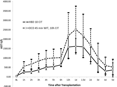
Figure 2. Aspartate aminotransferase (AST) (HBD n = 5, DCD n = 10). AST is a sensitive marker of hepatocellular injury. The peak after 24 hrs is lower in the HBD than in the DCD group, suggesting less hepatic reperfusion injury; the smaller standard deviation shows more homogeneous results in the HBD group.
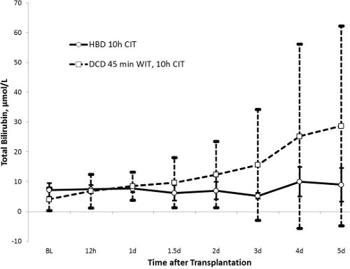
Figure 3. Total bilirubin (HBD n = 5, DCD n = 10). Total bilirubin, as a marker of biliary clearance and bile duct integrity, shows a stable and homogeneous trend with values below 10 µmol/L in the HBD group. The bilirubin curve in the DCD group increases gradually over time and shows a high standard deviation, suggesting biliary injury in only a portion of the experimental group.
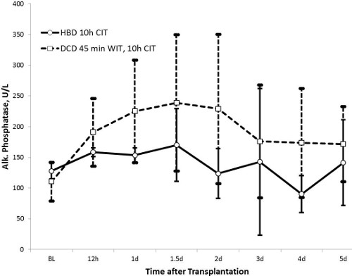
Figure 4. Alkaline phosphatase (HBD n = 5, DCD n = 10). Alkaline phosphatase is an indicator of biliary injury. The values for the HBD group are lower than that of the DCD group, which implies less biliary injury.
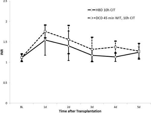
Figure 5. INR (HBD n = 5, DCD n = 10). A high INR value indicates diminished hepatocellular function due to decreased release of coagulation factors. In both HBD and DCD groups, the INR values return to normal values 5 days after transplantation, suggesting recovery of liver function. The values for the HBD group appear lower.
Discussion
Experimental porcine OLTx is a challenging procedure for a research setting without the intensive care resources of a clinical scenario. Possible complications include hemodynamic instability, hemorrhage, organ ischemia, hypothermia, and metabolic, as well as respiratory, decompensation. For any research group, sufficient procedural training of the surgical technique5 as well as the pig anesthesia14,15 is mandatory in order to achieve representative and reproducible results.
Many technical subtleties have been described in the literature, especially regarding the vascular reconstruction phase5. The OLTx protocol described above provides the required information for a cava-replacing model resembling human OLTx. The provided results demonstrate reliable animal survival and graft recovery in both HBD and DCD models. The protocol is applicable in short-term survival scenarios used in graft reperfusion experiments, for example, as well as in long-term survival models such as tolerance studies.
One great obstacle of porcine OLTx is the relatively poor tolerance of cava and portal vein cross-clamping. Splanchnic congestion during the anhepatic phase causes venous hypertension and capillary damage that can lead to major intestinal ischemia and hemodynamic instability to the point of an irreversible shock even after organ reperfusion7. Since the vena cava is completely embedded in liver parenchyma, a cava-preserving piggy-back procedure is not feasible. The total occlusion of the vena cava during the cava reconstruction phase impairs the hemodynamic stability of the pig. Although a few reports show that porcine OLTx can be accomplished during total cava and portal vein occlusion of less than 25 min16,17, a porto-cava-jugular bypass technique for the time of vascular reconstruction is the safer and more practical option7-9,18. In the authors’ experience, a passive porto-jugular bypass is not optimal to keep the pig hemodynamically stable during the anhepatic phase. The bypass model, including active decompression of both infrahepatic cava and portal vein, allows a calm reconstruction phase of the suprahepatic caval and portal anastomoses even with extended clamping time due to unforeseen complications. Contrary to earlier reports7, a splenectomy is not mandatory when the portal bypass catheter is removed. Both splenic artery and vein are closed about halfway along the spleen’s length leaving the proximal half sufficiently perfused. Complications like bleeding or air embolism due to bypass disconnection are avoidable by ensuring that the bypass is placed carefully and secured properly.
In long-term survival OLTx experiments, the bile duct anastomosis is considered a weak spot due to its high complication rate19. The biliary tissue is very fragile and needs special care when being handled. Many different anastomosis techniques have been described5,19. An end-to-end anastomosis is technically easy and associated with minimal complications19. A continuous suture with a non-cutting needle including big sections of peribiliary connective tissue appears to be superior to an interrupted suture. The bile duct is placed under unnecessary tension when the single stitches of the interrupted suture are knotted. This may result in tissue tears and consecutive bile leaks. The suture material – absorbable or non-absorbable – is usually not important, given its limited lifespan until the pig is terminated. For long-term survival models over several months, absorbable sutures – like in human OLTx – are preferable.
Specific care must be taken with the post-operative follow-up. Sufficient nutrition and fluid supply, a reliable pain relief protocol, and a proper immunosuppression regime are obligatory. For long-term experiments, immunosuppression appears particularly important. Compared to other mammals, pigs show a surprisingly low immunological rejection rate after OLTx20,21. Round cell infiltrations are maximal during the second week after transplantation and diminish spontaneously even without immunosuppression. Rejection is rarely the cause of death after porcine OLTx22. However, even with the immunosuppression protocol involving administering steroids i.v. and calcineurin inhibitors p.o. mentioned here, graft rejection is indicated by a mild increase of transaminases starting at about 4 days after OLTx and confirmed by apparent portal field round cell infiltration. Calcineurin inhibitors can be given either p.o.23,24 or i.v.25,26; both methods have disadvantages. Even with oral application aids, the actual amount reaching the gastro-intestinal tract remains elusive. On the other hand, continuous i.v. infusion in a pig’s pen with an active animal is difficult. Hence, the i.v. application must be performed as a bolus, which results in high drug concentration peaks along with potential toxic effects. Nonetheless, both methods of application appear to allow long-term survival.
Similar to a clinical setting, post-operative stress ulcer prophylaxis is recommended. Post-operative bleeding from peptic ulcers is a frequent problem and may be related to an impaired liver function27. After a few cases of gastrointestinal bleeding in both OLTx groups, the authors began regular prophylaxis with pantoprazole and did not experience any gastrointestinal bleeding ever since.
Strict maintenance of sterile conditions intraoperatively, comparable to the conditions in a clinical operating room, and consequent antibiotic prophylaxis, decreases the risk of infectious complications.
In conclusion, this article provides practical information for establishing a porcine OLTx program in a research setting. Sufficient dedication, practice, and teamwork is important in order to decrease the learning period, to produce reliable results, and to reduce costs and the number of research animals.
Disclosures
The authors have nothing to disclose.
Acknowledgements
The study was supported by research grants from the Roche Organ Transplant Research Foundation (ROTRF) and Astellas. Markus Selzner was supported by an ASTS Career Development Award. Matthias Knaak was supported by the Astellas Research Scholarship. We thank Uwe Mummenhoff and the Birmingham family for their generous support.
Materials
| Name | Company | Catalog Number | Comments |
|---|---|---|---|
| Atropine Sulphate 15 mg/30ml | Rafter 8 Products | 238481 | |
| Buprenorphine 0.3 mg/ml | RB Pharmaceuticals LDT | N/A | |
| Cefazolin 1 g | Pharmaceutical Partners of Canada Inc. | 2237138 | |
| Cyclosporin Oral Solution 5,000 mg/50 ml | Novartis Pharmaceuticals Canada Inc. | 2150697 | |
| Fentanyl Citrate 0.25 mg/5 ml | Sandoz Canada Inc. | 2240434 | |
| Heparin 10,000 iU/10 ml | Leo Pharma A/S | 453811 | |
| Isoflurane 99.9%, 250 ml | Pharmaceutical Partners of Canada Inc. | 2231929 | |
| Ketamine Hydrochloride 5000 mg/50 ml | Bimeda-MTC Animal Health Inc. | 612316 | |
| Lactated Ringer’s + 5% Dextrose, 0.5 L | Baxter Corporation | 61131 | |
| Lacteted Ringer’s, 1 L | Baxter Corporation | 61085 | |
| Metronidazole 500 mg/100 ml | Baxter Corporation | 870420 | |
| Midazolam 50 mg/10 ml | Pharmaceutical Partners of Canada Inc. | 2242905 | |
| Pantoprazole 40 mg | Sandoz Canada Inc. | 2306727 | |
| Potassium Chloride 40 mEq/20 ml | Hospira Healthcare Corporation | 37869 | |
| Propofol 1,000 mg/100 ml | Pharmascience Inc. | 2244379 | |
| Protamine Sulfate 50 mg/5 ml | Pharmaceutical Partners of Canada Inc. | 2139537 | |
| Saline 0.9%, 1 L | Baxter Corporation | 60208 | |
| Sodium Bicarbonate 50 mEq/50 ml | Hospira Healthcare Corporation | 261998 | |
| Solu-Medrol 500 mg | Pfizer Canada Inc. | 2367963 | |
| Tranexamic Acid 1,000 mg/10 ml | Pfizer Canada Inc. | 2064413 | |
| University of Wisconsin Solution, SPS-1 | Organ Recovery Systems | SPS-1 | |
| Xylocaine Endotracheal 10 mg/50 ml | AstraZeneca | 2003767 | |
| Appose ULC 35 W skin stapler | Covidien Canada | 803712 | |
| Maxon, 1 | Covidien Canada | 606173 | |
| Sofsilk, 0 | Covidien Canada | S606 | |
| Sofsilk, 2-0 | Covidien Canada | S405 | |
| Sofsilk, 3-0 | Covidien Canada | S404 | |
| Surgipro II, 4-0 | Covidien Canada | VP581X | |
| Surgipro II, 5-0 | Covidien Canada | VP725X | |
| Surgipro II, 6-0 | Covidien Canada | VP733X | |
| Catheter i.v, 18 G | BD Canada | 381147 | |
| Cook TPN catheter, 9.5 Fr | Cook Medical Company | C-TPNS-9.5-90 | |
| PSI Kit for sheath catheter, 8.5 Fr | Arrow International | ASK-09803-UHN | |
| Infusion Pump Line | Smith Medical ASD Inc. | 21-0442-25 | |
| Liver Admin Set (flush line) | CardioMed Supplies Inc | 17175 | |
| Mallinckrodt, Tracheal Tube, 6.5 mm | Covidien Canada | 86449 | |
| Med-Rx Suction Connecting Tube | Benlan Inc. | 70-8120 | |
| Organ Bag | CardioMed Supplies Inc | 2990 | |
| Suction Tip | Tyco Healthcare Group LP | 8888501023 | |
| Valleylab, Cautery Pencil | Covidien Canada | E2515H | |
| Valleylab, Patient Return Electrode | Covidien Canada | E7507 | |
| Bypass Connector 3/8” x 1/4“ | Raumedic AG | 955083-001 | |
| Bypass Connector 3/8” x 3/8” Luer Lock | Raumedic AG | 955163-001 | |
| Bypass Connector Y 3/8” x 3/8” x 1/4” | Raumedic AG | 961360-002 | |
| Bypass Tubing 1/4” x 1/16” | Raumedic AG | 039505-010 | |
| Bypass Tubing 3/8” x 3/32” | Raumedic AG | 039535-005 | |
| Rotaflow Centrifugal Pump | Maquet-Dynamed | HC 2821 | |
| Stainless Steel Hose Clamp Ring, 5mm | Oetiker | 16700007 | |
| Abdominal Retractor | Medite GmbH | N/A | |
| De Bakey – Beck, Infrahepatic Cava Clamp | Aesculap Inc. | FB519R | |
| Deithrich, Atraumatic Clamp (Portal Vein) | Aesculap Inc. | FB525R | |
| Gregory Bulldog Clamp, curved | Aesculap Inc. | FB382R | |
| Gregory Bulldog Clamp, straight | Aesculap Inc. | FB381R | |
| Potts – De Martel, Scissors | Aesculap Inc. | BC648R | |
| Satinsky, Suprahepatic Cava Clamp | Aesculap Inc. | FB605R | |
| Symetrical Tubing Clamp | Codman Instruments | 198010 | |
| Anesthesia Machine, Optimax | Moduflex Anesthesia Equipment | SN5180 | |
| Bypass Flow meter, HT 110 | Transonic Systems Inc. | HT110B11106 | |
| Flow meter probe, H6XL | Transonic Systems Inc. | H6Xl689 | |
| Heat Therapy Pump, T/Pump | Gaymar Industries Inc | TP500-G89D19 | |
| Infusion Pump 3,000 | SIMS Graseby LTD. | SN300050447 | |
| Isoflurane Vapor 19.1 | Draeger Medical Canada Inc. | N/A | |
| Monitor, Datex AS 3 | Instrumentarium Corp./ Hitachi | D-VHC14-23-02 | |
| Rotaflow Centrifugal Drive Unit | Maquet-Dynamed | 952301 | |
| Rotaflow Console | Maquet-Dynamed | 706035 | |
| Temperature Therapy Pad | Gaymar Industries Inc | TP26E | |
| Valleylab Force Tx | Valleylab Inc. | 216151480 | |
| Ventilator, AV 800 | DRE Medical Equipment | 40800AVV | |
| Warm Touch, Patient Warming System | Nellcor/ Covidien Canada | 5015300A |
References
- Mehrabi, A., Fonouni, H., Muller, S. A., Schmidt, J. Current concepts in transplant surgery: liver transplantation today. Langenbecks Arch. Surg. 393 (3), 245-260 (2008).
- Qiu, J., Ozawa, M., Terasaki, P. I. Liver transplantation in the United States. Clin. Transpl. , 17-28 (2005).
- Chalstrey, L. J. Technique of orthotopic liver transplantation in the pig. Br. J. Surg. 58 (3), 585-588 (1971).
- Esmaeilzadeh, M. Technical guidelines for porcine liver allo-transplantation: a review of literature. Ann. Transplant. 17 (2), 101-110 (2012).
- Calne, R. Y. Observations of orthotopic liver transplantation in the pig. Br. Med. J. 2 (5550), 478-480 (1967).
- Memsic, L., Quinones-Baldrich, W., Kaufman, R., Rasool, I., Busuttil, R. W. A comparison of porcine orthotopic liver transplantation using a venous-venous bypass with and without a nonpulsatile perfusion pump. J. Surg. Res. 41, 33-40 (1986).
- Torres, O. J. Hemodynamic alterations during orthotopic liver experimental transplantation in pigs. Acta. Cir. Bras. 23 (2), 135-139 (2008).
- Battersby, C., Hickman, R., Saunders, S. J., Terblanche, J. Liver function in the pig. 1. The effects of 30 minutes' normothermic ischaemia. Br. J. Surg. 61 (1), 27-32 (1974).
- Smith, A. C., Swindle, M. M. Preparation of swine for the laboratory. Ilar. J. 47 (4), 358-363 (2006).
- Swindle, M. M., Smith, A. C. Best practices for performing experimental surgery in swine. J. Invest. Surg. 26 (2), 63-71 (2013).
- Moench, C., Moench, K., Lohse, A. W., Thies, J., Otto, G. Prevention of ischemic-type biliary lesions by arterial back-table pressure perfusion. Liver Transpl. 9 (3), 285-289 (2003).
- Koski, E. M., Suhonen, M., Mattila, M. A. Ultrasound-facilitated central venous cannulation. Crit. Care Med. 20 (3), 424-426 (1992).
- Lange, J. J., Hoitsma, H. F., Meijer, S. Anaesthetic management in experimental orthotopic liver transplantation in the pig. Eur. Surg. Res. 16 (6), 360-365 (1984).
- Kaiser, G. M., Heuer, M. M., Fruhauf, N. R., Kuhne, C. A., Broelsch, C. E. General handling and anesthesia for experimental surgery in pigs. J. Surg. Res. 130 (1), 73-79 (2006).
- Heuer, M. Liver transplantation in swine without venovenous bypass. Eur. Surg. Res. 45 (1), 20-25 (2010).
- Gruttadauria, S. Porcine orthotopic liver autotransplantation: facilitated technique. J. Invest. Surg. 14 (2), 79-82 (2001).
- Falcini, F. Veno-venous bypass in experimental liver transplantation: portal-jugular versus caval-portal-jugular. G. Chir. 11 (4), 206-210 (1990).
- Filipponi, F., Benassai, C., Falcini, F., Martini, E., Cataliotti, L. Biliary tract complications in orthotopic liver transplantation: an experimental study in the pig. Ital. J. Surg. Sci. 19 (2), 131-136 (1989).
- Terblanche, J. Orthotopic liver homotransplantation: an experimental study in the unmodified pig. S. Afr. Med. J. 42 (20), 486-497 (1968).
- Calne, R. Y. Prolonged survival of liver transplants in the pig. Br. Med. J. 4 (5580), 645-648 (1967).
- Battersby, C., Egerton, W. S., Balderson, G., Kerr, J. F., Burnett, W. Another look at rejection in pig liver homografts. Surgery. 76 (4), 617-623 (1974).
- Net, M. The effect of normothermic recirculation is mediated by ischemic preconditioning in NHBD liver transplantation. Am. J. Transplant. 5 (10), 2385-2392 (2005).
- Guarrera, J. V. Hypothermic machine perfusion of liver grafts for transplantation: technical development in human discard and miniature swine models. Transplant Proc. 37 (1), 323-325 (2005).
- Minor, T. Hypothermic reconditioning by gaseous oxygen improves survival after liver transplantation in the pig. Am. J. Transplant. 11 (12), 2627-2634 (2011).
- Kelly, D. M. Porcine partial liver transplantation: a novel model of the 'small-for-size' liver graft. Liver Transpl. 10 (2), 253-263 (2004).
- Meijer, S., Hoitsma, H. F., Visser, J. J., de Lange, J. J. Long term survival following orthotopic liver transplantation in pigs; with special reference to gastric ulcer complications. Neth. J. Surg. 36 (6), 168-171 (1984).
Reprints and Permissions
Request permission to reuse the text or figures of this JoVE article
Request Permission