We describe an endpoint digital assay for quantifying nucleic acids with a simplified (analog) readout. We measure bulk fluorescence of droplet-based digital assays using a standard qPCR machine rather than specialized instrumentation and confirm our results by microscopy.
Digital assays are powerful methods that enable detection of rare cells and counting of individual nucleic acid molecules. However, digital assays are still not routinely applied, due to the cost and specific equipment associated with commercially available methods. Here we present a simplified method for readout of digital droplet assays using a conventional real-time PCR instrument to measure bulk fluorescence of droplet-based digital assays.
We characterize the performance of the bulk readout assay using synthetic droplet mixtures and a droplet digital multiple displacement amplification (MDA) assay. Quantitative MDA particularly benefits from a digital reaction format, but our new method applies to any digital assay. For established digital assay protocols such as digital PCR, this method serves to speed up and simplify assay readout.
Our bulk readout methodology brings the advantages of partitioned assays without the need for specialized readout instrumentation. The principal limitations of the bulk readout methodology are reduced dynamic range compared with droplet-counting platforms and the need for a standard sample, although the requirements for this standard are less demanding than for a conventional real-time experiment. Quantitative whole genome amplification (WGA) is used to test for contaminants in WGA reactions and is the most sensitive way to detect the presence of DNA fragments with unknown sequences, giving the method great promise in diverse application areas including pharmaceutical quality control and astrobiology.
Digital assays for nucleic acid quantification (digital PCR)1-4 and base order (sequencing) are strongly impacting the life sciences and medicine. Digital assays provide quantification of molecular counts on an absolute scale (not relative to a control), providing high sensitivity, enabling facile comparisons across experiments, and crucially, enabling the construction of large databases containing comparable data5 (Table 1).
Over the last 15 years, whole genome amplification (WGA) emerged alongside PCR as a general tool for nucleic acid amplification. Like PCR, WGA is useful for analytical and preparative applications by amplifying minute sample elements up to a level that can be easily detected or used for subsequent analyses like base sequencing. Unlike PCR, WGA is not specific to a particular DNA locus, rather allowing amplification of all sequences in the sample, including unknown sequences. This fundamental difference between PCR and WGA makes the methods complementary to one another and gives rise to different challenges in their application.
The high yield of WGA reactions6 enables routine amplification of genomic DNA from single molecules7, single cells8 and other low-biomass samples9 for quantification or further analysis. The major challenges associated with WGA chemistry are its extreme sensitivity to contaminants and the uneven amplification across individual template molecules6. However, WGA is gaining popularity as single-cell sequencing has emerged as the “killer application” of WGA technology10, and template quantification by WGA is important in many fields of application7.
Instrumentation for digital nucleic acid quantification has been previously described in a variety of valved and valveless microfluidic formats11-14, including droplet-based assays15,16 (Table 2). However, commercial microfluidic systems for digital analysis require specialized equipment for reaction setup and product detection17. Custom valved microfluidics are flexible, but require precision microfabrication and pneumatic control systems18. While it is relatively simple to make monodisperse micro-droplets for emulsion-based digital assays19,20, digital readout is technically burdensome, requiring either large-scale wide-field imaging (similar to popular next-generation sequencing technologies)21,22 or high-speed flow-based droplet detection23-25. Ideally, a digital assay would be simple from set-up to readout, reducing the need for complex instrumentation and allowing large numbers of samples to be read out quickly. Here we describe a simplified method for readout of digital droplet assays that uses a conventional real-time PCR instrument to measure bulk fluorescence of droplet-based digital assays.
While the new approach can be applied to digital PCR assays, it is particularly advantageous for digital WGA assays for which analog real-time amplification assays (that give excellent results for PCR) are problematic. WGA is commonly applied to samples with template molecules that are heterogeneous in sequence, length, and base content. These different template molecules are amplified at different rates6, necessitating the use of a reference (“standard”) sample with matching characteristics. Often, no such standard is available, or the characteristics of the incoming samples are unknown. The heterogeneity of the input material and length-dependent property of WGA chemistries26 also complicate the interpretation of results by creating ambiguity in what is being quantified–the input mass, input number of molecules, a combination of the two, or neither. Finally, the sequence-non-specificity renders quantitative multiple displacement amplification (MDA) more sensitive to contamination than quantitative PCR since contaminant molecules of any sequence have a potential to interfere. Microfluidic digital assays address contamination by segregating template molecules and reducing reaction volumes such that fewer contaminants are sampled.
Here we use a popular isothermal WGA method, MDA27. Of note, several other WGA chemistries including PicoPlex and MALBAC28 depend crucially on initial isothermal strand displacement steps. Isothermal steps exacerbate the challenge of applying analog real-time assays for quantitative WGA. WGA can neither be entirely prevented during setup, leading to unwanted variable pre-amplification, nor discretized (“cycled”) in a way where replication of heterogeneous molecules can be driven to completion and stopped prior to the next cycle like PCR11 (Figure 1). A digital assay format for WGA accomodates typical reaction setup procedures due to the segregation of each molecule for enumeration at the assay endpoint (so pre-amplification does not affect the results) original readout means accuracy would be largely independent of variation in amplification efficiency.
The assay depends on droplets produced in oil with uniform volumes, as the signal level per droplet at the assay endpoint will depend on the droplet volume, and we do not want the consistency of results to depend on averaging across a distribution of droplet sizes. Making monodisperse droplets is now a standard procedure (about 3,500 monodisperse droplet papers have been published since 2013), but requires microfluidic instrumentation25. In fact, more than six companies have developed independent commercial products that rely on production of such droplets, and droplet-making microfluidic chips are commercially available23,29. For this study, we used custom microfluidic devices produced in-house (see Protocol). Syringe pumps to drive flow through the devices are also commercially available, but alternatively, can be substituted with a single disposable syringe for vacuum-driven flow to reduce costs30.
Note: Fabricating the microfluidic device is not necessary for this assay, as droplets for this protocol can be formed with existing commercial droplet makers23,29.
1. Make the Droplet-forming Microfluidic Device
2. Prepare Reaction Mix for Bulk Droplet Readout
Note: The protocol can be used to quantify nucleic acids using many types of amplification reactions. As an example, the reagents needed for MDA of several Lambda DNA concentrations are given. Reagent details are listed in the Table of Specific Reagents. The distribution of template within the droplets depends on sufficient mixing of the sample.
3. Droplet Formation
Note: Droplets can be very sensitive to static electricity and shear stress. Remove clothes that may cause static electricity, and ground yourself before forming droplets. Handle tubes of emulsion from the top of the tube, as far from the emulsion as possible. Pipette emulsions very slowly, preferably with a wide bore pipet tip. When dispensing from a pipette tip, watch the emulsion on the side of the tip to determine pipetting speed.
4. Isothermal Amplification
5. Data Acquisition and Analysis
6. Producing Droplets for Proof-of-Principle Measurements
7. Proof-of-Principle MDA Experiment
8. PCR and MDA bulk reactions
While conventional bulk/real-time readouts can be used for both quantitative PCR and quantitative WGA assays (Figure 1), digital quantitative assays provide advantages (Table 1). In the method described, we read out digital assays in micro-droplet format with a simple bulk endpoint measurement (Figure 2). While this method is broadly applicable, we focus on quantitative WGA (MDA) because this method presents special challenges for conventional real-time assays.
To check whether our real-time PCR instrument could detect varying fractions of fluorescent microdroplets dispersed in oil, we measured the bulk fluorescence of synthetic mixtures of fluorescent and non-fluorescent droplets (Figure 2). The bulk fluorescence and positive droplet counts (independently assessed by fluorescence microscopy) scale linearly with the input fraction of fluorescent droplets as expected (Figure 3A). The experiment was performed three times, with independent droplet formation and mixing for each set.
To evaluate the theoretical quantification performance for “unknown” samples, we established linear standard curves using entirely positive and entirely negative control samples. With such droplet-lot-specific standard curves, we calculated the fraction of synthetic positive and negative droplet mixtures based on each sample’s bulk fluorescence. The results show good performance in droplet ratio quantification across two logs of dynamic range (R2 = 0.984; Figure 3B). The input analyte concentration is easily calculated from the fraction of positive or negative droplets38.
To test our method in a real quantitative WGA assay, we measured fluorescence levels and fraction of positive droplets of a digital droplet MDA assay with Lambda DNA across a wide range of concentrations (Figure 4). In Figure 4B, representative fluorescent images of the droplets are shown. Both bulk fluorescence and positive droplet fraction from the digital MDA samples scale as expected with the average template per droplet, indicating that the bulk readout can faithfully capture the result of a digital assay (R2 = 0.927). We repeated the experiment for each concentration, independent forming droplets and carrying out WGA for each replicate.
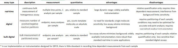
Table 1. Summary of real-time and digital assays. Please click here to view a larger version of this figure.
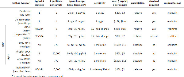
Table 2. Summary of existing methods for quantitating nucleic acids. Please click here to view a larger version of this figure.
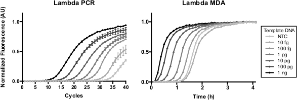
Figure 1. Real-time quantitative PCR and MDA of Lambda DNA. MDA is not discretized through temperature cycling like PCR, and is prone to preamplification if not prepared carefully on ice. Here, quantitative PCR and MDA were performed with increasing concentrations of Lambda DNA. Please click here to view a larger version of this figure.
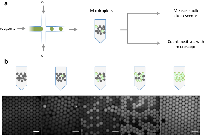
Figure 2. Schematic of analog readout of droplets and representative images. (A) Positive and negative droplets were generated from fluorescent and non-fluorescent reagents in a microfluidic device. The droplets were pre-mixed in different positive:negative ratios. Bulk fluorescence levels were measured with a standard real-time thermocycler, and fraction of positive droplets was determined by fluorescence microscopy. (B) Representative fluorescent overlaid images of increasing ratios of positive droplets (scale bar = 200 µm). Please click here to view a larger version of this figure.
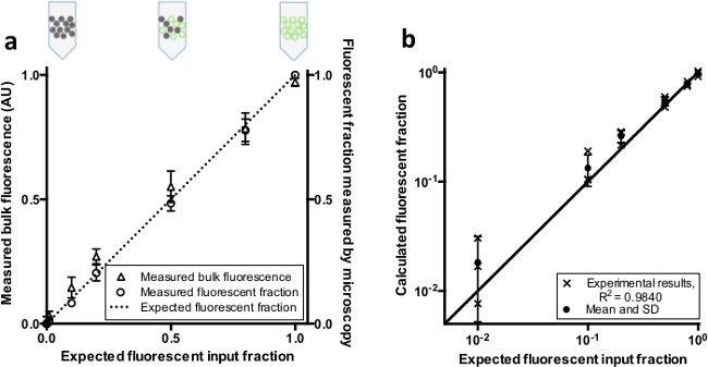
Figure 3. Bulk fluorescence and fluorescent fraction of combinations of separately prepared positive and negative droplets. (A) Fluorescent (positive) and non-fluorescent (negative) droplets were pre-mixed in different ratios. Comparison of the input fraction of positive droplets with the measured bulk fluorescence and measured positive droplet fraction (n = 3, bars represent +/- SD). The dotted line represents the expected fluorescent fraction given a linear relationship. (B) Comparison of predicted ratios based on standards to the expected fluorescent input fraction. The line indicates the expected value given a linear relationship. Please click here to view a larger version of this figure.
Figure 4. Bulk fluorescence and fluorescent fraction of a droplet digital MDA assay. Digital MDA was performed with increasing concentrations of template Lambda DNA (n = 3, error bars represent SD) and measured as described in Figure 2A. The line indicates the expected fluorescent fraction using the Poisson distribution to model the data from our experiment. b) Representative fluorescent overlaid images of droplets after reaction inactivation (scale bar = 200 µm) for increasing expected Lambda DNA molecules per droplet (NTC, 0.005, 0.05, 0.5, and 10). Please click here to view a larger version of this figure.
Digital assays are powerful methods that enable detection of rare cells and counting of individual of nucleic acid molecules. However, digital assays are still not routinely applied in analytical laboratories, due in part to the cost of specialized equipment associated with commercially available methods. Here we describe an endpoint digital assay for quantifying nucleic acids with a simplified analog readout using a standard real-time quantitative PCR machine. This method is quick to set-up, and readout is much simpler than droplet counting methods.
Our assay forfeits the single-molecule (single-droplet) sensitivity of individual droplet measurements for the simplicity of en masse endpoint measurement using standard instrumentation. In our digital MDA experiment, we could not distinguish less than 1 positive droplet per 200 negative droplets from background, despite confirmation by fluorescence microscopy that the actual fraction of positive droplets was as expected (Figure 4). The sensitivity and background in the bulk fluorescence measurement limit the sensitivity of the bulk measurement assay for low positive droplet fractions. If higher sensitivity is required, a digital readout is recommended. Differences in oil levels or bulk droplet volumes can greatly affect the sample fluorescence data. Even normalization with a reference dye cannot make up for the differences oil volumes make in fluorescence reads. It is possible that optimizing assay volume, instrument acquisition parameters, microwell geometry, or optical filters may improve assay performance.
The sensitivity and dynamic range of bulk digital assays can also be improved by optimizing the partition size. Larger droplets yield a greater quantity of fluorescent products from each template molecule, which can help overcome instrumental background and improve sensitivity for low input concentrations. On the other hand, smaller droplets allow the isolation of more molecules per volume assayed. If template concentration is highly variable across samples, both small and large droplet reactions can be carried out in parallel to expand the assay dynamic range39.
Consistent droplet volumes are also necessary for accurate measurement. Many custom microfluidic solutions exist for rapidly forming monodisperse droplets from a sample, both in series19,40 and in parallel41,42. While few enable the droplet generation from several separate samples in parallel, simple modifications to existing designs would allow for simultaneous droplet generation of any number of samples. For example, the Bio-Rad ddPCR system23 applies uniform pressure to the inlets of a series of independent droplet makers, enabling simultaneous droplet formation from multiple samples.
Our bulk readout methodology accesses key advantages of partitioned digital assays without the need for specialized readout instrumentation and is well-suited for quantitative WGA. Here, we focused on quantitative WGA, which is highly susceptible to background contaminants due to its lack of sequence specificity7. In addition, the unknown and potentially broad molecular weight distribution of products from real-time quantitative WGA assays make interpretation difficult in applications where the number of original templates is of interest. Digital assay formatting addresses both these problems. Contaminants are contained within individual reaction partitions, preventing the dominance of low-molecular-weight analytes by high-molecular-weight contaminants as would occur in a conventional real-time assay. In Figures 2B and 4B, possible contaminants, seen as bright spots within the droplets, are segregated and do not amplify or contribute substantially to overall fluorescence. Furthermore, the signal generated in digital assays is proportional to the number of templates, not the size of templates or their amplification rate. Although our method relies on standards to calibrate the bulk digital measurement, the requirement to match sample and standard characteristics can be relaxed significantly.
Quantitative WGA is commonly used to quantify contaminants in WGA reactions7,34, and is the most sensitive method known to quantify the presence of DNA of unknown sequence, giving the method great promise in application areas as diverse as pharmaceutical quality control, forensics, and astrobiology. Bulk digital readout can benefit other digital assay protocols such as digital PCR, by speeding up, parallelizing, and simplifying assay readout.
The authors have nothing to disclose.
The authors acknowledge Liyi Xu for providing MDA reagents and protocols. We also thank David Feldman and Navpreet Ranu for discussions relating to this work. This work was supported in part by a Burroughs Wellcome Career Award at the Scientific Interface to PCB.
Evagreen Dye | Biotium | 31000 | |
Bovine serum albumin | New England Biotechnologies | B9000S | |
Phi29 DNA Polymerase Reaction Buffer | New England Biotechnologies | B0269S | Vortex periodically until aggregate dissolves |
Lambda DNA | New England Biotechnologies | N3011S | Heated to 50C and quenched on ice prior to dilution |
Randomized MDA oligos | Integrated DNA Technologies | 5'-NNNNN*N-3' | |
PCR primers | Integrated DNA Technologies | 5’-CGGCAAACGGGAATGAAACGCC-3’ and 5’-TGCGGCAAAGACAGCAACGG-3’ | |
UltraPure Nuclease-free water | Life Technologies | 10977-015 | UV-treated for 30m in Stratalinker 2400 before use (optional) |
ROX reference dye | Life Technologies | 12223-012 | |
PCR optical strip caps | Life Technologies | 4323032 | |
PCR tubes | Agilent Technologies | 401428 | |
dNTP (25uM each) | Agilent Technologies | 200415 | |
Thermocycler – Mx3000P qPCR system | Agilent Technologies | 401403 | |
Barrier pipette tips | VWR | 89003-046 | |
Bio-rad oil for evagreen | Bio-rad | 186-4005 | |
JumpStart Taq ReadyMix | Sigma-Aldrich | P2893-100RXN |