Summary

Bepaling van neuromusculaire functie gebruiken percutane elektrische zenuwstimulatie

Published: September 13, 2015
doi:

Summary

We present a protocol to assess changes in neuromuscular function. Percutaneous electrical nerve stimulation is a non-invasive method that evokes muscular responses. Electrophysiological and mechanical properties of these responses permit the evaluation of neuromuscular function from brain to muscle (supra-spinal, spinal and peripheral levels).

Abstract

Percutaneous electrical nerve stimulation is a non-invasive method commonly used to evaluate neuromuscular function from brain to muscle (supra-spinal, spinal and peripheral levels). The present protocol describes how this method can be used to stimulate the posterior tibial nerve that activates plantar flexor muscles. Percutaneous electrical nerve stimulation consists of inducing an electrical stimulus to a motor nerve to evoke a muscular response. Direct (M-wave) and/or indirect (H-reflex) electrophysiological responses can be recorded at rest using surface electromyography. Mechanical (twitch torque) responses can be quantified with a force/torque ergometer. M-wave and twitch torque reflect neuromuscular transmission and excitation-contraction coupling, whereas H-reflex provides an index of spinal excitability. EMG activity and mechanical (superimposed twitch) responses can also be recorded during maximal voluntary contractions to evaluate voluntary activation level. Percutaneous nerve stimulation provides an assessment of neuromuscular function in humans, and is highly beneficial especially for studies evaluating neuromuscular plasticity following acute (fatigue) or chronic (training/detraining) exercise.

Introduction

Percutane elektrische zenuwstimulatie wordt wijd gebruikt om neuromusculaire functie 1 beoordelen. Het basisprincipe bestaat uit het induceren van een elektrische prikkel om een ​​perifere motorische zenuw een spiersamentrekking te roepen. Mechanische (koppel meting) en elektrofysiologische (elektromyografische activiteit) reacties worden simultaan opgenomen. Torque, opgenomen in de gezamenlijke beschouwd, wordt beoordeeld aan de hand van een ergometer. De elektromyografische (EMG) signaal opgenomen met oppervlakte-elektroden is aangetoond dat de activiteit van de spier 2 vertegenwoordigen. Deze niet-invasieve methode is niet pijnlijk en gemakkelijker geïmplementeerd dan intramusculaire opnames. Zowel monopolaire en bipolaire elektroden kunnen worden gebruikt. De monopolaire elektrode configuratie is aangetoond gevoeliger voor veranderingen in spieractiviteit 3, die nuttig kunnen zijn voor kleine spieren zijn. Echter, bipolaire elektrodes gebleken effectiever in het verbeteren van de signaal-ruis r zijnatie 4 en worden meestal gebruikt als opnamemethode en kwantificeren aandrijving activiteit. De hierna beschreven methode wordt geconcentreerd op bipolaire opnames. EMG-activiteit is een indicator van de effectiviteit en integriteit van het neuromusculaire systeem. Het gebruik van percutane zenuwstimulatie biedt verder inzicht in de neuromusculaire functie, dwz veranderingen op gespierd, spinale of supra-spinale niveau (figuur 1).

Figuur 1
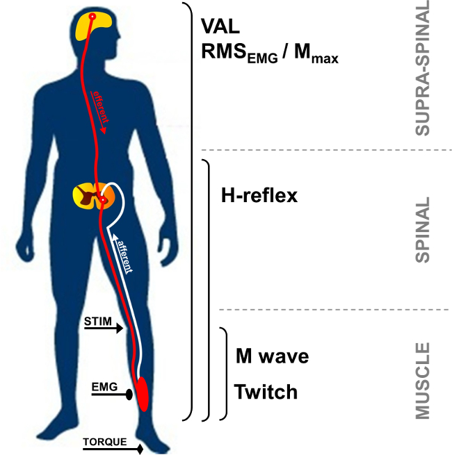
Figuur 1:. Overzicht van de neuromusculaire metingen STIM: zenuwstimulatie. EMG: Elektromyografie. VAL: Vrijwillige Activation Level. RMS: Root Mean Square. M max: Maximal M-wave amplitude.

In rust, de verbinding spier actiepotentiaal, ook wel M-wave, is de korte-latency respons waargenomen na stimulus artefact, en vertegenwoordigt prikkelbaar spiermassa door de directe activ atie van motorische axons die naar de spier (figuur 2, nummer 3). M-wave amplitude neemt toe met de intensiteit tot het bereiken van een plateau van de maximale waarde. Deze reactie, genaamd M max, vertegenwoordigt de synchrone samenvatting van alle motorische eenheden en / of spiervezels actiepotentialen geregistreerd onder de oppervlakte EMG elektroden 5. De evolutie van de amplitude of golven specifieke piek-tot-piek gebruikt om wijzigingen van neuromusculaire transmissie 6 identificeren. Veranderingen in de mechanische responsen geassocieerd met de M-golf, dat wil zeggen piek kramp torque / kracht kan te wijten zijn aan veranderingen in de spieren exciteerbaarheid en / of binnen de spiervezels 7. De vereniging van M max amplitude en piek twitch koppel amplitude (Pt / M ratio) biedt een index van elektromechanische efficiëntie van de spier 8, dat wil zeggen de mechanische respons voor een bepaalde elektrische motor commando.

52.974 / 52974fig2.jpg "/>
Figuur 2:. Motor en reflexieve trajecten geactiveerd door zenuwstimulatie Elektrische stimulatie van een gemengd (motorische / sensorische) zenuw (STIM) veroorzaakt een depolarisatie van zowel motorische axon en Ia afferente afvuren. Depolarisatie van Ia afferentia naar het ruggenmerg activeert een alfa motoneuron, wat op zijn beurt roept een H-reflex response (route 1 + 2 + 3). Afhankelijk van de stimulus intensiteit, motor axon depolarisatie roept een directe spierreactie: M-wave (route 3). Bij maximale M-wave intensiteit, is een antidromic huidige ook gegenereerd (3 ') en botst met reflex volley (2). Deze botsing geheel of gedeeltelijk annuleert de H-reflex reactie.

De H-reflex is een elektrofysiologische respons gebruikt om veranderingen in de Ia-α motorisch synaps 9 beoordelen. Deze parameter kan worden beoordeeld in rust of tijdens vrijwillige contracties. H-reflex een variante van de rekreflex (figuur 2, number 1-3). De H-reflex activeert motorische eenheden monosynaptische aangeworven door Ia afferente paden 10,11, en ​​kan worden onderworpen aan perifere en centrale invloeden 12. De werkwijze oproepen van een H-reflex is bekend dat een hoog intra- individuele betrouwbaarheid spinale prikkelbaarheid beoordelen 13,14 in rust en tijdens isometrische contracties 15 heeft.

Tijdens vrijwillige contractie, kan de grootte van de vrijwillige neurale schijf beoordeeld met behulp van de amplitude van het EMG-signaal, gewoonlijk gekwantificeerd met behulp van de Root Mean Square (RMS). RMS EMG wordt algemeen gebruikt middel kwantificeren van het excitatie van het motorsysteem tijdens vrijwillige contractie (figuur 1). Vanwege de intra- en inter-subject variabiliteit 16, RMS EMG moet worden genormaliseerd met behulp van de EMG opgenomen tijdens een spier-specifieke maximale vrijwillige contractie (RMS EMGmax). Bovendien, omdat veranderingen in EMG signaal be als gevolg van veranderingen op perifeer niveau, normalisatie via een randapparaat parameter, zoals M-wave is nodig om alleen de centrale component van EMG signaal te beoordelen. Dit kan door het verdelen van de RMS EMG de maximale amplitude of RMS Mmax van de M-wave. Normalisering met RMS Mmax (dwz RMS EMG / RMS Mmax) is de voorkeur omdat het rekening houdt met de mogelijke verandering van de M-wave duur 17.

Motor commando's kunnen ook worden geëvalueerd door het berekenen van de vrijwillige activering niveau (VAL). Deze methode gebruikt de kramp interpolatietechniek 18 door superpositie van elektrische stimulatie Mmax intensiteit gedurende een maximale vrijwillige contractie. De extra draaimoment veroorzaakt door stimulering van de zenuw wordt vergeleken met een controle twitch door identieke zenuwstimulatie in een ontspannen spier 19 gepotentieerd. Om maximale VAL, de oorspronkelijke twitch interpo evaluerenning techniek beschreven door Merton 18 omvat een enkele prikkel geïnterpoleerd over een vrijwillige contractie. Onlangs is het gebruik van gepaarde stimulatie populairder geworden omdat de stappen opgewekte koppel groter, gemakkelijker ontdekt en minder variabel dan enkelvoudige stimulatie reacties 20. De VAL verschaft een index van de capaciteit van het centrale zenuwstelsel maximaal te activeren werkende spieren 21. Momenteel VAL geëvalueerd met behulp van de twitch interpolatie techniek is de meest waardevolle methode voor het beoordelen van het niveau van spieractivatie 22. Bovendien maximumkoppel beoordeeld met behulp van een ergometer is de meest goed onderzochte sterkte testen parameter van toepassing voor gebruik in onderzoek en klinische settings 23.

Elektrische zenuwstimulatie kunnen worden gebruikt in verschillende spiergroepen (bijv buigers elleboog, pols flexors, kniestrekkers, plantairflexoren). Echter, de zenuwen bereikbaarheid maakt hettechniek moeilijk bepaalde spiergroepen. De plantaire flexor spieren, vooral triceps surae (soleus en gastrocnemii) spieren, worden vaak onderzocht in de literatuur 24. Inderdaad, deze spieren die betrokken zijn bij de motoriek, hun bijzonder belang rechtvaardigen. De afstand tussen de stimulatie zelf en registrerende elektroden maakt de identificatie van de verschillende opgewekte golven van de triceps surae spieren. De oppervlakkige deel van de nervus tibialis posterior in de knieholte en het grote aantal assen gemakkelijker te reflexreacties opnemen vergelijking met andere spieren 24. Om deze redenen is de thans gepresenteerde reflex methode is gericht op de triceps surae spiergroep (gastrocnemius en soleus). Het doel van dit protocol is derhalve percutane zenuwstimulatie techniek beschrijven neuromusculaire functie in de triceps surae onderzoeken.

Protocol

De experimentele procedures geschetst ontvangen Institutionele ethische goedkeuring en in overeenstemming zijn met de Verklaring van Helsinki. De gegevens werden verzameld uit een vertegenwoordiger deelnemer die op de hoogte van de procedures was en gaf zijn schriftelijke toestemming. 1. Instrument Voorbereiding Reinig de huid op de elektrode plaats door het scheren, en verwijder het vuil met alcohol om lage impedantie (<5 kOhm) te verkrijgen. Plaats twee AgCl-elektro…

Representative Results

Vergroting stimulusintensiteit tot een andere ontwikkeling van de respons amplitudes tussen H- en M-golven. In rust, de H-reflex maximum waarde bereikt voordat volkomen afwezig EMG signaal, terwijl M golf geleidelijk toeneemt tot het bereiken van een plateau aan maximale intensiteit (zie figuur 4 een grafische weergave van de M-wave en figuur 6 de evolutie M-golven en H-reflex met intensiteit). Voor de soleus spier, de wachttijd tussen de stimulus onset en M-wave ongeve…

Discussion

Percutane zenuwstimulatie maakt de kwantificering van talrijke kenmerken van het neuromusculaire systeem niet alleen het begrijpen van de fundamentele controle van neuromotorische functie bij gezonde mensen, maar ook kunnen acute of chronische aanpassingen doorgelicht vermoeidheid of opleiding 17. Dit is zeer gunstig vooral vermoeiend protocols, waarbij metingen zo snel mogelijk moeten worden uitgevoerd na de oefening einde het effect van snelle herstel 42 voorkomen.

Ho…

Disclosures

The authors have nothing to disclose.

Acknowledgements

The authors have no acknowledgements.

Materials

Biodex dynamometer Biodex Medical System Inc., New York, USA www.biodex.com
MP150 Data Acquisition System Biopac Systems Inc., Goleta, USA
Acknowledge 4.1.0 software Biopac Systems Inc., Goleta, USA www.biopac.com
DS7A constant current high voltage stimulator Digitimer, Hertfordshire, UK www.digitimer.com
Silver chloride surface electrodes Control Graphique Medical, Brie-Comte-Robert, France
Computer
1 Cable for connecting the Biodex to the MP150
1 Cable for connecting the Digitimer to the MP150
1 Cable for connecting the MP150 to the computer

References

  1. Desmedt, J. E., Hainaut, K. Kinetics of myofilament activation in potentiated contraction staircase phenomenon in human skeletal muscle. Nature. 217 (5128), 529-532 (1968).
  2. Bouisset, S., Maton, B. Quantitative relationship between surface EMG and intramuscular electromyographic activity in voluntary movement. American Journal of Physical Medicine. 51 (6), 285-295 (1972).
  3. Gabriel, D. A. Effects of monopolar and bipolar electrode configurations on surface EMG spike analysis. Medical Engineering and Physics. 33 (9), 1079-1085 (2011).
  4. Merletti, R., Rainoldi, A., Farina, D. Surface electromyography for noninvasive characterization of muscle. Exercise and Sport Sciences Reviews. 29 (1), 20-25 (2001).
  5. Lepers, R. Aetiology and time course of neuromuscular fatigue during prolonged cycling exercises. Science, & Motricité. 52, 83-107 (2004).
  6. Baudry, S., Klass, M., Pasquet, B., Duchateau, J. Age related fatigability of the ankle dorsiflexor muscles during concentric and eccentric contractions. European Journal of Applied Physiology. 100 (5), 515-525 (2007).
  7. Place, N., Yamada, T., Bruton, J. D., Westerblad, H. Muscle fatigue From observations in humans to underlying mechanisms studied in intact single muscle fibres. European Journal of Applied Physiology. 110 (1), 1-15 (2010).
  8. Scaglioni, G., Narici, M. V., Maffiuletti, N. A., Pensini, M., Martin, A. Effect of ageing on the electrical and mechanical properties of human soleus motor units activated by the H reflex and M wave. The Journal of Physiology. 548 (Pt. 2), 649-661 (2003).
  9. Schieppati, M. The Hoffmann reflex a means of assessing spinal reflex excitability and its descending control in man. Progress in Neurobiology. 28 (4), 345-376 (1987).
  10. Pierrot Deseilligny, E., Burke, D. . The circuitry of the human spinal cord: its role in motor control and movement disorders. , (2005).
  11. Duclay, J., Pasquet, B., Martin, A., Duchateau, J. Specific modulation of corticospinal and spinal excitabilities during maximal voluntary isometric shortening and lengthening contractions in synergist muscles. The Journal of Physiology. 589 (Pt. 11), 2901-2916 (2011).
  12. Grosprêtre, S., Papaxanthis, C., Martin, A. Modulation of spinal excitability by a sub threshold stimulation of M1 area during muscle lengthening. Neuroscience. 263, 60-71 (2014).
  13. Mynark, R. G. Reliability of the soleus H reflex from supine to standing in young and elderly. Clinical Neurophysiology. 116 (6), 1400-1404 (2005).
  14. Palmieri, R. M., Hoffman, M. A., Ingersoll, C. D. Intersession reliability for H reflex measurements arising from the soleus peroneal and tibialis anterior musculature. The International Journal of Neuroscience. 112 (7), 841-850 (2002).
  15. Chen, Y. S., Zhou, S., Cartwright, C., Crowley, Z., Baglin, R., Wang, F. Test retest reliability of the soleus H reflex is affected by joint positions and muscle force levels. Journal of Electromyography and Kinesiology. 20 (5), 987-987 (2010).
  16. Lehman, G. J., McGill, S. M. The importance of normalization in the interpretation of surface electromyography A proof of principle. Journal of Manipulative and Physiological Therapeutics. 22 (7), 444-446 (1999).
  17. Lepers, R. Interest and limits of percutaneous nerve electrical stimulation in the evaluation of muscle fatigue. Science, & Motricité. 70 (70), 31-37 (2010).
  18. Merton, P. A. Voluntary strength and fatigue. The Journal of Physiology. 123, 553-564 (1954).
  19. Gandevia, S. C. Spinal and supraspinal factors in human muscle fatigue. Physiological Reviews. 81 (4), 1725-1789 (2001).
  20. Shield, A., Zhou, S. Assessing voluntary muscle activation with the twitch interpolation technique. Sports Medicine. 34 (4), 253-267 (2004).
  21. Rozand, V., Pageaux, B., Marcora, S. M., Papaxanthis, C., Lepers, R. Does mental exertion alter maximal muscle activation. Frontiers in Human Neuroscience. 8, 755 (2014).
  22. Place, N., Maffiuletti, N. A., Martin, A., Lepers, R. Assessment of the reliability of central and peripheral fatigue after sustained maximal voluntary contraction of the quadriceps muscle. Muscle and Nerve. 35 (4), 486-495 (2007).
  23. Kannus, P. Isokinetic evaluation of muscular performance: implications for muscle testing and rehabilitation. International Journal of Sports Medicine. 15, S11-S18 (1994).
  24. Tucker, K. J., Tuncer, M., Türker, K. S. A review of the H reflex and M wave in the human triceps surae. Human Movement Science. 24 (5-6), 667-688 (2005).
  25. Taylor, N. A., Sanders, R. H., Howick, E. I., Stanley, S. N. Static and dynamic assessment of the Biodex dynamometer. European Journal of Applied Physiology and Occupational Physiology. 62 (3), 180-188 (1991).
  26. Sale, D., Quinlan, J., Marsh, E., McComas, A. J., Belanger, A. Y. Influence of joint position on ankle plantarflexion in humans. Journal of Applied Physiology. 52 (6), 1636-1642 (1982).
  27. Cattagni, T., Martin, A., Scaglioni, G. Is spinal excitability of the triceps surae mainly affected by muscle activity or body position. Journal of Neurophysiology. 111 (12), 2525-2532 (2014).
  28. Gerilovsky, L., Tsvetinov, P., Trenkova, G. Peripheral effects on the amplitude of monopolar and bipolar H-reflex potentials from the soleus muscle. Experimental Brain Research. 76 (1), 173-181 (1989).
  29. Schieppati, M. The Hoffmann reflex a means of assessing spinal reflex excitability and its descending control in man. Progress in Neurobiology. 28 (4), 345-376 (1987).
  30. Hermens, H. J., Freriks, B., Disselhorst Klug, C., Rau, G. Development of recommendations for SEMG sensors and sensor placement procedures. Journal of Electromyography and Kinesiology. 10 (5), 361-374 (2000).
  31. Kamen, G., Sison, S. V., Du, C. C., Patten, C. Motor unit discharge behavior in older adults during maximal effort contractions. Journal of Applied Physiology. 79 (6), 1908-1913 (1995).
  32. Neyroud, D., Rüttimann, J., et al. Comparison of neuromuscular adjustments associated with sustained isometric contractions of four different muscle groups. Journal of Applied Physiology. 114, 1426-1434 (2013).
  33. Rupp, T., Girard, O., Perrey, S. Redetermination of the optimal stimulation intensity modifies resting H-reflex recovery after a sustained moderate-intensity muscle contraction. Muscle and Nerve. 41 (May), 642-650 (2010).
  34. Zehr, E. P. Considerations for use of the Hoffmann reflex in exercise studies. European Journal of Applied Physiology. 86 (6), 455-468 (2002).
  35. Gondin, J., Duclay, J., Martin, A. Soleus and gastrocnemii evoked V wave responses increase after neuromuscular electrical stimulation training. Journal of Neurophysiology. 95 (6), 3328-3335 (2006).
  36. Rochette, L., Hunter, S. K., Place, N., Lepers, R. Activation varies among the knee extensor muscles during a submaximal fatiguing contraction in the seated and supine postures. Journal of Applied Physiology. 95 (4), 1515-1522 (2003).
  37. Fuglevand, A. J., Zackowski, K. M., Huey, K. A., Enoka, R. M. Impairment of neuromuscular propagation during human fatiguing contractions at submaximal forces. The Journal of Physiology. 460, 549-572 (1993).
  38. Vandervoort, A. A., McComas, A. J. Contractile changes in opposing muscles of the human ankle joint with aging. Journal of Applied Physiology. 61 (1), 361-367 (1986).
  39. Grosprêtre, S., Martin, A. Conditioning effect of transcranial magnetic stimulation evoking motor evoked potential on V wave response. Physiological Reports. 2 (11), e12191 (2014).
  40. Allen, G. M., Gandevia, S. C., McKenzie, D. K. Reliability of measurements of muscle strength and voluntary activation using twitch interpolation. Muscle and Nerve. 18 (6), 593-600 (1995).
  41. Cooper, M. A., Herda, T. J., Walter Herda, A. A., Costa, P. B., Ryan, E. D., Cramer, J. T. The reliability of the interpolated twitch technique during submaximal and maximal isometric muscle actions. Journal of Strength and Conditioning Research. 27 (10), 2909-2913 (2013).
  42. Froyd, C., Millet, G. Y., Noakes, T. D. The development of peripheral fatigue and short term recovery during self paced high intensity exercise. The Journal of Physiology. 591 (Pt 5), 1339-1346 (2013).
  43. Pierrot Deseilligny, E., Morin, C., Bergego, C., Tankov, N. Pattern of group I fibre projections from ankle flexor and extensor muscles in man. Experimental Brain Research. 42 (3-4), 337-350 (1981).
  44. Brooke, J. D., McIlroy, W. E., et al. Modulation of H reflexes in human tibialis anterior muscle with passive movement. Brain Research. 766 (1-2), 236-239 (1997).
  45. Hultborn, H., Meunier, S., Morin, C., Pierrot Deseilligny, E. Assessing changes in presynaptic inhibition of I a fibres a study in man and the cat. The Journal of Physiology. 389, 729-756 (1987).
  46. Meunier, S., Pierrot Deseilligny, E. Cortical control of presynaptic inhibition of Ia afferents in humans. Experimental Brain Research. 119 (4), 415-426 (1998).
  47. Aymard, C., Baret, M., Katz, R., Lafitte, C., Pénicaud, A., Raoul, S. Modulation of presynaptic inhibition of la afferents during voluntary wrist flexion and extension in man. Experimental Brain Research. 137 (1), 127-131 (2001).
  48. Abbruzzese, G., Trompetto, C., Schieppati, M. The excitability of the human motor cortex increases during execution and mental imagination of sequential but not repetitive finger movements. Experimental Brain Research. 111 (3), 465-472 (1996).
  49. Garland, S. J., Klass, M., Duchateau, J. Cortical and spinal modulation of antagonist coactivation during a submaximal fatiguing contraction in humans. Journal of Neurophysiology. 99, 554-563 (2008).
  50. Rodriguez Falces, J., Place, N. Recruitment order of quadriceps motor units Femoral nerve vs direct quadriceps stimulation. European Journal of Applied Physiology. 113, 3069-3077 (2013).
  51. Rodriguez Falces, J., Maffiuletti, N. A., Place, N. Spatial distribution of motor units recruited during electrical stimulation of the quadriceps muscle versus the femoral nerve. Muscle and Nerve. 48 (November), 752-761 (2013).
  52. Bathien, N., Morin, C. Comparing variations of spinal reflexes during intensive and selective attention (author’s transl). Physiology, & Behavior. 9 (4), 533-538 (1972).
  53. Earles, D. R., Koceja, D. M., Shively, C. W. Environmental changes in soleus H reflex excitability in young and elderly subjects. The International Journal of Neuroscience. 105 (1-4), 1-13 (2000).
  54. Paquet, N., Hui Chan, C. W. Human soleus H reflex excitability is decreased by dynamic head and body tilts. Journal of Vestibular Research Equilibrium, & Orientation. 9 (5), 379-383 (1999).
  55. Miyahara, T., Hagiya, N., Ohyama, T., Nakamura, Y. Modulation of human soleus H reflex in association with voluntary clenching of the teeth. Journal of Neurophysiology. 76 (3), 2033-2041 (1996).
  56. Pinniger, G. J., Nordlund, M. M., Steele, J. R., Cresswell, a. GH reflex modulation during passive lengthening and shortening of the human triceps surae. Journal of Physiology. 534 (Pt 3), 913-923 (2001).
  57. Tallent, J., Goodall, S., Hortobágyi, T., St Clair Gibson, A., French, D. N., Howatson, G. Repeatability of corticospinal and spinal measures during lengthening and shortening contractions in the human tibialis anterior muscle). PLoS ONE. 7 (4), e35930 (2012).
  58. Grospretre, S., Martin, A. H. reflex and spinal excitability methodological considerations. Journal of Neurophysiology. 107 (6), 1649-1654 (2012).
  59. Hugon, M. Methodology of the Hoffmann reflex in man. New Developments in Electromyography and Chemical Neurophysiology. 3m, 277-293 (1973).
  60. Bigland Ritchie, B., Zijdewind, I., Thomas, C. K. Muscle fatigue induced by stimulation with and without doublets. Muscle and Nerve. 23 (9), 1348-1355 (2000).
  61. Kent Braun, J. A., Le Blanc, R. Quantitation of central activation failure during maximal voluntary contractions in humans. Muscle and Nerve. 19 (7), 861-869 (1996).
  62. Herbert, R. D., Gandevia, S. C. Twitch interpolation in human muscles mechanisms and implications for measurement of voluntary activation. Journal of Neurophysiology. 82, 2271-2283 (1999).
  63. Miller, M., Downham, D., Lexell, J. Superimposed single impulse and pulse train electrical stimulation A quantitative assessment during submaximal isometric knee extension in young healthy men. Muscle and Nerve. 22 (8), 1038-1046 (1999).
  64. Button, D. C., Behm, D. G. The effect of stimulus anticipation on the interpolated twitch technique. Journal of Sports Science and Medicine. 7 (4), 520-524 (2008).
  65. Goss, D. a., Hoffman, R. L., Clark, B. C. Utilizing Transcranial Magnetic Stimulation to Study the Human Neuromuscular System. Journal of Visualized Experiments. (59), e3387 (2012).
  66. Sartori, L., Betti, S., Castiello, U. Corticospinal excitability modulation during action observation. Journal Of Visualized Experiments: Jove. (82), 51001 (2013).
  67. Rozand, V., Lebon, F., Papaxanthis, C., Lepers, R. Does a mental training session induce neuromuscular fatigue. Medicine and Science in Sports and Exercise. 46 (10), 1981-1989 (2014).
  68. Rozand, V., Cattagni, T., Theurel, J., Martin, A., Lepers, R. Neuromuscular fatigue following isometric contractions with similar torque time integral. International Journal of Sports Medicine. 36, 35-40 (2015).
  69. Belanger, A. Y., McComas, A. J. Extent of motor unit activation during effort. Journal of Applied Physiology. 51 (5), 1131-1135 (1981).
  70. Morse, C. I., Thom, J. M., Davis, M. G., Fox, K. R., Birch, K. M., Narici, M. V. Reduced plantarflexor specific torque in the elderly is associated with a lower activation capacity. European Journal of Applied Physiology. 92 (1-2), 219-226 (2004).
  71. Dalton, B. H., McNeil, C. J., Doherty, T. J., Rice, C. L. Age related reductions in the estimated numbers of motor units are minimal in the human soleus. Muscle and Nerve. 38 (3), 1108-1115 (2008).
  72. Hunter, S. K., Todd, G., Butler, J. E., Gandevia, S. C., Taylor, J. L. Recovery from supraspinal fatigue is slowed in old adults after fatiguing maximal isometric contractions. Journal of Applied Physiology. 105 (4), 1199-1209 (2008).
  73. Jakobi, J. M., Rice, C. L. Voluntary muscle activation varies with age and muscle group. Journal of Applied Physiology. 93 (2), 457-462 (2002).
  74. Lepers, R., Millet, G. Y., Maffiuletti, N. a Effect of cycling cadence on contractile and neural properties of knee extensors. Medicine and Science in Sports and Exercise. 33 (11), 1882-1888 (2001).
  75. Duchateau, J., Hainaut, K. Isometric or dynamic training differential effects on mechanical properties of a human muscle. Journal of Applied Physiology. 56 (2), 296-301 (1984).
  76. Millet, G. Y., Martin, V., Martin, A., Vergès, S. Electrical stimulation for testing neuromuscular function From sport to pathology. European Journal of Applied Physiology. 111, 2489-2500 (2011).
  77. Cattagni, T., Scaglioni, G., Laroche, D., Van Hoecke, J., Gremeaux, V., Martin, A. Ankle muscle strength discriminates fallers from non fallers. Frontiers in Aging Neuroscience. 6, 336 (2014).
  78. Horstman, A. M., Beltman, M. J., et al. Intrinsic muscle strength and voluntary activation of both lower limbs and functional performance after stroke. Clinical Physiology and Functional Imaging. 28 (4), 251-261 (2008).
  79. Sica, R. E., Herskovits, E., Aguilera, N., Poch, G. An electrophysiological investigation of skeletal muscle in Parkinson’s disease. Journal of the Neurological Sciences. 18 (4), 411-420 (1973).
  80. Knikou, M., Mummidisetty, C. K. Locomotor Training Improves Premotoneuronal Control after Chronic Spinal Cord Injury. Journal of Neurophysiology. 111 (11), 2264-2275 (2014).
check_url/52974?article_type=t

Play Video

Cite This Article
Rozand, V., Grosprêtre, S., Stapley, P. J., Lepers, R. Assessment of Neuromuscular Function Using Percutaneous Electrical Nerve Stimulation. J. Vis. Exp. (103), e52974, doi:10.3791/52974 (2015).

View Video