This manuscript describes the protocols for prostate micro-dissection and surgical castration in the laboratory mouse. We also depict representative results produced by these protocols. Finally, we discuss the advantages and utilization of these protocols.
Method Article
Murine Prostate Micro-dissection and Surgical Castration
In This Article
Summary
Abstract
Mouse models are used extensively to study prostate cancer and other diseases. The mouse is an excellent model with which to study the prostate and has been used as a surrogate for discoveries in human prostate development and disease. Prostate micro-dissection allows consistent study of lobe-specific prostate anatomy, histology, and cellular characteristics in the absence of contamination of other tissues. Testosterone affects prostate development and disease. Androgen deprivation therapy is a common treatment for prostate cancer patients, but many prostate tumors become castration-resistant. Surgical castration of mouse models allows for the study of castration resistance and other facets of hormonal biology on the prostate. This procedure can be coupled with testosterone reintroduction, or hormonal regeneration of the prostate, a powerful method to study stem cell lineages in the prostate. Together, prostate micro-dissection and surgical castration opens up a multitude of opportunities for robust and consistent research of prostate development and disease. This manuscript describes the protocols for prostate micro-dissection and surgical castration in the laboratory mouse.
Introduction
The prostate is the most common site of cancer in men in the US. Nearly 220,000 men each year will be diagnosed with prostate cancer, and approximately 27,000 men will succumb to their disease 1. Men have a 1 in 7 lifetime risk of a prostate cancer diagnosis in the US 1. Benign prostate hyperplasia (BPH), an age-related noncancerous enlargement of the prostate, is also a widespread condition, affecting over 80% of men over 80 2. As such, the prostate is the focus of considerable research.
Mouse models have been used widely to study diseases of the prostate 3. Overall, the mouse prostate is an excellent representative of the human gland 4, but there are similarities and differences between mouse and human prostate anatomy and physiology 5. In both species, the prostate is located at the base of the bladder, surrounding the urethra. The human prostate gland is a single lobe, split into four zones: central, transition, peripheral, and anterior fibromuscular stroma. In contrast, the mouse prostate consists of three paired lobes at different positions around the urethra: anterior, ventral, and dorsolateral. On a cellular level, the main difference is that the basal cell to luminal cell ratio in human is about 1:1, but it is 1:4 in mice 6. The murine stroma is also different in mice relative to humans – humans have more extensive smooth muscle, while the muscle layer is much thinner in murine prostate. The proper identification, dissection, and handling of the mouse prostate are essential to mouse prostate research.
Hormone levels drastically affect the development and homeostasis of the prostate gland in both humans and mice. While estrogen seems to play a role in prostate development 7, the most important hormone in the prostate is testosterone. Testosterone is essential for glandular development and maintenance. Following either physical or chemical castration, approximately 90% of the luminal cells in the adult prostate apoptose, and the gland shrinks. If testosterone is reintroduced to an individual under castration conditions, the prostate is able to regenerate itself to full capacity. Testosterone also seems to drive prostate cancer, which is why androgen deprivation therapy is a commonly used therapeutic strategy. However, many prostate cancers become resistant to androgen deprivation. In addition, men undergoing androgen deprivation therapy for treatment of prostate cancer undergo cycles of castrated and regenerated conditions. In the mouse, surgical castration, along with hormonal regeneration of the prostate by reintroducing testosterone, is an important tool with which to study castration resistance and the effects of testosterone cycling on the prostate.
In this article, we will discuss and demonstrate the proper techniques by which to locate and micro-dissect a mouse prostate, as well as to surgically castrate a mouse.
Protocol
This protocol meets and follows the guidelines set by the Johns Hopkins University Institutional Animal Care and Use Committee.
1. Micro-dissecting a Mouse Prostate
- Euthanize the mouse by carbon dioxide asphyxiation or an alternative approved method in accordance with institutional animal care and use guidelines.
- Pin the mouse in a supine position to a dissection board by putting a pin through each of the four paws.
- Spray the abdomen with 70% ethanol.
- Hold the skin of the lower abdomen with forceps, and use scissors to cut through the skin and peritoneum approximately 1 cm anterior to the opening of the penis, careful not to cut any organs.
- Cut away the skin and peritoneum from the lower abdomen, up both sides of the abdomen to the rib cage, exposing the peritoneal cavity.
- Identify the bladder and urogenital tract, and expose them by gently moving fat and other organs to the side.
- Using forceps, grip the vas deferens at the base near the urethra, and tear it away.
- Repeat with the opposite vas deferens.
- Using forceps, carefully grip the bladder and pull it up, while simultaneously using scissors to cut through the urethra below the bladder and ventral prostate (approximately 1 cm below the base of the bladder).
Note: As the bladder is pulled up, the entire urogenital tract (containing bladder, a section of the urethra, all prostate lobes, and the seminal vesicles) will come with it. There may be some resistance. - Place the urogenital tract (UGT) into a 60-100 mm Petri dish containing approximately 5-10 ml phosphate buffered saline (PBS).
- Perform the remaining part of the protocol under a dissection microscope. Use two fine forceps, one in each hand. Hold the forceps "like a pencil" and rest the forearms on the bench top so as to steady them.
- Use fine forceps to position the UGT such that the bladder is on top, the urethra is pointed down, and the seminal vesicles are positioned on either side of the bladder.
- Grip the urethra with one forceps and carefully pull away fat with the other forceps without tearing away any prostate tissue. Note: Fat will appear "shiny" relative to the surrounding tissue.
- Once the fat is removed, use forceps to tear the connective tissue between the two ventral lobes of the prostate and separate them.
- To remove one of the ventral lobes, use one forceps to grip a single ventral lobe near the tip, and use the other forceps to grip that lobe at the base as close to the urethra as possible. While still gripping the base of the lobe, use the other forceps to grip the urethra. Pull the lobe away from the urethra with a firm, smooth motion. Make sure no prostate tissue remains attached to the urethra. Place the lobe into the appropriate medium, depending on the next experimental step.
- To fix and paraffin-embed the sample for histological analysis, place the lobe in 10% neutral-buffered formalin (NBF) 8.
- Place the lobe in freezing medium, such as OCT, for cryopreservation and sectioning 8.
- Place the lobe in cold PBS for single cell isolation for culture or flow cytometry 9.
- Repeat step 1.15 with the other ventral lobe.
- To remove one of the anterior lobes, use forceps to gently pull the lobe away from the seminal vesicle, making sure not to puncture the seminal vesicle. Once the lobe is independent of the seminal vesicle, remove it in the same fashion as the ventral lobe in step 1.15.
- Repeat step 1.17 with the other anterior lobe.
- Flip the remaining urogenital tract over such that the dorsal prostate is visible.
- To remove one of the dorsolateral lobes, use forceps to tear the connective tissue between the two dorsolateral lobes and also the connective tissue between the lobes and the posterior region of the seminal vesicles. Remove the lobe in a similar fashion as the ventral lobe in step 1.15.
- Repeat step 1.20 with the other dorsolateral lobe.
2. Surgical Castration
Note: This is a survival surgery, so asepsis, anesthesia, and pain management are important to the ethical and successful completion of this protocol. Follow all institutional animal care and use guidelines. Use an external heat source, such as a recirculating water blanket, during the surgical procedure to prevent hypothermia. Do not leave an animal unattended until it has regained sufficient consciousness to maintain sternal recumbency. Do not return an animal that has undergone surgery to the company of other animals until fully recovered. Autoclave all surgical instruments before use in surgery.
- On a clean, sterile surface anesthetize the mouse using appropriate measures (e.g. 2% inhaled isoflurane) and position the mouse in a supine position. Note: Alternative anesthesia methods, such as ketamine, may also be used, depending on the approved animal protocol.
- Check the mouse's reflexes by pinching the toes. If the mouse does not react, proceed to the next step. If it does react, wait 1-3 min and check again. Shave the surgical area with an electric razor.
- Aseptically prepare the abdomen of the mouse using alternating scrubs of 70% ethanol and iodine or procedures recommended by the institutional veterinary staff or IACUC.
- Using a sterile scalpel, make a 1 cm vertical incision through the skin in the midline of the lower abdomen, approximately 1.5 cm anterior to the penis.
- Make a small incision (<1 cm) through the peritoneum, careful not to cut any organs.
- Using sterile forceps, grip the cut edge of the peritoneum and lift it up gently so as to be able to see the peritoneal cavity beneath.
- Using another sterile forceps, reach into the peritoneal cavity and grip either the left or right testicular fat pad, lateral to the bladder. Pull the fat pad through the opening in the peritoneum and skin until the testis comes out as well. Be careful not to injure any other tissues or organs during this step.
- Using a cautery pen, cut through the fat pad that is holding the testis. Cut slowly through the testicular artery with the cautery pen Note: If the testicular artery is cut too fast, there may be too much bleeding and the mouse may have to be euthanized.
- Once the fat pad and artery are cut, remove the testis and fat pad.
- Repeat steps 2.6-2.9 with the other testis. Note: An alternative method for removal of the testes is to surgically ligate the testicular artery, followed by cutting the vessel with scissors.
- Suture the peritoneum closed with absorbable suture.
- Staple the skin closed with surgical wound clips.
- Administer an analgesic to manage pain, and place the mouse in a clean cage on a cage warmer for 5-10 min. Monitor the mouse for signs of pain or bleeding.
Results
All six prostate lobes were removed from a mouse via prostate micro-dissection (Figure 1). The complete urogenital tract (UGT) is composed of all prostate lobes, the bladder, seminal vesicles, and the urethra (Figure 1a). The vas deferens attaches to the urethra, but is unnecessary for prostate micro-dissection, and can thus be detached before removal of the UGT by cutting the urethra (Figure 1b-1c). The urogenital tract will remain together once removed from the abdomen (Figure 1d). At this point, the UGT remains covered in fatty tissue, which needs to be removed to gain access to the prostate lobes (Figure 1e). All six lobes (ventral, anterior, and dorsolateral pairs) can be seen surrounding the urethra at the base of the bladder. Pure prostate tissue can then be removed following the protocol above (Figure 1f). These lobes may be placed in the appropriate medium for further analysis.
Castration is the removal of the testes, which are the primary producers of testosterone. Testosterone is present in the mouse until castration occurs, at which point the prostate regresses (Figure 2a). Hormonal regeneration via reintroduction of testosterone can follow castration (Figure 2b). We performed castration, followed by two cycles of hormonal regeneration, and testosterone levels were monitored in mouse serum by ELISA (Figure 3). Under castration conditions, testosterone levels virtually disappear. Alternatively, tamoxifen (a selective estrogen receptor modulator) shows no significant difference to the hormonally normal condition. After castration, testosterone was re-introduced to the mouse, causing testosterone levels to spike dramatically (Figure 3). We then micro-dissected the prostate lobes of the mice and imaged them as individual anterior lobes (Figure 4a). We also formalin-fixed, paraffin-embedded, and sectioned the tissue. Figure 4b shows hematoxylin and eosin stained anterior prostate tissue under normal, castrated, and regenerated conditions. Castrated prostate lobes shrink to approximate one-tenth the size of a normal prostate, and that hormonal regeneration restores the prostate to its original size.
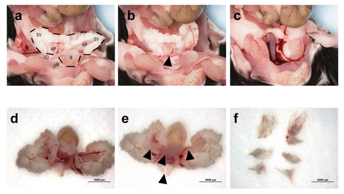
Figure 1: Gross Histology of the Prostate Micro-dissection Protocol. (a) The urogenital tract (UGT) in its natural state, exposed in the murine abdomen. The dotted lines encompass the various parts of the UGT, including the seminal vesicles (SV), prostate lobes (anterior prostate (AP) and ventral prostate (VP) are visible here), bladder (B), and urethra (located beneath the bladder). The vas deferens (VD) is also visible. (b) The vas deferens have been removed. The arrow points to the region from which the vas deferens has been removed. (c) The UGT has been removed from the abdomen by cutting the urethra below the ventral prostate lobes. (d) The UGT has been placed in PBS and is seen beneath a dissection microscope. (e) Fat has been removed from the UGT, allowing for the visibility of all six lobes of the prostate. The arrows point to the two anterior lobes, the ventral lobes, and the dorsolateral lobes. (f) All six individual prostate lobes have been removed. Please click here to view a larger version of this figure.
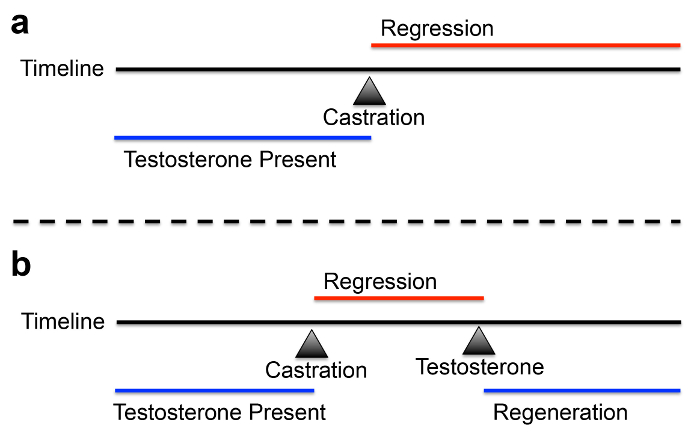
Figure 2: Timeline for Castration and Hormonal Regeneration. (a) Testosterone is present at physiological levels until castration, when testosterone disappears and regression of the prostate begins. (b) Castration removes testosterone, until testosterone is reintroduced, causing hormonal regeneration of the prostate. Please click here to view a larger version of this figure.
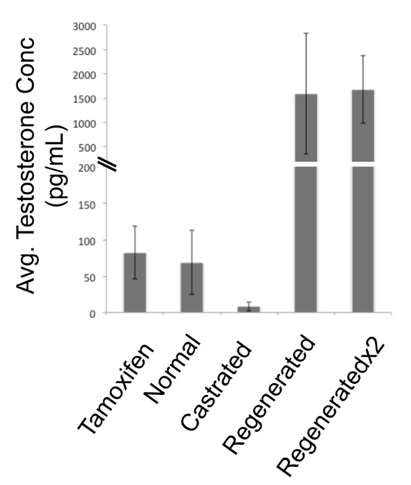
Figure 3: Testosterone Levels in Various Hormonal Conditions. Testosterone ELISA under tamoxifen treated, hormonally normal, castrated, and hormonally regenerated conditions. Regenerated x 2 refers to implantation and removal of synthetic testosterone, followed by implantation of a second round of synthetic testosterone. Error bars represent standard deviation. This figure has been modified from 5. Please click here to view a larger version of this figure.
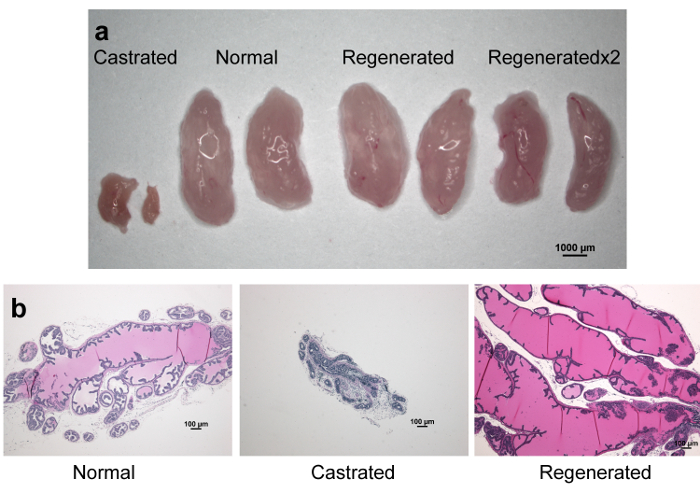
Figure 4: Prostate Appearance under Castration and Regeneration Conditions. (a) Gross appearance of anterior prostate lobes under castrated, hormonally normal, and hormonally regenerated conditions. Regenerated x 2 refers to implantation and removal of synthetic testosterone, followed by implantation of a second round of synthetic testosterone. (b) Formalin-fixed, paraffin-embedded anterior prostate lobes sectioned and stained with hematoxylin and eosin. Please click here to view a larger version of this figure.
Discussion
Prostate micro-dissection allows for lobe-specific experimentation and analysis of the mouse prostate (Figure 1). In genetically engineered mouse models, phenotypes may be seen in one lobe that is not seen in others. Also, for histological analysis, this protocol assures that the maximum amount of pure prostate tissue can be sectioned and stained, without other urogenital tract tissues present in the section. Finally, for single-cell experiments, this protocol allows for isolation of almost pure prostate cells. Epithelial cell enrichment kits can further purify prostate cells from stromal cells. Limitations to micro-dissection include the use of a dissection microscope, and the learning curve necessary to use fine forceps while looking through the dissection microscope. Alternatively, prostate tissue may be sectioned for histology as part of the UGT, making micro-dissection unnecessary. However, the downside to sectioning the entire UGT is that other tissues will be present in the section, making it not as clean. In addition, for generating single prostate cell populations, the lobes must be separated from the UGT to avoid contamination.
Surgical castration is a fundamental surgery for many prostate research labs and is essential for the study of the effects of testosterone on prostate development and disease. This protocol is quick (typical time of surgery for an experienced user is about 5 min) and removes almost 100% of testosterone from circulation (Figure 3). Of note, testosterone levels in mice differ significantly, and may vary by as much as 30-fold from mouse to mouse 10. Castration removes this variation. Following castration, researchers may decide to add testosterone back in to regenerate the prostate (Figures 3-4). This can be done by subcutaneously implanting a source of testosterone, such as an osmotic pump, a testosterone pellet, or a semi-permeable silastic tube containing testosterone powder 8. Depending on the amount of testosterone added, this procedure can provide physiological to superphysiological testosterone levels in the circulation (Figure 3). This protocol can also be used to study prostate cell lineage and stem cell biology 11. Another option is chemical castration, or androgen deprivation therapy, via treatment of mice with compounds that inhibit the production of testosterone, such as dutasteride 12. This also mimics a common treatment of prostate cancer patients, who are rarely surgically castrated. However, surgical castration provides a reliable, permanent method for removal of virtually all testosterone. It is important to consider that the adrenal glands also synthesize androgen species, such as androstenedione and dehydroepiandrosterone, which can be converted into testosterone, though in much smaller amounts, relative to the testes 13. In some cases, the adrenal glands can also be removed surgically, though this procedure is more difficult.
Overall, these two protocols are useful for a myriad of research into prostate diseases, as well as hormonal studies. These protocols are a reliable and consistent way to analyze prostate biology in hormonally normal, castrated, or hormonally regenerated conditions.
Disclosures
The authors declare that they have no competing financial interests.
Acknowledgements
Some of the figures in this manuscript were generously provided by the laboratory of Bart O. Williams. The authors are supported by National Cancer Institute grants U54CA143803, CA163124, CA093900, AND CA143055.
Materials
| Name | Company | Catalog Number | Comments |
|---|---|---|---|
| Surgical tools | Roboz | Various | |
| Formaldehyde | Sigma | F8775 | |
| OCT medium | VWR | 25608-930 | Must be placed on dry ice to freeze, and stored at -80 °C |
| PBS | Life Technologies | 10010 | |
| Isoflurane | Johns Hopkins | Also available from vendors online | |
| Cautery Pen | Medline | ESCT002 | |
| Silk suture | AD Surgical | M-S330R19 | |
| Surgical Wound Clips | Roboz | RS-9265 |
References
- Siegel, R. L., Miller, K. D., Jemal, A. Cancer statistics. CA Cancer J Clin. 65 (1), 5-29 (2015).
- Roehrborn, C. G. Pathology of benign prostatic hyperplasia. Int J Impot Res. 20 (suppl 3), 11-18 (2008).
- Valkenburg, K. C., Williams, B. O. Mouse models of prostate cancer. Prostate Cancer. , 895238(2011).
- Shappell, S. B., et al. Prostate pathology of genetically engineered mice: definitions and classification. The consensus report from the Bar Harbor meeting of the Mouse Models of Human Cancer Consortium Prostate Pathology Committee. Cancer Res. 64 (6), 2270-2305 (2004).
- Valkenburg, K. C., Pienta, K. J. Drug discovery in prostate cancer mouse models. Expert Opin Drug Discov. , 1-14 (2015).
- El-Alfy, M., Pelletier, G., Hermo, L. S., Labrie, F. Unique features of the basal cells of human prostate epithelium. Microsc Res Tech. 51 (5), 436-446 (2000).
- Prins, G. S., Huang, L., Birch, L., Pu, Y. The role of estrogens in normal and abnormal development of the prostate gland. Ann N Y Acad Sci. 1089, 1-13 (2006).
- Valkenburg, K. C., et al. Activation of Wnt/beta-catenin signaling in a subpopulation of murine prostate luminal epithelial cells induces high grade prostate intraepithelial neoplasia. Prostate. 74 (15), 1506-1520 (2014).
- Lukacs, R. U., Goldstein, A. S., Lawson, D. A., Cheng, D., Witte, O. N. Isolation, cultivation and characterization of adult murine prostate stem cells. Nat Protoc. 5 (4), 702-713 (2010).
- Michiel Sedelaar, J. P., Dalrymple, S. S., Isaacs, J. T. Of mice and men--warning: intact versus castrated adult male mice as xenograft hosts are equivalent to hypogonadal versus abiraterone treated aging human males, respectively. Prostate. 73 (12), 1316-1325 (2013).
- Wang, X., et al. A luminal epithelial stem cell that is a cell of origin for prostate cancer. Nature. 461 (7263), 495-500 (2009).
- Pascal, L. E., et al. 5alpha-Reductase inhibition coupled with short off cycles increases survival in the LNCaP xenograft prostate tumor model on intermittent androgen deprivation therapy. J Urol. 193 (4), 1388-1393 (2015).
- Shen, M. M., Abate-Shen, C. Molecular genetics of prostate cancer: new prospects for old challenges. Genes Dev. 24 (18), 1967-2000 (2010).
Reprints and Permissions
