This protocol demonstrates murine islet isolation and seeding onto a decellularized scaffold. Scaffold-supported islets were transplanted into the epididymal fat pad of streptozotocin (STZ)-induced diabetic mice. Islets survived at the transplantation site and reversed the hyperglycemic condition.
A subscription to JoVE is required to view this content. Sign in or start your free trial.
Method Article
Scaffold-supported Transplantation of Islets in the Epididymal Fat Pad of Diabetic Mice
* These authors contributed equally
In This Article
Summary
Abstract
Islet transplantation has been clinically proven to be effective at treating type 1 diabetes. However, the current intrahepatic transplantation strategy may incur acute whole blood reactions and result in poor islet engraftment. Here, we report a robust protocol for the transplantation of islets at the extrahepatic transplantation site-the epididymal fat pad (EFP)-in a diabetic mouse model. A protocol to isolate and purify islets at high yields from C57BL/6J mice is described, as well as a transplantation method performed by seeding islets onto a decellularized scaffold (DCS) and implanting them at the EFP site in syngeneic C57BL/6J mice rendered diabetic by streptozotocin. The DCS graft containing 500 islets reversed the hyperglycemic condition within 10 days, while the free islets without DCS required at least 30 days. The normoglycemia was maintained for up to 3 months until the graft was explanted. In conclusion, DCS enhanced the engraftment of islets into the extrahepatic site of the EFP, which could easily be retrieved and might provide a reproducible and useful platform for investigating the scaffold materials, as well as other transplantation parameters required for a successful islet engraftment.
Introduction
Type 1 diabetes mellitus (T1D) is an autoimmune endocrine disorder in which islet cells are ablated by the immune system, rendering patients dependent upon the injection of exogenous insulin for their whole lives. The Edmonton protocol represents a milestone in clinical studies of islet transplantation; islets were infused through the portal vein and transplanted at the intrahepatic site1. However, two main obstacles-inadequate sources of donor islets and poor islet engraftment-prevent the wide success of the islet transplantation2. Usually, islets need to be collected from three cadaveric donors to reverse the hyperglycemic condition of one patient; this is due to the low yield of islet isolation procedures and the islet loss after transplantation. In particular, although the post-transplantation islets were bathed in oxygen-rich blood, the direct contact with blood often evoked the instant blood-mediated inflammatory reaction (IBMIR), which could cause the acute loss of the islets. In the long term, it is thought that the gradual loss of islets in patients accounted for the drop of diabetes reversal rates in the clinical groups, which could reach 90% in the first year and declined to 30% and 10% by 2 and 5 years post-transplantation, respectively3.
Islet transplantation at extrahepatic sites has been an attractive strategy to reduce the direct contact of islets with blood while confining the transplants to more definable locations compared to intrahepatic infusion. Studies have been carried out in the kidney capsule, eye, muscle, fat pads, and subcutaneous spaces over the past years, showing that islets at these sites are able to survive and function to restore normoglycemia4. In addition, the islets at these sites are retrievable, making it possible for biopsy or even for further replacement procedures. Extrahepatic sites therefore demonstrate great potential for clinical transplantation5.
Biomaterial-based scaffolds have been intensively investigated for cell transplantation and tissue engineering. Three-dimensional (3D) scaffolds usually contain porous structures and can serve as cellular templates to generate spatial structure/organization of cells or as reservoirs to provide the controlled release of bioactive cues. Scaffolds have also been fabricated from polymeric materials, such as poly(glycolide-L-lactide)6, poly(dimethylsiloxane)7, and thermoplastic poly(urethane)8, to transplant islets in the EFP. Compared to the direct transplantation of islets, the use of scaffolds was found to reduce islet loss by preventing the leakage of islets into the intraperitoneal cavity9,10, providing mechanical protection and modulating the local inflammatory reaction. The scaffolds thus may be developed to promote islet engraftment at the transplantation sites7.
In this study, we intend to demonstrate a paradigm of islet transplantation in the EFP, carried out in mice models using a DCS. Scaffolds derived from extracellular matrices have attracted great interest in recent years due to the superior biocompatibility and more natural porous structures compared to synthetic products. Here, we describe a robust isolation protocol to obtain pancreatic islets at high yields from C57BL/6J mice. DCSs processed from the bovine pericardium were then seeded with islets, and the grafts were transplanted to the EFP in syngeneic diabetic models. Normoglycemia in mice was achieved within 10 days and was maintained for up to 100 days, until the removal of the grafts.
Access restricted. Please log in or start a trial to view this content.
Protocol
All experiments were approved by Peking University Institutional Animal Care and Use Committee (IACUC, IACUC no. COE-LuoY-1).
1. Islet Isolation
- Preparation of reagents and equipment.
- Reconstitute collagenase P powder (2 U/mg) in HBSS to make a 5 mg/mL solution and filter it through a 0.22 µm filter to remove the bacteria. Prepare 0.6 mL-aliquot solutions of collagenase P in 15-mL conical tubes and store at -20 °C.
NOTE: During use, each aliquot is diluted with HBSS to give 6-mL working solutions with final concentrations of 0.5 mg/mL, or 1 U/mL (enough for treating 3 mice). The working solution is kept on ice for immediate use within 1 h. The working solution should not be restored or re-frozen for additional usage. - Prepare neutralization solution by adding FBS (2.5%) and P/S (1%) into HBSS; keep the solution on ice. Prepare 60 mL of neutralization solution to treat 6 mice.
- For the islet culture medium, add D-glucose (7 mM), FBS (10%), and P/S (1%) to RPMI 1640 medium.
- Autoclave the surgery instruments at 115 °C for 30 min and 15 psi of pressure.
- Reconstitute collagenase P powder (2 U/mg) in HBSS to make a 5 mg/mL solution and filter it through a 0.22 µm filter to remove the bacteria. Prepare 0.6 mL-aliquot solutions of collagenase P in 15-mL conical tubes and store at -20 °C.
- Inflation of the pancreas.
- Prepare 12 mL of collagenase working solution, as specified in step 1.1.1, for 6 mice (12 weeks old). Fill the 10 mL syringe with 9 mL of working solution and connect the syringe to the 27 G intravenous needle. Store the syringe on ice and use the solution within 1 h.
- Euthanize the mouse by cervical dislocation. Place the mouse in supine position on a paper towel, with the tail pointing towards the operator. Spray the whole body with 70% ethanol, making it completely wet.
- Make a V-incision, starting from the genital area and extending to the diaphragm, using dissection scissors and forceps. Fold the skin over the chest to completely reveal the abdominal cavity.
- Move the bowel to the right side of the mouse and expose the pancreas and common bile duct. Grab the duodenum carefully with forceps and pull it until the bile duct is taut (Figure 1A-1 and A-2).
- Find the portal vein and bile duct leading into the liver. Clamp the portal vein and bile duct with a microscopic hemostatic clamp.
NOTE: The clamping prevents excessive bleeding and the entry of the collagenase into the liver. - While still holding the intestine with the forceps, find the location of the ampulla that connects the bile duct and the duodenum. Insert the 27G intravenous needle into the common bile duct through the ampulla (Figure 1B-1 and B-2).
- Dispense about 200 µL of collagenase working solution to check if the cannulation is all the way through bile duct. If the collagenase solution begins to fill the duct ( Figure 1C-1 and C2), the needle is in the lumen of the duct; clamp the duct segment containing the needle using an artery hemostatic clamp and dispense the rest of the 2 mL of solution at a slow and constant rate within 1 min (Figure 2A-1, A-2, and B-1).
NOTE: If the tissue surrounding the duct begins to inflate (Figure 2B-2), the needle has poked through the wall of the bile duct, making it necessary to reposition the needle and try cannulating the bile duct again. Similarly, if the duodenum begins to inflate (Figure 2C-1 and C-2), the artery hemostatic clamp needs to be adjusted and the bile duct re-cannulated. As the collagenase solution fills up the pancreas, the tissue near the duodenum inflates first, followed by the region near the pancreatic tail. The perfusion of the pancreatic tail (the splenic lobe) is important to maximize the islet yield. - After the complete inflation of the pancreas (Figure 2C-1), push the bowel to the left side of the mouse and remove the pancreas by starting at the descending colon. Use the forceps to lift the bowel and separate it from the pancreas with another pair of forceps. Continue to remove the pancreas until it is unattached from the top of the stomach. Finally, lift the pancreas out of the abdomen and cut it free from the remaining spleen.
NOTE: The whole separation should be performed quickly, as the digestion continues during the removal process. - Place the pancreas in an empty 15-mL conical tube and leave it on ice. Repeat the above procedure for the remaining mice. All pancreas should be further processed within 1 h by promptly following step 1.3 to prevent over-digestion by collagenase P.
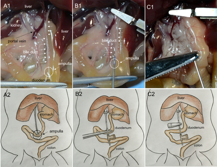
Figure 1: Photographs showing the cannulation of the bile duct and the perfusion of the pancreas with collagenase solutions. (A1) Pulling the duodenum until the bile duct is taut. (ampulla: the triangular, milky area on the surface of the duodenum; bile duct: the cord-like milky structure on the surface). (B1) Inserting the needle into the bile duct from the ampulla. (C1) Inflating the pancreas with the injection of enzyme. (A2, B2, and C2) Cartoon images of the procedures shown in A1, B1, and C1, respectively. Please click here to view a larger version of this figure.
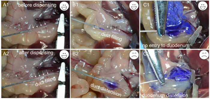
Figure 2: Troubleshooting for the cannulation. (A1) The needle tip inserted in the lumen of the bile duct. (A2) The duct filled with enzyme solutions. (B1) The needle inserted in the lumen of the bile duct, and the duct filled with a blue dye. (B2) Due to inappropriate cannulation, the needle is beneath the bile duct, and only an inflated capsule is observed after dispensing the blue dye. (C1) A successful cannulation is evidenced by the distension of pancreas. (C2) Due to inappropriate clamping, blue dye enters the duodenum and causes distention. Please click here to view a larger version of this figure.
- Digestion and purification of islets.
- Incubate the conical tubes containing the perfused pancreas at 37 °C for 17 min.
NOTE: The incubation time may vary with the age and species of the animal. - Terminate the digestion by adding 7 mL of neutralization solution and putting the tubes on ice.
- Dissociate the tissue by shaking the tubes vigorously (e.g., 20 times in 10 s) until fine tissue particles are obtained.
NOTE: The islet yield will be low if the pancreas fails to dissociate completely. - Filter the digested tissue samples through 0.5-mm wire mesh to remove any non-digested tissue chunks. Collect the islet suspensions in a new 50-mL conical tube.
- Centrifuge the tubes for 3 min at 230 x g and 4 °C and pour off the supernatant carefully, without disturbing the tissue pellets.
- Resuspend the pellets from 3 mice in 4 mL of polysucrose density gradient by vortexing gently or pipetting the suspension up and down a few times. Slowly pipette 4 mL of HBSS down the side of the tube to the top of the polysucrose solutions.
NOTE: The two solutions should be well-separated layers with a sharp interface.
NOTE: The addition of HBSS should be performed with care, without disturbing the polysucrose density gradient at the bottom. - Centrifuge the suspension for 20 min at 900 x g and 4 °C by selecting a very slow acceleration rate. End the centrifugation with no brake.
NOTE: This is a step to purify the islets from the exocrine cells, with most of the islets migrating to the interface between the layers of polysucrose and HBSS and most exocrine cells settling at the bottom. - Remove the entirety of the 8-mL supernatant solutions using a large-bore 15-mL pipette. Pass the solutions through an inverted 70-µm strainer.
NOTE: The islets will be further purified and retained by the strainer, while the exocrine cells will pass through the filter.
Caution: The polysucorse density gradient is cell-toxic; the filtration will help islets get rid of the polysucrose. - Pipette 2 mL of cold neutralization solution into a 35-mm Petri dish. Invert the cell strainer right-side-up, dip the surface retaining the islets in the solution, and gently shake to release the islets.
- Hand-pick the islets under a microscope using a 20-µL pipette with a white tip.
NOTE: The islets are yellowish, compact cell aggregates (Figure 3A), while the contaminating exocrine tissue or cells have a blackish and loose structure under the dissecting microscope. - Place about 200 islets in a 35-mm dish with 2 mL of culture medium and incubate at 37 °C in an incubator supplied with 5% CO2 for 12 h.
NOTE: Usually 150-300 islets can be harvested from one 12-week-old C57BL/6J mouse. Islets are prone to forming aggregates during culture, and the big islets (>300 µm) are susceptible to central necrosis (Figure 1B, inset). Shake the suspension well to evenly distribute the islets in the dish and to reduce clumping.
- Incubate the conical tubes containing the perfused pancreas at 37 °C for 17 min.
2. Islet Culture on the Scaffold
NOTE: DCS has a porosity of about 79%, a thickness of about 0.6 mm, and a pore size ranging from 12 to 300 µm.
- Cut the scaffolds into 7-mm disks, soak them in 70% ethanol, and wash them with HBSS. Place the scaffolds in the 24-well tissue culture inserts.
NOTE: When fresh islets are recovered after overnight culture, the islets appear bright and tight, with smooth borders (Figure 3B). - Swirl the dish and collect the islets from the center of the dish using a 20 µL pipette with a white tip. Transfer the islets to the scaffolds (250 islets/scaffold) using a pipette and add 2 mL of culture medium to the well. Culture the islets for 12 h before transplantation.
3. Islets Transplantation at the EFP Site
- Diabetes induction in recipient mice.
- Place C57BL/6J mice (more than 10 weeks old) in a fresh cage with a water supply but no food. Fast the mice for 10 h before the induction of diabetes.
- Prepare buffer solutions with pH values of 4.2-4.5 by mixing 0.1 M citric acid with 0.1 M sodium citrate. Dissolve STZ (10 mg/mL) in the freshly prepared buffer and sterilize the solution by passaging it through a 0.22 µm filter.
NOTE: STZ is light-sensitive and loses activity within 10 min; always prepare the fresh STZ solution right before injection. - Inject each mouse intraperitoneally with STZ at a dose of 140-150 mg/kg for each C57BL/6J mouse.
NOTE: The dose varies depending upon the age and species of the animal. It is recommended to perform a small-scale dose optimization test for the given species before starting the formal experiment. - Collect the tail vein blood and monitor the blood glucose with a glucose meter on days 2, 3, and 4 post-STZ injection.
NOTE: When the animals are hyperglycemic (non-fasting blood glucose > 16.7 mM) on two consecutive days, they are ready for islet transplantation.
- Transplantation of islets to the EFP site.
- Anesthetize the mice with pentobarbital delivered intraperitoneally (50 mg/kg). Place the mouse in supine position on a paper towel, with the tail pointing towards the operator. Shave the abdomen extensively to remove the fur from around the incision site. Tape down the four limbs and swab the skin completely with alternating alcohol and iodophor wipes moved in a circular fashion to sterilize the incision site. Drape the mouse with a sterile drape, only allowing access to the incision area. Make a 7 mm incision through the peritoneal wall in the midline, close to the genital area.
NOTE: All instruments used during the surgery, including gloves, should be sterile. - Gently grab and remove the EFP from the abdominal cavity using forceps. Spread the EFP on wetted sterile gauze. Place the scaffold containing islets on the EFP and fold the EFP to wrap the transplant. Secure the direct contact between the islets and EFP by suturing the EFP with absorbable 6-0 sutures. Wet the surface of the EFP with sterile saline using a soaked cotton swab to prevent the EFP from drying out.
- Gently place the EFP back in the abdominal cavity. Close the incision by suturing the peritoneal wall and clamping the dermal layer with wound clips.
- Inject buprenorphine (0.1 mg/kg) subcutaneously as an analgesic.
- Inject 1 mL of saline subcutaneously to prevent dehydration.
- Place the mouse in a cage on a heating pad until the mouse recovers from anesthesia.
- Check the non-fasting glucose levels two days later by collecting blood from the tail vein. If retrieving the graft, repeat the above transplantion procedures, gently pull out the EFP, use 3-0 suture to ligate the end of the fat pad adjacent to the epididymis, occlude the blood vessels, explant the EFP graft, and preserve it for histology.
NOTE: Explanting the islets should render the recipient mice hyperglycemic again within 3 days, confirming the function of the graft.
- Anesthetize the mice with pentobarbital delivered intraperitoneally (50 mg/kg). Place the mouse in supine position on a paper towel, with the tail pointing towards the operator. Shave the abdomen extensively to remove the fur from around the incision site. Tape down the four limbs and swab the skin completely with alternating alcohol and iodophor wipes moved in a circular fashion to sterilize the incision site. Drape the mouse with a sterile drape, only allowing access to the incision area. Make a 7 mm incision through the peritoneal wall in the midline, close to the genital area.
Access restricted. Please log in or start a trial to view this content.
Results
Our clamping method, performed using a microscopic hemostatic clamp, is straightforward and time-saving compared with the suture ligation technique. It took roughly 4 h to isolate and purify about 1,200 islets from 6 mice. The freshly isolated islets typically had a rough periphery under an optical microscope (Figure 3A). Once the islets recovered from the isolation process, they looked bright and tight and acquired a smooth surface. However, the stressful is...
Access restricted. Please log in or start a trial to view this content.
Discussion
Pancreas perfusion and digestion time are two key parameters that affect islet yield and quality. Moskalewski first reported the use of a crude collagenase mixture to digest minced guinea pig pancreas11. Lacy et al. reported the injection of enzymes into the duct system to perfuse the pancreas, which greatly increased the islet yield12. The ductal perfusion of enzyme allows for the maximal exposure of pancreatic surface area to the enzyme, resulting in a more homog...
Access restricted. Please log in or start a trial to view this content.
Disclosures
The authors have nothing to disclose.
Acknowledgements
The authors would like to thank Wei Zhang from Guanhao Biotech for providing the decellularized scaffolds. We thank Xiao-hong Peng for the helpful discussions. This research was financially supported by the National Natural Science Foundation of China (Project No.31322021).
Access restricted. Please log in or start a trial to view this content.
Materials
| Name | Company | Catalog Number | Comments |
|---|---|---|---|
| Dissecting scissor | Ningbo Medical | ||
| Forceps | Ningbo Medical | ||
| 0.5 mm diameter wire mesh | Ningbo Medical | ||
| 70 μm cell strainer | Falcon | 352350 | |
| Artery hemostatic clamp | Ningbo Medical | ||
| Microscopic hemostatic clamp | Ningbo Medical | ||
| Hemostatic forceps | Ningbo Medical | ||
| Absorbable 6-0 PGLA sutures | JINHUAN | With needle | |
| Wound clip | Ningbo Medical | ||
| Cotton swab | Ningbo Medical | ||
| Gauze | Ningbo Medical | ||
| Sterile drapes | Ningbo Medical | ||
| 10mL syringe | JINGHUAN | ||
| 1 mL syringe | JINGHUAN | ||
| 27G intravenous needle | JINGHUAN | 0.45x15 RWSB | |
| 1.5 mL Eppendorf tube | Axygen | ||
| 15mL conical tube | Corning | 430791 | |
| 50mL conical tube | Corning | 430829 | |
| 35mm Non-treated Peri-dishes | Corning | 430588 | |
| Transwell | Corning | 3422 | |
| 0.22 μm filter | Pall | PN4612 | |
| 10 mL serological pipet | Corning | 4488 | |
| Pipet filler S1 | Thermo Scientific | 9501 | |
| Pipette (2-20μL) | Axygen | AP-20 | AXYPETTM |
| Dissecting microscope | Olympus | SZ61 | |
| Centrifuge | Eppendorf | 5810R | |
| Hank’s balanced salt solution | Gibco | C14175500CP | |
| Collagenase P | Roche | COLLP-RO | |
| Histopaque 1077 | Sigma | 10771 | |
| RPMI 1640 | Gibco | 11879-20 | |
| FBS | Gibco | 16000-044 | |
| D-glucose | Gibco | A24940-01 | |
| Glucose meter | Roche | ACCU-CHEK | |
| Penicillin-streptomycin | Gibco | 15140-122 | |
| Streptozotocin | Sigma | V900890 | VetecTM |
| Chloral hydrate | J&K | C0073 | |
| Sodium citrate | Sigma | 71497 | |
| Citric acid | Sigma | C2404 | |
| Iodophors | Ningbo Medical | ||
| C57BL/6J, 10-12 weeks old | VitalRiver | Beijing, China | |
| Decellularized scaffold | Guanhao Biotec | 131102 | Guangzhou, China |
References
- Shapiro, A. M., et al. Islet transplantation in seven patients with type 1 diabetes mellitus using a glucocorticoid-free immunosuppressive regimen. N Engl J Med. 343, 230-238 (2000).
- Shapiro, A. M. J., et al. International Trial of the Edmonton Protocol for Islet Transplantation. N Engl J Med. 355, 1318-1330 (2006).
- Ryan, E. A., et al. Five-year follow-up after clinical islet transplantation. Diabetes. 54, 2060-2069 (2005).
- Merani, S., Toso, C., Emamaullee, J., Shapiro, A. M. Optimal implantation site for pancreatic islet transplantation. Br J Surg. 95, 1449-1461 (2008).
- Schmidt, C. Pancreatic islets find a new transplant home in the omentum. Nat Biotechnol. 35 (1), (2017).
- Dufour, J. M., et al. Development of an ectopic site for islet transplantation, using biodegradable scaffolds. Tissue Eng. 11, 1323-1331 (2005).
- Weaver, J. D., et al. Controlled Release of Dexamethasone from Organosilicone Constructs for Local Modulation of Inflammation in Islet Transplantation. Tissue Eng Part A. 21, 2250-2261 (2015).
- Wang, K., et al. From Micro to Macro: The Hierarchical Design in a Micropatterned Scaffold for Cell Assembling and Transplantation. Adv Mater. 29, (2017).
- Blomeier, H., et al. Polymer Scaffolds as Synthetic Microenvironments for Extrahepatic Islet Transplantation. Transplantation. 82, 452-459 (2006).
- Gibly, R. F., et al. Extrahepatic islet transplantation with microporous polymer scaffolds in syngeneic mouse and allogeneic porcine models. Biomaterials. 32, 9677-9684 (2011).
- Moskalewski, S. Isolation and Culture of the Islets of Langerhans of the Guinea Pig. Gen Comp Endocrinol. 5, 342-353 (1965).
- Lacy, P. E., Kostianovsky, M. Method for the isolation of intact islets of Langerhans from the rat pancreas. Diabetes. 16, 35-39 (1967).
- Zmuda, E. J., Powell, C. A., Hai, T. A Method for Murine Islet Isolation and Subcapsular Kidney Transplantation. J Vis Exp. (50), (2011).
- Li, D. S., Yuan, Y. H., Tu, H. J., Liang, Q. L., Dai, L. J. A protocol for islet isolation from mouse pancreas. Nat Protoc. 4, 1649-1652 (2009).
- Stull, N. D., Breite, A., McCarthy, R., Tersey, S. A., Mirmira, R. G. Mouse Islet of Langerhans Isolation using a Combination of Purified Collagenase and Neutral Protease. J Vis Exp. (67), (2012).
- Sakata, N., Yoshimatsu, G., Tsuchiya, H., Egawa, S., Unno, M. Animal models of diabetes mellitus for islet transplantation. Exp Diabetes Res. , 256707(2012).
- Schmidt, C. Pancreatic islets find a new transplant home in the omentum. Nat Biotech. 35, 8-8 (2017).
- Londono, R., Badylak, S. F. Biologic scaffolds for regenerative medicine: mechanisms of in vivo remodeling. Ann Biomed Eng. 43, 577-592 (2015).
Access restricted. Please log in or start a trial to view this content.
Reprints and Permissions
