The study was approved by the data protection officials at Oslo University Hospital and the Regional Committee for Medical and Health Research Ethics, Southern Norway 2419/2011. All participants signed a written informed consent at inclusion.
1. Preparations
NOTE: A timeline for the procedures is outlined in Figure 1.
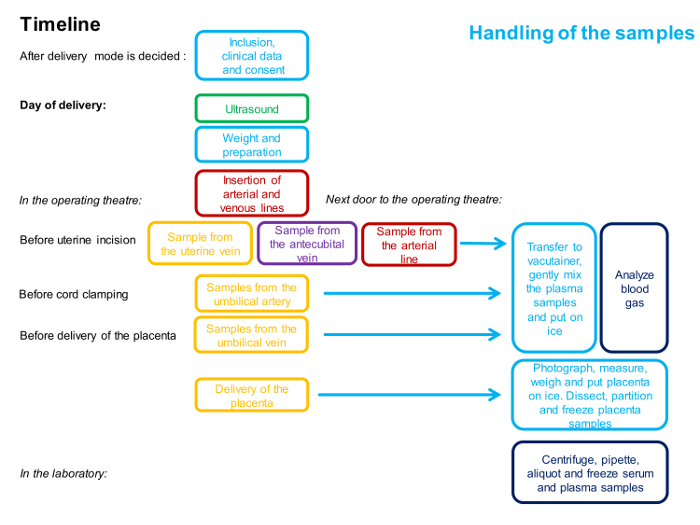
Figure 1: Flowchart Describing the Timing and the Personnel Involved in the 4-vessel Sampling Procedure.
One color represents one person. Detailed description of the method is given in the protocol. Please click here to view a larger version of this figure.
- Staff
- Make sure all required personnel are available: a highly skilled Fetal Medicine specialist conducting the ultrasound measurements, two obstetricians conducting the surgery, one of the obstetricians and two nurses collecting the samples, one assistant handling the blood gas analyses and one assistant handling the other samples consecutively and immediately following the collection.
NOTE: In cases of more advanced collection of placental tissue, an additional person is required.
- Make sure all required personnel are available: a highly skilled Fetal Medicine specialist conducting the ultrasound measurements, two obstetricians conducting the surgery, one of the obstetricians and two nurses collecting the samples, one assistant handling the blood gas analyses and one assistant handling the other samples consecutively and immediately following the collection.
- Equipment
- Prepare the equipment, 50 mL of ice cold 1 M phosphate buffered saline (PBS), 25 mL of cold RNA stabilizing solution and 5 x 0.5 mL of optimal cutting temperature compound (OCT). Label the vacutainers and tubes. See tentative list of equipment.
2. Maternal Characteristics
- Record the maternal clinical and non-clinical characteristics at inclusion and repeat relevant questions and measurements, including weight, at the time of delivery. Record the duration of the fasting period prior to the cesarean section, and any hypotensive episodes occurring during the surgery.
Note: Include the minimal maternal clinical dataset reported in a recent publication from Global Pregnancy CoLaboratory (COLAB). This article also includes some very important aspects in choosing study populations and should be addressed while planning the study 18. - Consider recording paternal characteristics, including ethnicity, age and body mass index (BMI).
3. Ultrasound
- Perform the Doppler ultrasound examination on the day of the delivery, with the women in a fasting state. Perform the examination during a period of fetal quiescence, with the woman in semi-supine position, tilted slightly laterally opposite to the region of interest in order to avoid compression of the aorta and vena cava. Monitor the output intensity by the mechanical and thermal indices on the display.
- Umbilical vein
- Visualize the umbilical vein in a sagittal or oblique transection of the fetal abdomen. Measure the internal vessel diameter in the straight portion of the intra-abdominal umbilical vein, before any visible branches. Use regular B-mode and visualize the vessel in a perpendicular insonation angle for diameter measurements and keep several optimal frames for later measurements to minimize the effect of pulsatile diameter changes.
- Repeat the measurements five to ten times 19.
- At the same site, use Doppler ultrasound and adjust the probe to get an insonation angle as low as possible (always <30°) in order to measure the time-averaged maximum velocity (TAMX). Obtain the velocity over a period of 3 – 5 s (non-pulsating flow).
- Visualize the umbilical vein in a sagittal or oblique transection of the fetal abdomen. Measure the internal vessel diameter in the straight portion of the intra-abdominal umbilical vein, before any visible branches. Use regular B-mode and visualize the vessel in a perpendicular insonation angle for diameter measurements and keep several optimal frames for later measurements to minimize the effect of pulsatile diameter changes.
- Uterine artery
- Use Doppler ultrasound to visualize the uterine artery as it crosses the external iliac artery, immediately after it branches from the internal iliac artery. Adjust the probe at this site to get a low insonation angle (always <30°) and measure TAMX. Obtain the velocity as the mean velocity of three heart cycles.
- As it is unlikely to get a perpendicular angle at the same site as TAMX is measured, follow the vessel distally to get a correct angle for diameter measurements as close to the sites of diameter measurements as achievable. Exclude the diameter measurements if any visible vessels branch off before this site as evaluated by color Doppler ultrasound.
- Use regular B-mode and visualize the vessel in a perpendicular insonation angle for diameter measurements and keep several optimal frames for later measurements to minimize the effect of pulsatile diameter changes.
- Repeat the measurements five to ten times 19.
- Note the position of the placenta.
4. 4-vessel Blood Sampling
NOTE: The timeline for the procedures is outlined in Figure 1 and an overview of the samples is illustrated in Figure 2.
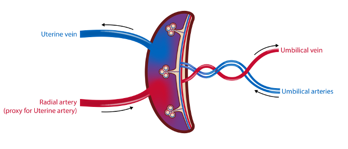
Figure 2: Schematic Illustration of the Placental Vasculature and the Sampling Sites.
In the 4-vessel sampling method blood samples are drawn from the uterine vein, the radial artery (as a proxy for the uterine artery) and the umbilical arteries and vein. Blood flow in the uterine artery and the umbilical vein is measured by ultrasound. Tissue samples from the placenta are collected. Illustration: Øystein H. Horgmo, University of Oslo. Please click here to view a larger version of this figure.
- Safety procedures
- Provide all personnel in the operation theater with gloves, surgical scrub suits, masks and headwear.
- Provide the surgeons and research personnel in contact with the operation field with surgical scrub suits, masks, headwear, gowns and double gloves. Glasses are optional.
- Provide personnel handling the blood samples with gloves.
- Provide personnel handling the placenta samples with gloves and surgical mask. Homogenization requires the use of hoods.
- Preparation in the operation theater
- Give a briefing and hand the equipment to all personnel that will assist the sampling before onset of surgery.
- Address the anesthesiologist and anesthesiology nurse who will assist with the necessary peripheral arterial and venous access, and ensure that no liquids are given intravenously before sampling.
- Give three syringes (10 mL) without needles to the person assisting with the antecubital vein sample and two syringes (one 20 mL and one 10 mL) and one blood gas syringe (with heparin) to the person assisting with the radial artery.
- Prepare two sterile syringes (20 mL), five sterile syringes (10 mL), three "butterfly needles" and two blood gas syringes for the operation field.
- Access to blood vessels.
- Follow standard procedure before the cesarean section to assure peripheral intravenous (iv) access.
NOTE: The antecubital vein is preferable because it is easier to draw samples from this site. - Localize the radial artery at the wrist by ultrasound or by palpation. Following 0.5 mL of subcutaneous lidocaine analgesia, place an arterial line into the radial artery. Abandon the sampling from this site in case of three failed insertions, or if the woman experiences pain during the insertion.
NOTE: Perform the surgical procedure of cesarean section according to standard procedure. Only the adjustments needed for the sampling procedure are underlined below.
- Follow standard procedure before the cesarean section to assure peripheral intravenous (iv) access.
- Maternal blood samples
NOTE: Obtain all three maternal blood samples (uterine vein, radial artery and antecubital vein) simultaneously before the uterine incision.- For the uterine vein, after opening the abdominal cavity, use a retractor to lift the abdominal wall and expose the main branches of the uterine veins on the anterolateral sides of the uterus. Obtain blood from uterine vein branches at the same side as the placenta whenever possible or use the most prominent vein plexus if the placenta is located in the uterine midline.
- Insert a butterfly needle on a blood gas syringe in the uterine vein at an angle of approximately 30 degrees and collect blood through gentle aspiration to avoid hemolysis. While carefully securing the iv position of the butterfly needle, replace the filled blood gas syringe by a 20 mL and a 10 mL syringe consecutively.
NOTE: Optimal access is best ensured when standing on the contralateral side of the chosen uterine vein.
- Insert a butterfly needle on a blood gas syringe in the uterine vein at an angle of approximately 30 degrees and collect blood through gentle aspiration to avoid hemolysis. While carefully securing the iv position of the butterfly needle, replace the filled blood gas syringe by a 20 mL and a 10 mL syringe consecutively.
- For the radial artery, aspirate from the intra-arterial line. Discard the first 5 mL, and then aspirate 3 mL in heparin syringe for blood gas analyses, followed by 3 mL in two syringes (20 + 10 mL).
- For the antecubital vein, aspirate gently from the intravenous catheter. Discard the first 5 mL, and then aspirate 30 mL in three syringes (10 mL).
- Perform a final inspection of the sampling site on the uterine vein before starting to close the abdomen.
- For the uterine vein, after opening the abdominal cavity, use a retractor to lift the abdominal wall and expose the main branches of the uterine veins on the anterolateral sides of the uterus. Obtain blood from uterine vein branches at the same side as the placenta whenever possible or use the most prominent vein plexus if the placenta is located in the uterine midline.
- Fetal blood samples
- When the child is born, immediately aspirate blood from the umbilical artery, without clamping the umbilical cord or delivering the placenta. Start with the syringe for blood gas analysis, and follow with three 10 mL syringes if possible.
- When the arterial samples are secured, clamp the cord and hand the child to the midwife before sampling from the umbilical vein (blood gas and 20 + 10 mL syringes).
NOTE: Obtain all umbilical samples within seconds of delivery and with the placenta in situ unless it has detached spontaneously. - Follow the Norwegian recommendations on late cord clamping. In case of a distressed child, clamp and cut the cord immediately and the hand the child to the midwife and neonatologist.
- Handling of blood samples
- Put the blood gas syringes on ice while preparing the rest of the blood samples, and analyze them in a blood gas analyzer within 5 min.
- Transfer the blood samples immediately to vacutainers and place the plasma tubes on a rocker for 1 – 2 min before putting them on ice. Leave the serum tubes on the laboratory bench to settle for 30 minutes.
NOTE: This is a critical step in the procedure that needs extra attention because samples from all five sites have to be prepared simultaneously to ensure good quality. - Centrifuge the plasma samples as soon as possible, and within 30 min, at 6 °C, 2,500 x g for 20 min.
- After 30 min, centrifuge the serum samples at room temperature for 10 min at 2,500 x g.
- Aliquot the supernatants carefully to 2 mL cryo tubes, leaving 0.5 mL of the supernatant above the pellet to ensure platelet free plasma.
- Store the samples at -80 °C.
5. Collection of Placental Tissue
- Place the placenta flat down on an ice chilled dissection tray as soon as possible after it has been delivered. Photographand measure the longest diameter and the diameter at 90 degrees.
- Weigh the placenta.
- Record the weight, the two diameters, any gross pathology, number of vessels in the cord and the time interval from delivery to when the placenta was placed on ice.
NOTE: Send the placenta to pathological examination if clinically indicated. - Place the placenta with the maternal surface facing up and identify 4 – 5 sampling sites randomly located in each quadrant of the placenta, avoiding areas of frank pathology. Remove the decidua using scissors to cut away 3 – 5 mm from the maternal surface. Collect a 1 – 2 cm3 piece of villous tissue from each site.
- Wash the collected tissue gently in 50 mL of cold 1M PBS. Divide into several pieces from each sampling site and aliquot.
Note: The size of the placenta pieces will depend on the planned analyzes. - Add aliquots of 0.1 – 0.5 cm3 tissue samples to 5 cryo tubes and snap freeze in liquid nitrogen.
- Add small pieces of 0.1 – 0.2 cm3 to the tube with 25 mL of RNA stabilization solution. Store at 4 °C for 24 h, discard the RNA stabilization solution and replace it. Freeze.
- Add pieces of 0.5 cm3 to the 5 cryo tubes with 0.5 mL of OCT, top up with OCT, mix and freeze.
- Store the samples at -80 °C until analysis.
NOTE: Burton et al. provides an excellent overview of practical aspects of placental sampling depending on the analyses planned. 20 Consider to prepare the remaining tissue for isolation of the microvillous and basal membranes, and to collect decidual tissue by vacuum suction technique. 21,22
6. Neonatal Characteristics
- Record the neonatal characteristics, including Apgar-score (1, 5 and 10 min), sex, weight, length, gestational age and admission to Newborn Intensive Care Unit (length and outcome of stay).
- Consider measuring neonatal body composition by anthropometric measurements, air-displacement plethysmograph or dual X-ray absorptiometry.23,24
7. Calculations
- Assume similar blood composition in the radial and uterine artery and calculate the uteroplacental arteriovenous concentration difference.
Uteroplacental arteriovenous concentration difference = CA – CV
Umbilical venous – arterial concentration difference = Cv – Ca
Where C is concentration with subscripts: A, the radial artery; V, the uterine vein; v the umbilical vein and a, the umbilical artery. - Calculate the volume blood flow, mL/min (Q):
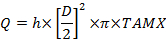
Where D is the vessel diameter (cm), TAMX is time averaged maximum velocity and h is the coefficient for the spatial blood velocity profile. Use 0.5 as the coefficient for the umbilical vein and 0.6 for the uterine artery25,26. - Calculate the placental uptake and release according to Fick's principle:
Uteroplacental uptake = (CA – CV) x Qm
Fetal uptake = (Cv – Ca) x Qf
Subscripts: m, maternal and f, fetal.
The 4-vessel sampling method is applicable in clinical practice and we have successfully obtained blood samples from 209 mother/infant-pairs. In 128 of these we also achieved to measure volume blood flow. Complete 4-vessel sampling and good quality flow measurements of both maternal and fetal vessels were obtained in 70 mother-fetus pairs (Figure 3). In addition, we have so far collected blood and placenta samples from 30 preeclamptic patients. We have previously published articles on human placental transfer of nutrients, as well as placental release of vasoactive factors, demonstrating two applications of the method 14,15,16.
Example of how the 4-vessel method is used to study placental transfer
There are significant arteriovenous glucose differences on both sides of the placenta demonstrating an in vivo uteroplacental and fetal uptake of glucose (Table 1). The placental transfer of glucose is dependent on the maternal-fetal glucose gradient, and thereby on the maternal glucose levels. However, we have previously demonstrated that this gradient, and thus the glucose transfer, is significantly influenced also by fetal insulin levels and glucose consumption. This is an example of how this method illustrates important maternal-fetal interplay 14.
| Vessel | Glucose mmol/L | p-value* |
| Radial artery | 4.49 [4.22, 4.84] | |
| Uterine vein | 4.23 [3.94, 4.53] | |
| Umbilical vein | 3.78 [3.52, 4.06] | |
| Umbilical artery | 3.24 [2.95, 3.56] | |
| Paired differences | ||
| Radial artery – uterine vein | 0.29 [0.13, 0.41] | <0.001 |
| Umbilical artery – umbilical vein | 0.54 [0.29, 0.76] | <0.001 |
| Radial artery – umbilical artery | 1.25 [1.03, 1.51] | <0.001 |
Table 1: Median [Q1, Q3] Concentrations and Arteriovenous Differences of Glucose
* Wilcoxon Signed-Rank test
The fetal glucose uptake from (placental release to) the umbilical circulation is believed to be dependent not only on the maternal-fetal gradient, but on placental blood flow 5. Likewise, it may be relevant to study the fetal glucose uptake as a function of placenta weight or birth weight. In n= 128 we found a median [Q1, Q3] total umbilical venous flow of 196.2 [158.3, 232.2] mL/min, and calculated a median [Q1, Q3] fetal glucose uptake from (placental release to) the umbilical circulation of 0.10 [0.05, 0.15] mmol/min. Normalized for birthweight this equals 0.03 [0.02, 0.04] (mmol/min)/kg. The placenta releases 0.16 [0.10, 0.26] (mmol/min) per kg placenta.
Example of how the 4-vessel method is used to study placental uptake
Animal studies suggest that glutamic acid is important both in interconversion of amino acids in the placenta and the fetal liver, and as an oxidative fuel in other metabolic pathways 27. Using the placenta 4-vessel sampling method we studied the uteroplacental and umbilical arteriovenous differences of glutamic acid in humans (Table 2). We found a placental uptake (thus a fetal release) of glutamic acid from the umbilical circulation. Further we found a placental uptake of glutamic acid from the maternal circulation. This placental uptake from both circulations is an example of how the 4-vessel method can be used to demonstrate in vivo in the human that placental metabolism of nutrients is a part of the regulation of the transplacental transfer.
| Vessel | Glutamic acid µmol/L | p-value* |
| Radial artery | 61.5 [51.0, 77.7] | |
| Uterine vein | 51.0 [36.3, 65.0] | |
| Umbilical vein | 39.3 [24.7, 52.8] | |
| Umbilical artery | 44.7 [33.1, 59.3] | |
| Paired differences | ||
| Radial artery- uterine vein | 10.4 [1.6, 21.2] | <0.001 |
| Umbilical artery –umbilical vein | -8.7 [-16.0, 0.2] | <0.001 |
Table 2: Median [Q1, Q3] Concentrations and Arteriovenous Differences of Glutamic Acid
* Wilcoxon Signed-Rank test
Example of how the 4-vessel method is used to study placental release
It is established that the placenta secretes progesterone and in order to validate our 4-vessel method on the maternal side of the placenta, we measured the in vivo release of progesterone at term 28. We found a significant placental release of progesterone to the maternal circulation (Table 3). The observed arteriovenous difference demonstrates how the placental 4-vessel sampling method can be used to detect substances released by the placenta, and is highly relevant when studying pathological pregnancies.
| Vessel | Progesterone nmol/L | p-value* |
| Radial artery | 678 [514, 971] | |
| Uterine vein | 1852 [1059, 2786] | |
| Paired differences | ||
| Radial artery- uterine vein | -1187 [-1855, -404] | p<0.001 |
Table 3: Median [Q1, Q3] concentrations and uteroplacental arteriovenous difference of progesterone
* Wilcoxon Signed-Rank test
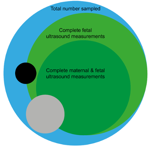
Figure 3: Flowcharts and illustration of included and lost participants.
A. Shows the inclusion of the participants, demonstrating that participants were lost mainly due to start of labor prior to caesarean section or lack of sufficient personnel to conduct the study. B. Of the 179 women with normal pregnancies (blue) complete 4-vessel blood samples were obtained in 162 (91%) (incomplete fetal blood samples: black, incomplete maternal blood samples: grey). Fifty-one (28%) participants were not included for ultrasound measurements due to logistical limitations. Of the 128 participants (72%) subjected to ultrasound, blood flow measurements at the fetal side of the placenta were obtained in all participants (light green), whereas complete blood flow measurements at both the maternal and fetal side were obtained in 77 (60%) (dark green). Illustration: Øystein H. Horgmo, University of Oslo. Please click here to view a larger version of this figure.
