This protocol first describes the surgical procedure of the permanent implantation of a urinary bladder catheter combined with external urethral sphincter electrodes, and second, the measurement of the function of the urinary bladder and external urethral sphincter in implanted awake animals.
Lower urinary tract function is mainly assessed by means of cystometric bladder function analysis in rodents. Conventional cystometries are usually performed as terminal analysis under urethane anesthesia. It is well known that anesthetic drugs can influence bladder function. Hence, the aim of this technique is to perform cystometric measurements of the urinary bladder and external urethral sphincter in lightly restrained awake rats. For this purpose, a bladder catheter is implanted into the bladder dome. Subsequently, two electrodes are implanted bilateral to the external urethral sphincter and a ground electrode is sutured to a non-responsive skeletal muscle. The bladder catheter and the three electrodes are finally tunneled subcutaneously to the neck region and affixed to a harness. With this technique, the lower urinary tract can be measured at multiple time points in the same animal to assess lower urinary tract function. The main application of this technique is the follow-up of simultaneous urinary bladder and external urethral sphincter function in awake healthy rats and after induction of a disease or injury. Moreover, subsequent lower urinary tract monitoring can be performed during evaluation of the disease/injury and to monitor treatment efficacy.
To analyze urinary storage and voiding function and dysfunction, most studies have used rodent models. Through sequential activation of reflexes, micturition is produced. The coordination of these reflexes is essential for efficient voiding1. Cystometric recording techniques provide valuable tools for analyzing the urinary bladder function under its neural control1.
Most conventional cystometries in rats are done as a single, final analysis in anesthesia, mainly urethane2, and focus on the urinary bladder solely. However, in some pathologies like neurogenic lower urinary tract dysfunction (NLUTD), not only the urinary bladder, but also the bladder outlet, the external urethral sphincter, is dysfunctional3,4. This makes NLUTD difficult to follow-up, if only the bladder is examined in a single cystometric measurement. To get reliable results that are comparable to humans, it is essential to accurately measure both the urinary bladder and the external urethral sphincter function and its interactions2. Furthermore, it is crucial to perform functional analyses in awake rats as anesthesia is very likely to alter bladder function2,5,6. A good cystometric recording in awake animals is the basis for the identification of bladder function and malfunction7.
The small animal cystometry station used (e.g., Catamount cystometry station (CCS)) is a unit to perform cystometric analyses in small awake animals8. By means of a permanent bladder catheter and implanted external urethral sphincter electrodes, repetitive measurements can be performed over a longer time periods2. Thus, the CCS provides a valuable tool for non-neurogenic and NLUTD evaluations in the rodent model, in which the pathomechanisms can change during short- or medium-term follow-up. Additionally, this method includes an artefact-reduced cystometric analysis by using a restrainer to conduct bladder measurements in awake rats.
In this paper, we describe the surgical approach to permanently implant a bladder catheter and external urethral sphincter electrodes, along with cystometric measurements in awake rats.
All procedures described here were approved by the Austrian Governmental Ethics committee for animal research (Bundesministerium für Wissenschaft, Forschung und Wirtschaft, WF / V /3b) and were in compliance with the Association for Assessment of Laboratory Animal Care guidelines for animal use. Rats used for this approach were female, 12-week-old Lewis rats. Use sterile instruments throughout the protocol.
1. Material Preparation
2. Animal Preparation
3. Bladder Catheter Implantation
4. Urethral Sphincter Electrode Implantation
5. Tunneling
6. Harness Fitting
7. Manufacturing of the Electrode Connector
8. Post-surgery Care
9. Preparation for Cystometric Measurement
10. Calibration
11. Animal Database (Animal DB)
12. Measurement Settings Prior to Recording
13. Animal Preparation
14. Recording
A schematic showing the process of awake cystometric measurements is presented in Figure 1 and the internal anatomy for bladder catheter implantation is shown in Figure 2. Surgery takes about 2 h. Postoperative analgesia and antibiotics, as described in the protocol, cover pain and infections over five days after surgery. No signs of any pain were noticed thereafter. Twice daily careful inspection of the abdomen, abdominal suture, and the neck suture is necessary to maintain the animal's health. Harness control (position and tightness) should be conducted once daily in the first five days and later on a regular basis. Abdominal sutures can be removed at the 10th post-operative day.
We use soft, non-woody bedding for the first 10 days after surgery to avoid inflammation. Bedding is changed at least twice a week to further lower the risk of inflammation. Animals are kept in single housing, as group housing increases the risk of harness, catheter, or electrode cable biting by cage mates.
The catheter should be flushed at least once per week, either in the course of a cystometric measurement or by flushing the catheter manually with 1-3 mL of sterile 0.9% sodium chloride at a low infusion speed (Figure 3). Regular antibiotic coverage of the animals further reduces the risk of infections and urinary stone formation. Monitoring fluid uptake is a further major point to prevent urinary stone formation. Citric acid in low concentrations (2-3%) is administered either intravesically via the catheter or into the drinking water to prevent calculi formation.
The success rate of the surgical procedure, as well as maintaining the bladder catheter and electrodes intact, is around 80%. In the remaining 20% of cases, the main problem was detachment of the electrode wires from the plug. Thus, a careful attachment of the electrode wires to the harness is crucial to avoid electrode loss.
Cystometric measurements are usually done until three consecutive voiding cycles are recorded per measurement, which takes between 20 to 40 min, depending on the anxiety and handling status of the rat. The first cystometric measurement is usually done one week after catheter implantation surgery.
Main read out parameters of the cystometric recording are the baseline pressure, threshold pressure, maximum detrusor pressure, voided volume, average flow, voiding time, average pressure, compliance of the urinary bladder, and simultaneous read out of the external urethral sphincter EMG-activity (Figure 4).
Consecutive cystometric measurements in the follow-up period can be performed for at least four weeks after surgery. If the catheter line is regularly flushed, catheter blockage is no problem. Regular handling and optical control of the rats should be carried out during the whole follow-up period.
If the catheter is kinked or blocked, the intravesical pressure will increase linearly up to very high pressures (above 100 cm H2O). In this case, filling should be stopped and the visible catheter end should be checked for kinking. If no kinking is seen, the catheter should be checked for a blocked outlet. For this purpose, the catheter can be flushed manually via the catheter. If fluid is not easily flowing into the bladder, pulling back and forward lightly can be tried. For one last attempt, an acidic flushing solution (citric acid 2-3%) can be used to try to clear the obstructed region within the catheter. This solution might have a higher chance to dissolve the blockade, yet, the bladder will be irritated after successful flushing and consecutive measurements should only be performed two days after flushing with acidic solution. If no fluid can be flushed into the bladder, the catheter is permanently blocked and no further measurements are possible, and the animal is lost for follow-up.
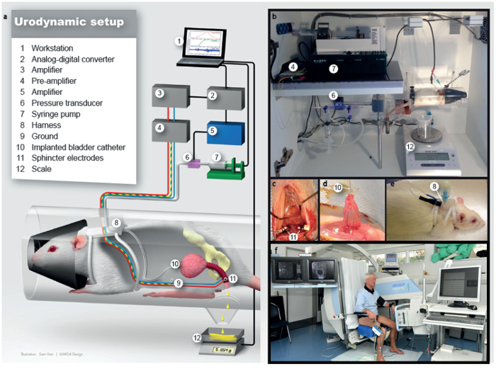
Figure 1: Schematic drawing of the cystometric measurements in awake rats. This figure has been adapted from2. (a) Illustration of the urodynamic setup. (b) Lab station for urodynamic examination. (c) Implantation of the external urethral sphincter electromyography electrodes lateral to urethra, intraoperative view. (d) View of the bladder dome at the moment of bladder catheter implantation, intraoperative view. (e) After implantation of electrodes and catheter, the rat will be fitted with a harness to safely store plugs and connectors. (f) Human urodynamics. Numbers in b-e relate to the legend in a. Please click here to view a larger version of this figure.
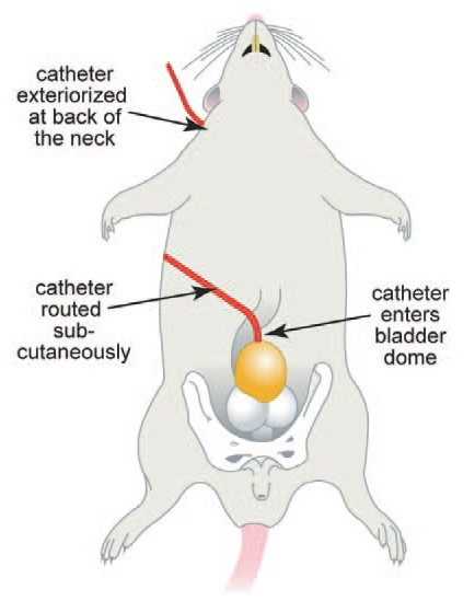
Figure 2: Internal anatomy of the rat for bladder catheter implantation. This figure has been modified from8.
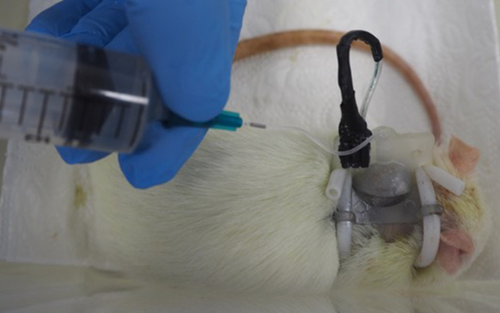
Figure 3: Flushing of the catheter line of a rat. Please click here to view a larger version of this figure.
Figure 4: Urodynamic tracings in an animal 12 day after catheter implantation. (a) Representative urodynamic tracing from a naïve rat. On top is shown the bladder pressure tracing, in the middle the secreted urine weight tracing, and on the bottom the external urethral sphincter EMG tracing. (b) Zoom window from a naïve animal of 60 s, taken from (a). An important remark is that there is less external urethral sphincter EMG activity during voiding than before and after voiding. On top is shown the bladder pressure tracing, in the middle the secreted urine weight tracing, and on the bottom the external urethral sphincter EMG tracing. At the bottom, a heat plot is shown with time matched frequency spectrogram (corresponding to frequency at the current time point). Red represents a high power and blue represents low power. Please click here to view a larger version of this figure.
This protocol describes the surgical procedure of a permanent catheter and urethral sphincter electrodes implantation and the cystometric recording technique in awake, lightly restrained rats including both the simultaneous analysis of the urinary bladder and external urethral sphincter.
Critical steps during surgery are the careful implantation of the bladder catheter, avoiding leakage and extensive manipulation. Moreover, a precise implantation of the electrodes bilateral to the urethral external sphincter is crucial for a sound measurement of the external urethral sphincter. A close inspection of the surgical fields after implantation is also essential to maintaining the animal's health. Antibiotic coverage during follow-up helps in preventing infections along the catheter line, as well as the occurrence of urinary tract infections.
During cystometric measurement, a handled rat will be calmer and more relaxed than a rat which was not handled previously. Thus, the cystometric recording might differ in its outcome. Moreover, the restrainer offers the rat a confining space with a dark front area to feel more comfortable and thus, reduces stress levels. In other published awake cystometric measurements, rats can freely move in the measuring cage. However, this bears a higher artefact risk during the measurement and might increase the time of recording, and also the stress level in the animal. In healthy rats, the optimal cystometric measurement can be replicated at multiple measuring time points during follow-up. During the cystometric measurement, problems that commonly occur are kinked catheters or a mistake in the step-wise conduction of the protocol. If a technical or software error occurs, a re-initialization of the cystometric measurement and stepwise repeat of the protocol is highly recommended for troubleshooting.
Limitations of this technique are the inter-animal variability of the cystometric recordings, structural changes in the urinary bladder tissue due to the implanted catheter, which might hamper histological or molecular biological examinations of this tissue, and the single housing of the animals during the follow-up period. Furthermore, this technique has only been tested in female rats, the applicability and outcomes for male rats have not yet been examined.
The main advantage of this technique is the simultaneous measurement of the urinary bladder and the external urethral sphincter, as well as the awake measurement setting. Thus, a more translational examination of the lower urinary tract in awake animals is available, in comparison to the single, terminal cystometric analysis in anesthesia5,6,9,10. Moreover, with this approach, the progression of a lower urinary tract dysfunction or pathology can be followed in the same animal over time, as well as treatment successes. Especially, NLUTD can be examined in a time course, which was not possible to a full extent with the common cystometric technique2.
In conclusion, the presented surgery and cystometric measurement are used for multiple, artefact-reduced analyses of the lower urinary tract, including the interaction of the urinary bladder and the external urethral sphincter in awake rats.
The authors have nothing to disclose.
The authors do not have any acknowledgements.
Polyethylene tubing PE 50 | Becton Dickinson | 427411 | Catheter |
Prolene 6-0 (BV-1, 9.3 mm, 3/8c) | Ethicon | EH7403H | Suture |
Teflon coated steel wire | Cooner wire | AS631 | Electrode material |
Silver wire 0.250 mm | World Precision Instruments | AGW1030 | Electrode material |
Rotilabo – PVC tube | Carl Roth | 97241 | Harness |
Vicryl rapide 4-0 (P-3, 13 mm, 3/8c) | Ethicon | V4940H | Suture |
Quick Connect Single Harness | SAI Infusion Technologies | QCH-23CW | Harness |
Shrinking tubes | ChiliTec | 17894 | Electrode soldering |
Soldering wire Pb60 Sn40 | Stannol | LD0029 | Electrode soldering |
Fluxing agent 157 | Castolin Eutectin | 157 0150 | Electrode soldering |
Conn Unshrouded Header HDR 3 POS, 2.54mm Solder ST Thru-Hole Box | Preci-dip | 801-87-050-10-001101 | Electrode soldering |
Conn Socket Strip SKT 50 POS 2.54mm, Solder ST Thru-Hole Box | Preci-dip | 890-18-003-10-001101 | Electrode soldering |
Rat Cystometry Package (contains pump, scale, pressure transducer, hardware for cystometric analysis) | Catamount Research and Development Inc. | CAT-CYT-R | |
Differential amplifier with active headstage | AD instruments | DP-311 | EMG amplifier |
Restrainer Medium size for rats 200-300 g | emka Technologies | HLD-RM | |
Uro Dyn Software | Zürich of University | MTA-based | |
Female rats (Strain Lewis) 12 weeks of age | Charles River, Sulzfeld, Germany | animals |