This protocol describes fabrication of a cell culture system to allow seeding of stem cells on a conductive polymer scaffold for in vitro electrical stimulation and subsequent in vivo implantation of the stem cell-seeded scaffold using a minimally invasive technique.
Stem cell therapy has emerged as an exciting stroke therapeutic, but the optimal delivery method remains unclear. While the technique of microinjection has been used for decades to deliver stem cells in stroke models, this technique is limited by the lack of ability to manipulate the stem cells prior to injection. This paper details a method of using an electrically conductive polymer scaffold for stem cell delivery. Electrical stimulation of stem cells using a conductive polymer scaffold alters the stem cell’s genes involved in cell survival, inflammatory response, and synaptic remodeling. After electrical preconditioning, the stem cells on the scaffold are transplanted intracranially in a distal middle cerebral artery occlusion rat model. This protocol describes a powerful technique to manipulate stem cells via a conductive polymer scaffold and creates a new tool to further develop stem cell-based therapy.
Stroke is the second leading cause of death in the world and the fifth leading cause of death in the United States. Despite these high death rates, treatments for stroke recovery currently remain a challenge with no viable medical options currently available1. There are currently about 300 clinical trials dealing with ischemic strokes, of which only 40 utilize stem cells. Previous studies have shown that stem cell therapies have a beneficial effect on stroke repair2,3. Paracrine factors such as brain-derived neurotrophic factor (BDNF) and thrombospondin-1 (THBS-1) released from transplanted human neural progenitor cells (hNPCs) have shown improved functional recovery through mechanisms associated with an increase in synapse formation, angiogenesis, dendritic branching and new axonal projections, as well as modulating the immune system4,5,6. However, the optimal delivery methods of the stem cells remain elusive.
Successful stem cell delivery to the brain remains a challenge. Currently, injectable hydrogel and polymeric scaffolding systems have been introduced to deliver stem cells. These delivery methods protect stem cells during transplantation as well as offer protection from the harsh post-stroke environment including the host's inflammatory response and hypoxic conditions7,8,9,10. However, the most commonly used materials are inert, which limits the use of continuous modulation (i.e., electrical stimulation) of the cells11. Electrical stimulation is a cue that influences differentiation, ion channel density, and neurite outgrowth of stem cells12. As compared to inert polymers, conductive polymers can carry a current allowing for electrical stimulation and manipulation of stem cells2. However, the precise mechanism by which electrical stimulation modulates neurotrophic factor release (i.e., BDNF and THBS-1) is still not fully explored.
In this protocol, we describe the steps to construct a cell culture system consisting of a conductive polymer scaffold, polypyrrole (PPy), that allows for in vitro electrical stimulation. Because of the manner in which the cell culture system is fabricated, subsequent implantation of the stem cell-seeded scaffold onto the peri-infarct cortex is possible. For this system, we electrically precondition stem cells on the scaffold for a short time period prior to implantation. Following electrical stimulation, the conductive polymer scaffold carrying the cells is successfully implanted intracranially using a minimally invasive method.
All stem cell and animal procedures were approved by Stanford's Stem Cell Research Oversight committee and by Stanford University's Administrative Panel on Laboratory Animal Care (SCRO-616 and APLAC-31909).
1. Etching of ITO Glass
2. Preparation of Pyrrole Solution
3. Electroplating of Polypyrrole on ITO glass
4. Preparation of Polydimethylsiloxane (PDMS)
5. Fabrication of the In Vitro Electrical Stimulation Chamber
6. Plating human Neural Progenitor Cells (hNPCs) on PPy
7. Electrical Stimulation of hNPCs
8. In Vivo PPy Implantation
The schematic shown in Figure 1 represents the overall workflow of the electrical stimulation of hNPCs and potential downstream applications. A current limitation in stem cell therapy is that stem cells are exposed to a harsh post-transplantation environment including inflammation and ischemic conditions. These difficult conditions likely limit their therapeutic efficacy14,15. The use of a conductive scaffold to protect hNPCs from this environment may augment hNPCs therapeutic benefits through electrical preconditioning. The first step in this stem cell delivery technique is the development of a conductive scaffold using an electroplating approach2,16. We characterized the scaffolds biocompatibility and optimized electrical preconditioning characteristics with hNPCs. Controls were defined as unstimulated stem cells grown on a tissue culture plate.
Various voltages were evaluated to determine the safety of electrical stimulation and to maximize the preconditioning efficacy. To ensure that the applied field is same in each chamber, the resistivity of assembled chamber was measured using a multimeter (Resistances (Ohm, Ω) of chambers were approximately 10 kΩ, and resistivity≈3 Ωm). According to the commercial vendor, the hNPCs used have shown no significant cytotoxicity in normal culture systems. hNPCs with or without exposure to electrical stimulation were stained with cell viability assays (Live: green; Dead: red) (Figure 2). These results indicate that hNPCs were viable after the electrical stimulation (±400 mV, 100 Hz for 1 h). To validate the cell viability assay, we performed cell viability testing using a resazurin assay. The results also demonstrate no significant cytotoxicity of electrical stimulation on hNPCs.
Our previous in vivo data demonstrated that paracrine factors released from hNPCs (SD56, NPCs derived from embryonic stem cells) with an exposure to electrical stimulation improve the recovery after stroke2. To explore further candidate factors known to be important in stroke recovery that are released from hNPC (Aruna Biomedical, NPCs derived from embryonic stem cells), BDNF and THBS-1 were evaluated. These factors have been extensively studied due to their role in neuronal outgrowth and increase in cell-to-cell interactions17,18. To investigate the efficacy of electrical stimulation on transcriptome changes for BDNF and THBS-1, qRT-PCR was performed including BDNF and THBS-1 genes with GAPDH as a housekeeping gene. Approximately 1 µg of total RNA was reverse-transcribed into cDNA according to the manufacturer's protocol. Normalized fold-change ratios were calculated by ΔΔCt method comparing gene expression in hNPCs on PPy and hNPCs on PPy with an exposure to electrical stimulation. Statistical analysis showed significant upregulations in gene expression of BDNF and THBS-1 between groups that were electrically stimulation dependent (p ≤0.01) (Figure 3). This data suggests that optimization is possible to maximize hNPC efficacy without significant cell death.
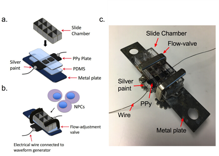
Figure 1. In vitro conductive polymer scaffold system for electrical stimulation. (A) A slide chamber is placed on top of PPy plate. (B) The schematic shows the fabrication of the in vitro electrical stimulation chamber with hNPCs plated on the surface of PPy. Flow valves are used to hold the slide chamber, PDMS, PPy, and metal plate together. (C) Image of the chamber. Please click here to view a larger version of this figure.
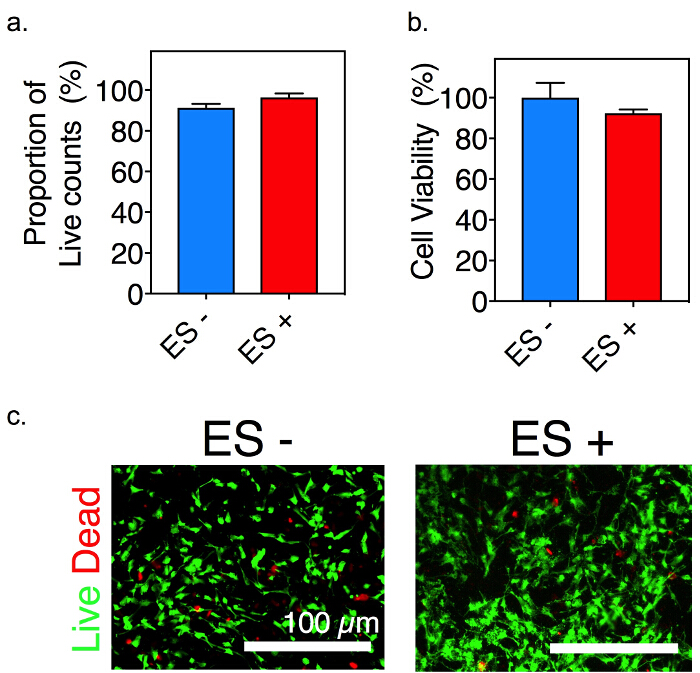
Figure 2. Cell viability assay in hNPCs with or without electrical stimulation. (A) Bar graph demonstrating live cell stained with Calcein-am. Data presented are mean ± S.D.; n = 4. (B) Cell viability assay using resazurin. Bar graph shows there was no cytotoxicity of electrical stimulation on hNPCs; n = 4. (C) Images of cell viability assay before and after electrical stimulation treatment. Green signal indicates live cells (Calcein-am), whereas red signal indicates dead cells (EtHD-1). ES indicates electrical stimulation. ES + or – indicates the presence or absence of electrical stimulation on cells. Please click here to view a larger version of this figure.
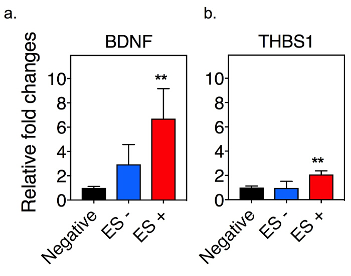
Figure 3. Gene expression changes with electrical stimulation. Bar graph demonstrating fold changes in gene expressions of BDNF (A) and THBS1 (B) in hNPCs (** indicates statistically significant between electrically preconditioned and all other groups, p <0.01, error bars show S.D.; n = 4, one-way ANOVA). Negative indicates the control where cells were cultured on a regular tissue culture plate. ES + or – indicates the presence or absence of electrical stimulation on cells. Please click here to view a larger version of this figure.
Growing evidence has demonstrated the promise of stem cells as a novel stroke therapy. This promise has resulted in a major effort to advance stem cell therapeutics to the bedside with at least 40 ongoing or completed clinical trials. Stroke pathology offers a unique neurological disorder that lends itself to stem cell therapy because after the acute insult, there is no neurodegenerative process preventing recovery. The exact mechanism of stem cell-enhanced stroke recovery remains unclear. Angiogenesis, synaptogenesis, and synaptic remodeling have all been shown to be important. When important molecules for synapse formation such as BDNF and THBS-1 are removed, stroke recovery is impaired6,19. hNPCs have improved functional recovery after stroke in numerous animal models20,21. The mechanisms of the hNPCs effect remains not fully understood, and the ideal delivery method for these cells is unknown. By developing a system that allows one to manipulate the important molecules released from hNPCs, one can help differentiate the integral recovery mechanisms and optimize stem cell therapy.
Successful cell delivery to the brain remains a challenge. Currently, a number of technologies have been developed using different biomaterial scaffolding systems to deliver stem cells to improve the survival and integration of stem cells9,10. However, due to the inert characteristics of these materials, it is difficult to modulate stem cells after their transplantation.
This article describes a method using a PPy scaffold to electrically manipulate the transcriptome of hNPCs as shown by the upregulation of BDNF and THBS-1 in our qRT-PCR data. Our previous study revealed that an increase in VEGFA expression on hNPCs after exposure to electrical stimulation improved functional recovery after stroke2. Here we demonstrate that additional neurotrophic factors such as BDNF and THBS-1 are also modulated after exposure to electrical stimulation in a different hNPC cell line, showing that our system can be used with numerous cell lines.
During the fabrication of the in vitro electrical stimulation chamber, it is critical that the metal wire is completely connected to PPy plate using silver paint and epoxy. If this connection is not sound, it causes varying electric fields even with the same parameters. Moreover, at times media may leak after the assembly of the scaffolding system. To resolve this issue, vacuum grease can be applied to seal the space between the PDMS and the PPy plate with care not to apply to the cell-seeding surface. One limitation due to the non-transparent nature of the PPy plate is that checking cells’ confluency with standard light microscopy is limited.
The advantage of the method described in this protocol allows for modulation of hNPCs to help determine the mechanistic changes of the cells. Currently, electrical modulation has not been approached using conventional hydrogel or inert scaffolding systems. After electrical stimulation using the PPy scaffold, the hNPCs can be delivered without removal from the polymer surface to an in vivo model. Developing methods to further study disease models of stem cell applications allows us to better design translational applications. The electrical preconditioning approach, made possible by the conductive polymer system, can be applied to different cell types and allows for better understanding of the impact of electrical stimulation on a cell. The ability to optimize the cells in vitro and then without further manipulation (such as passaging or removal) allows for testing of these in vitro manipulations in in vivo disease states such as stroke. The ultimate aim is to utilize new techniques, such as the described conductive polymer scaffold, to advance stroke therapeutics and improve understanding of stroke recovery mechanisms.
The authors have nothing to disclose.
We thank Dr. Kati Andreasson (Department of Neurology and Neurological Science, Stanford University) for use of the qRT-PCR machine. The work was supported by National Institutes of Health Grants K08NS098876 (to P.M.G.) and Stanford School of Medicine Dean’s Postdoctoral Fellowship (to B.O.).
FGF-Basic | Invitrogen | CTP0261 | 20 ng/mL for working media |
Matrigel | Corning | cb40234a | 1:200 dilution |
LIF Protein, Recom. Hum. (10 µg/mL) | EMD Millipore | LIF1010 | 10 ng/mL for working media |
Sylgard 184 silicone 3.9 kg | Fisherbrand | NC0162601 | |
Hydrochloric acid | Fisherbrand | SA56-4 | |
Nitric Acid Concentrate (Certified) ACS, Fisher Chemical | Fisherbrand | SA95 | |
ITO Glass | Delta Technologies | CG-40IN-0115 | |
Sodium dodecylbenzenesulfonate | Sigma | 289957-1KG | |
Pyrrole | Sigma | 131709-500ML | Protect pyrrole solution from light and room temperature |
8 well glass slide chambers | Thermo Sci Nuc | 125658 | Detach the cell chamber and keep it under sterilized conditions |
Flat-Surface Bracket, 3"x1" | McMaster-Carr | 1030A4 | |
TWO PART SILVER PAINT 14G | Electron Microscopy Sciences | 1264214 | Mix two parts (1:1) in plastic plate |
DPBS, 1x, with Ca and Mg, No Phenol Red | Genesse | 25-508C | |
AB2 ArunA Neural Cell Culture Media Kit | Aruna Biomedical | ABNS7013.2 | |
hNP1 Human Neural Progenitor Expansion Kit | Aruna Biomedical | hNP7013.1 | |
Noncontact Flow-Adjustment Valve, Nickel-Plated Brass, for 3/32" to 5/8" Tube OD | McMaster-Carr | 5330K22 | |
Multimeter | Keysight | E3641A | |
Wavefoam generator | Keysight | 33210A-10MHz | |
Pt meshes | Sigma-Aldrich | 298107-425MG | Reference electrode with dimensions, 1×1 cm |
LIVE/DEAD Viability/Cytotoxicity Kit, for mammalian cells | Thermo Fisher Scientific | L3224 | |
BDNF | Thermo Fisher Scientific | Hs02718934_s1 | |
THBS1 | Thermo Fisher Scientific | Hs00962908_m1 | |
GAPDH | Thermo Fisher Scientific | Hs02758991_g1 | |
RNeasy Mini Kit (250) | Qiagen | 74106 | |
QIAshredder (250) | Qiagen | 79656 | |
RNase-Free DNase Set (50) | QIAGEN | 79254 | |
iScript cDNA Synthesis Kit, 100 x 20 µL rxns | BIORAD | 1708891 | |
TaqMan Gene Expression Master Mix | Thermo Fisher Scientific | 4369510 | |
7-8 Week Old, male, RNU Rats | NCI-Frederick | ||
4-0 Ethicon Silk Suture | eSutures.com | 683G | |
Isoflurane | Henry Schein | 29405 | |
V-1 Tabletop Laboratory Animal Anesthesia System | VetEquip | 901806 | |
Surgicel Original Absorbable Hemostat | Ethicon | 1952 | |
Lab Standard Stereotaxic Instrument, Rat | Stoelting | 51600 | |
Kimberly-Clark Professional Safeskin Purple Nitrile Sterile Exam Gloves | Fisherbrand | 19-063-130 | |
Sterile Drape | Medline | DYNJSD1092 | |
Thermo Scientific Shandon Stainless-Steel Scalpel Blade Handle, Holds No. 20-25 Blades | Fisherbrand | 53-34 | |
Walter Stern Scalpel Blade Series 300 | Fisherbrand | 17-654-456 | |
QuantStudio 6 Flex Real-Time PCR System | Thermo Fisher Scientific | 4484642 | |
Frazier Micro Dissecting Hook | Harvard Apparatus | 52-2706 |