Here, we describe a simple method to induce clinically relevant skin pressure ulcers (PUs) in a mouse model of spinal cord injury (SCI). This model can be used in pre-clinical studies to screen for different therapeutics for healing PUs in SCI patients.
Pressure ulcers (PUs) are common debilitating complications of traumatic spinal cord injury (SCI) and tend to occur in soft tissues around bony prominences. There is, however, little known about the impact of SCI on skin wound healing in the context of animal models in controlled experimental settings. In this study, a simple, non-invasive, reproducible and clinically relevant mouse model of PUs in the context of complete SCI is presented. Adult male mice (Balb/c, 10 weeks old) were shaved and depilated. Post-depilation (24 h), mice were subjected to laminectomy followed by complete spinal cord transection (T9-T10 vertebrae). Immediately after, a skin fold on the back of the mice was lifted and sandwiched between two magnetic discs held in place for next 12 h, thus creating an ischemic area that developed into a PU over the following days. The wounded areas demonstrated tissue edema and epidermal disappearance by day 3 post-magnet application. PUs spontaneously developed and healed. Healing was, however, slower in the SCI mice compared to control non-SCI mice when the wound was created below the level of SCI. Conversely, no difference in healing was seen between SCI and control non-SCI mice when the wound was created above the level of SCI. This model is a potentially useful tool to study the dynamics of skin PU development and healing after SCI, as well as to test therapeutic approaches that may help heal such wounds.
Pressure ulcers (PUs) are major secondary complications of traumatic SCI1. PUs are localized injuries to the skin and/or underlying tissues that usually occur over bony prominences where body weight is concentrated while the patient is sitting or lying1. The skin, fat, and muscle are exposed to this constant pressure that leads to the development of localized ischemia, tissue inflammation, mechanical damage, and necrosis2,3.
The development of PUs is affected by several local factors, including the magnitude of pressure and shear, loading duration, skin moisture and temperature, injury longevity, and general skin hygiene. There are also systemic factors that play a role, such as general physical condition, bone and muscle tissue morphology and strength4, patient age, hematological measures, gender, and even socio-economic factors including marital status, education, and income4,5.
The prevention and treatment of PUs remain significant challenges in SCI patients. SCI patients develop PUs in ~30-40% of cases, with a re-occurrence rate of 60-85%, possibly due to weak scar tissue and lack of protective sensation1. Thus, PUs often leads to re-hospitalization of SCI patients, and overall pose a significant financial burden (80% more vs. SCI only) to the health care system5,6,7,8,9,10.
To the best of our knowledge, there have been no studies in controlled experimental settings to investigate the impact of SCI on the PU healing process because of the lack of suitable animal models. Here, a reproducible and clinically relevant mouse model of PU in the skin is described. This model can be used to study the dynamics of PU onset and subsequent healing, as well as to test potential therapeutic approaches to prevent PU or improve PU healing in the context of SCI.
All animal handling and surgical procedures were performed in accordance with a protocol approved by the Rutgers University Institutional Animal Care and Use Committee. Mice were fed standard diet and water ad libitum.
1. Preparation of Surgical and Non-surgical Instruments
2. Preparation of Animals and Performing the T9-T10 Spinal Laminectomy
3. Performing the T9-T10 Complete Spinal Cord Injury
4. Induction of Skin Pressure Ulcer after Complete SCI
5. Post-operative Animal Care, Euthanasia, and Tissue Collection for Histology
This protocol creates a PU in the setting of complete SCI. Briefly (as illustrated in Figure 1), all mice with or without complete SCI tolerated the magnets very well, which remained in their original position for the full 12 h (Figures 1c, 1d, 1f, 1h). All the mice developed two circular wounds separated by a bridge of normal tissue (Figure 1e, 1g, 1i). The initial wounding response was similar in mice without SCI (Figure 1e), or with SCI below the SCI site (Figure 1g) or above the SCI site (Figure 1i).
Figure 2 depicts the disappearance of the epidermis by day 3 of post magnet removal, and its reappearance by day 7, albeit with a significantly slower migration in wounds created below the level of the SCI in SCI mice.
The pressure wounds were graded as per previously published criteria12. Figure 3 and Figure 4 depict the healing features in SCI and control non-SCI mice. The SCI group exhibited slower healing, larger scar area, thinner epidermis and dermis, and lower density of proliferating cells (Ki67+ cells), blood vessels (CD31+ cells), and alpha-smooth muscle actin (α-SMA+) in the skin wounds11,12.
When the skin wounds were created above the level of SCI, as shown in Figure 5, no change in healing time, epidermal and dermal thicknesses or scar area was seen when compared to non-SCI controls.
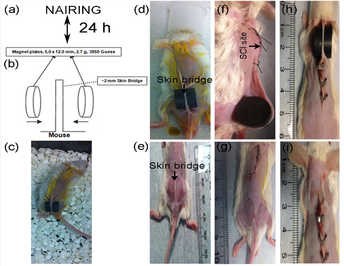
Figure 1: Experimental procedure for creating pressure wounds in non-SCI (n=3) and complete-SCI mice (n = 3). After 1-week habituation in the laboratory, mice were shaved and depilated (a). Schematic of magnetic disc (M) placement (b, modified from Stadler et al.15). Placement of magnetic discs on skin dorsum of a normal mouse and its activity after recovery from anesthesia in the cage (d, c). In SCI mice, discs can be placed below (f) or above (h) the SCI site. The area of magnet-induced skin injury is visible immediately after 12 h of M application, which shows 2 wounds separated by an intact skin bridge in the non-SCI (e) and SCI mice (g, i). Please click here to view a larger version of this figure.
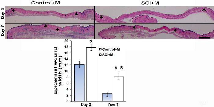
Figure 2: Skin wound histology (H & E stain) and epidermal wound width. On day 3 and day 7 after M application, a set of mice were sacrificed in the non-SCI (Control + M) and complete-SCI (SCI + M) groups to study the early effects M-induced ischemia and then reperfusion. Arrows indicate the epidermal wound edges, located where the epithelial lining thins out and disappears. Scale bar = 1 mm. Epidermal wound width as measured in each group (n = 3 at each time point) is represented in the lower panel bar diagram. Statistical significance determined by Student t-test. *p < 0.05 and **p < 0.01. Data are presented as mean ± SEM (standard error of the mean). This figure has been modified with permission from the Journal of Neurotrauma, 35, 6, 815-824, (2018), published by Mary Ann Liebert, Inc., New Rochelle, NY12. Please click here to view a larger version of this figure.
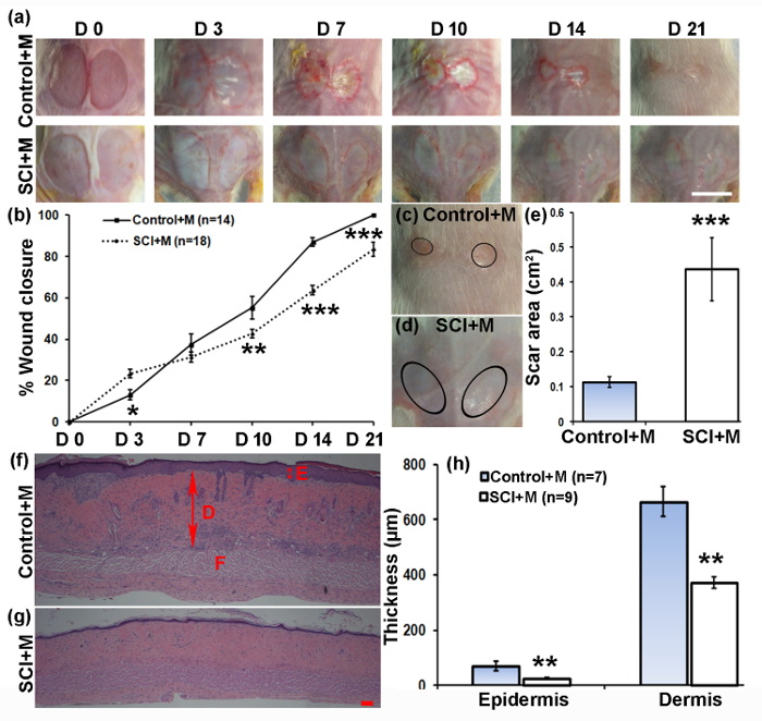
Figure 3: Impact of complete SCI on M-induced PU development and healing. (a) Representative images of skin wounds in non-SCI (Control + M) and complete-SCI (SCI+M) mice after PU induction over the period of 21 days. Scale bar = 1 cm. (b) Quantified wound images showing the fraction of wound closure as a function of time. (c, d) Representative images of healed wounds showing the scar area in non-SCI and SCI mice. (e) Quantified scar area in non-SCI versus SCI mice. (f, g) Representative histology of healed wound skin (H&E staining) showing epidermis (E, double arrows), dermis (D, large double arrows), and fat layer (F). Scale bar = 100 µm. (h) Quantification of epidermal and dermal thicknesses of healed wounds at time of wound closure (21 days for non-SCI, and 35 days for SCI mice). Data are presented as mean±SEM. Statistical significance determined by ANOVA followed by post-hoc Fisher's LSD test and Student's t-test. *p < 0.05, **p < 0.01, and ***p < 0.001. This figure has been reprinted with permission from the Journal of Neurotrauma, 35, 6, 815-824, (2018), published by Mary Ann Liebert, Inc., New Rochelle, NY12. Please click here to view a larger version of this figure.
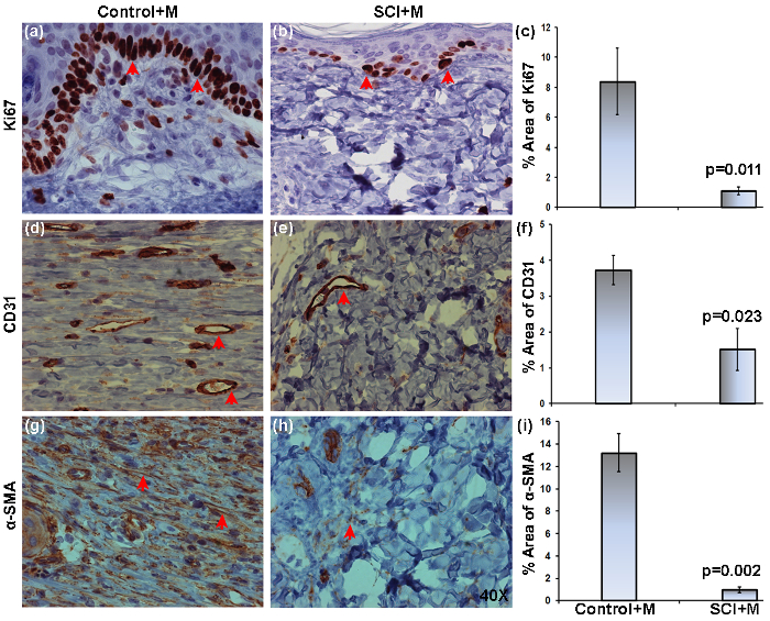
Figure 4: Expression of Ki67, CD31, and a-SMA in skin ulcers of non-SCI and SCI mice. Wound tissues were harvested after wound closure, namely on days 21 (Control+M) and 35 (SCI+M) post-PU induction. Representative images of 5 µm thick sections stained with anti-Ki67, anti-CD31, or anti-α-SMA and visualized with a 40x objective. Representative images of non-SCI (a, d, g) and SCI (b, e, h) mice show the distribution of Ki67+, CD31+, and α-SMA+ (brown stain, red arrows point to some stained areas). The % positive area of expression as obtained by image analysis is compared between the groups (c, f, i). The area was averaged from three 40x fields (two from wound edges and one from wound center) per section (2/mouse, 3 mice in each group). Data are presented as mean ± SEM. Statistical significance was determined by Student's t-test. This figure has been reprinted with permission from the Journal of Neurotrauma, 35, 6, 815-824, (2018), published by Mary Ann Liebert, Inc., New Rochelle, NY12. Please click here to view a larger version of this figure.
Figure 5: Impact of SCI on development and healing of ulcers above the SCI site. Representative images of ulcers in the non-SCI (Control + M) and SCI (SCI + M) mice over the observation period (a). Scale bar = 1 cm. The scar area on day 21 is represented by circles (a) and values were averaged in non-SCI and SCI mice (b). Quantified wound images showing the fraction of wound closure as a function of time (c). Representative histology of healed skin ulcers showing epidermis (E, small double arrow) and dermis (D, large double arrow). Scale bar = 100 µm (d). Quantification of epidermal and dermal thicknesses of healed skin ulcers at wound closure time (day 21). Data are presented as mean ± SEM. Statistical significance determined by ANOVA followed by posthoc Fisher's LSD test, or Student t-test. NS-statistically non-significant. This figure has been modified and reprinted with permission from the Journal of Neurotrauma, 35, 6, 815-824, (2018), published by Mary Ann Liebert, Inc., New Rochelle, NY12. Please click here to view a larger version of this figure.
Supplementary Figure 1: Motor function in SCI mice was assessed using the BMS score on post-injury day 2 and then weekly. The BMS score at day 2 and week 5 were 0.058 ±0.058 (median 0-no movement, n = 17) and 0.35±0.12 (n = 17, median-0). Data are represented as mean±SEM. This figure has been modified and reprinted with permission from the Journal of Neurotrauma, 35, 6, 815-824, (2018), published by Mary Ann Liebert, Inc., New Rochelle, NY12. Please click here to download this figure.
The protocol in this study describes a novel experimental model of PUs to evaluate the impact of SCI on wound healing. The skin PUs were induced via a 12 h application of two 12 mm diameter disc magnets on a dorsal skin fold, either set above or below the SCI site. Data show that SCI slows down skin wound healing in mice. Importantly, these observations were specifically made in skin wounds below the innervation level of the SCI, as wounds made above the SCI level, and thus that remained innervated, were largely unaffected in their healing pattern compared to the non-SCI mice.
The PU protocol described herein is based on a previously published method used in mice, where weaker magnets (1000 Gauss) were applied in three cycles of 12 h magnet application separated by 12 h without magnet15. Initially, we compared using one, two, and three 12 h cycles of magnet application. A disadvantage of using repeated applications is that each time special care must be taken to detach the magnet from the skin fold and reapply it at the exact same location. A single application is therefore much simpler and was sufficient to create a PU in the mouse dorsal skin fold. Otherwise, this methodology does not require a complex device, does not impair animal movements, and is very reproducible. Studies using different magnet sizes, shapes and or strengths, or attempting to create PUs in different anatomical locations may need to reoptimize the protocol. As in all skin wound healing studies, it is important to properly apply and regularly change the transparent film covering to avoid drying and contamination of the wound bed.
The mechanism of PU formation in this model relies on the compressive force between two magnets, which is expected to substantially exceed capillary and venous perfusion pressures in the skin, and has previously been well documented to induce lesions in larger animals16. Using a single 12 h cycle created a skin ulcer that had injury extending deep into the dermis, which corresponds to stages I and II according to standard criteria15. One limitation of this technique is that we were not able to get a stage III-IV PU. Therefore, one could not study the involvement of muscles and bones in PU development and healing without significant modifications to our current model. Furthermore, the PUs we generate heal spontaneously and, therefore, do not faithfully represent a chronic PU as might be seen in human patients. Interestingly, however, the method resulted in a PU of the initial similar size that closed with similar dynamics as commonly used 1 cm x 1 cm full-thickness excisional skin wounds. The PU induction method spares the underlying panniculus carnosus, while it is completely removed in the excisional model. Thus, it appears that the panniculus carnosus does not play a major role in the wound closure process, which, in mice and other rodents, primarily occurs via contraction mediated by fibroblasts and myofibroblasts in viable dermis surrounding the wound site, with little scar formation17.
The site of PU induction, above or below SCI, does not interfere with the skin incision used to access the site of SCI. Therefore, the method can be readily applied below or above the level of SCI, which enables studies that can differentiate the local vs. systemic effects of SCI on wound healing. While non-SCI animals continue to gain weight after PU induction, the growth of SCI animals is stunted. In spite of this profound systemic change, wound healing was not affected when the PU was created above the SCI level. This model can, therefore, be used to better understand the role of the local innervation in wound healing. As in all studies that involve SCI, it is important to provide special care and monitoring to the post-SCI mice, which require manual draining of the bladder, monitoring of bowel conditions, and prophylactic antibiotic treatment11,12.
SCI patients are substantially challenged even with the best hospital care and education, and skin PUs impart significant costs to the US healthcare system. This model allows side-by-side evaluation of the effect of SCI on skin PU development and healing, which provides a platform for testing various therapeutic strategies that may help PU healing in SCI patients.
The authors have nothing to disclose.
This work was partially supported by the New Jersey Commission on Spinal Cord Research (CSCR15IRG010), the U.S. Department of Defense (SC160029), and the Yale Department of Surgery Ohse Research Grant Program. We thank Sean O’ Leary from the W.M. Keck Center for Collaborative Neuroscience, Rutgers for technical assistance.
Magnets | Master Magnetcs, Inc., Castle Rock, CO | CD14C | 3800 G Magnetic force |
Mice standard diet | PMI Nutrition International, Brentwood, MO | Standard Food Pellet | |
Isoflurane | HENRY SCHEIN Animal Health | SKU 029405 | |
ImageJ | NIH, Bethesda, MD | Image Analysis Software | |
BETADINE Surgical Scrub | HENRY SCHEIN Animal Health | ||
Ophthalmic Ointment | HENRY SCHEIN Animal Health | SKU 008897 | |
NAIR-Hair Remover Lotion | Church & Dwight Co., Inc. Princeton, NJ | ||
ELOXIJECT (meloxicam) Injection | HENRY SCHEIN Animal Health | SKU 049755 | 5 mg/mL, 10 mL |
Cefazolin Sodium | HENRY SCHEIN Animal Health | SKU 054846 | 1 g, 10 mL bottle |
Buprenorphine-SR | ZooPharm, Windsor, CO | – | – |
0.9% Sodium Chloride Injection USP | BRAUN, Irvine, CA | S8004-5384 | |
10% Neutral Buffered Formalin | VWR, Radnor, PA | 16004-130 | |
BALB/C Male Mouse | Charles River Lab., Wilmington, MA | 28 | |
Sterile Cotton Tipped Applicator | Puritan, Guilford, ME | SKU#: 25-806 | |
Michel Suture Clips | Fine Science Tools (USA) Inc., Foster City, CA | 12040-01 | |
Surgical Suture, U.S.P. | Henry Schein Animal Health | 101-2636 |