1. Construction of electrical stimulation cell culture chamber
- To build the EStim chamber, collect two lids of standard 6-well cell culture plates; 99.99% platinum wire, 60 cm in length with a diameter of 0.5–1 mm; silver-coated copper wire, 70 cm in length with a diameter of 0.6 mm; cutting pliers; soldering iron kit; one tube of superconductive glue; one wire terminal block connector, six small 2.2 V LEDs (optional); one tube of noncorrosive silicone adhesive coating (optional); one roll of black electrical insulation tape; standard, flexible, insulated copper electric wire (0.14 mm2), 2 m in length (Table of materials).
- In a 6-well plate lid, mark and then drill two holes (with a diameter of ~1 mm), 25 mm apart, near the outer edge of each of the six wells (12 holes in total), as shown in Figure 1A,B.
- Cut twelve 5 cm lengths of platinum wire with cutting pliers. Bend each of the wires manually into an L-shape, leaving one end 3 cm long and the other 2 cm. Cut the silver-coated wire into two 35 cm lengths.
- Insert the longer (3 cm) bent end of one platinum wire (from inside to out) into each drilled hole, leaving 1–2 mm protruding from the outside of the lid, bend it using forceps. Secure the platinum wires in the lid holes with superconductive glue and leave it to dry for around 6 h.
- Solder the tips of all six platinum wires (that will later serve as cathodes, see Figure 1A) protruding from the lids to one of the silver-coated wires. Repeat the same procedure, soldering the remaining six platinum wires (that will later serve as anodes) to the other silver-coated wire.
- Add LEDs in the circuit between each of the six anode-cathode platinum electrode pairs, to confirm functionality during the experiments (optional). Place a piece of black insulation tape under each LED to prevent exposing the cells in the culture plates to the LED light (Figure 1C).
- Glue the wire terminal block connector to the top left corner of the 6-well plate lid and connect both silver-coated wires to the input terminals, as shown in Figure 1C.
- Cut out a 20 mm x 20 mm section from the top left corner of a second 6-well plate lid (Figure 1B, Lid Nr. 2) to accommodate the terminal block connector on the first lid. Cover the first lid, equipped with the electrodes, with the second lid and tape them together with adhesive tape.
- To improve the bonding of the two lids, use silicone adhesive coating (optional). To do so, cover the lid with the silver electrodes with a 3–5 mm layer of silicone adhesive and cover it with the other lid, allowing 12 h for the adhesive to dry.
- Connect one end of the two standard insulated copper wires to the output terminals of the wire connector and the other ends to banana male connectors (4 mm).
NOTE: The length of these wires depends on the distance from the shelf in the incubator, where the cells will be kept, to the power supply, outside the incubator (Figure 1D). - Adjust the dosage (voltage) and regimen of EStim delivered to the cells by regulating the DC power supply.
- Turn on the power supply by pressing the ON/OFF button on the front panel. Activate channel 1 by pressing button 1.
- Press button Nr. 4 (V-set) to set the voltage. Press buttons 2, . and 5 to set the load output at 2.5 V. Press Enter.
- Ensure that the load output of 2.5 V (2,500 mV) corresponds to an EStim of 100 mV/mm, according to the following, simplified equation.
VEStim = ; or
; or
V = VEStim × d
Here, V = the power supply voltage output in millivolts; d = the distance between electrodes in millimeters; VEStim = the stimulation voltage in millivolts per millimeter.
NOTE: This simplification applies only in case of constant DC voltage. The resistance between the medium and the cells is negligible as electrodes are not in direct contact with the cells; instead, EStim is delivered to the cells through the medium.
2. Mesenchymal stem cell culture in osteogenic medium
- Purchase and store commercially available rat MSCs (see Table of Materials) in liquid nitrogen until the day of the experiment. Alternatively, isolate MSCs from other animals according to protocols published elsewhere7,8 in accordance with local institutional regulations for the use of experimental animals.
- On the day of the experiment, remove one vial (1 x 10⁶ cells) of MSCs from the liquid nitrogen storage, and quickly (within 1 min) thaw the cells in a water bath preheated to 37 °C.
- Under sterile conditions in a laminar flow hood, pipette the vial content into a 50 mL falcon tube and add 9 mL of normal medium (NM) prewarmed to 37 °C, consisting of Dulbecco’s modified Eagle’s medium (DMEM; 1x) with 10% heat-inactivated fetal bovine serum (FBS) and 1% penicillin/streptomycin solution. Pellet the cells for 5 min by centrifugation at 300 x g.
- In a laminar flow hood, remove the supernatant and carefully resuspend the cell pellet in 12 mL of NM prewarmed to 37 °C. Transfer the resuspended cells to a T-75 cell culture flask.
- Culture the cells at 37 °C, 5% CO2, 5% O2 until they reach an 80%–90% confluence (after approximately 3–5 days).
- Passage the cells.
NOTE: Perform all operations except centrifugation and incubation under sterile conditions in a laminar flow hood.- After reaching an 80%–90% confluence, retrieve the cells with cell detachment solution. Aspirate the cell culture medium, wash 2x with 1x phosphate-buffered saline (PBS), add 5 mL of 1x cell detachment solution, and return the cells to the incubator for 5 min.
- Once the cells are detached, add an equal amount of culture medium to inactivate the detachment solution. Collect the cells in a 50 mL falcon tube and spin them at 300 x g for 5 min.
- Discard the medium and resuspend cells in 1 mL of fresh normal medium. Assess the number of viable cells with trypan blue stain.
- Seed 1 x 10⁶ cells in a new T-75 flask with 12 mL of prewarmed NM. Culture the cells at 37 °C, 5 % CO2, 5 % O2 until they reach an 80%–90% confluence.
NOTE: Cell passaging (steps 2.4.1–2.4.4) can be repeated a few times until the needed number of cells is obtained. Do not use cells older than passage 8.
- Seed 9 x 10⁴ cells in 3 mL of NM (10% heat-inactivated fetal bovine serum, 1% penicillin/streptomycin, DMEM) in each well of a 6-well culture plate. Incubate the cells for 1 day at 37 °C, 5 % CO2, 5 % O2.
- The next day, aspirate culture medium and apply 3 mL of osteogenic differentiation medium (OM; normal medium supplemented with 10-7 M dexamethasone, 10 mM β-glycerophosphate, and 0.05 mM ascorbic acid-2-phosphate).
- Place the 6-well plate with cells in the incubator and incubate at 37 °C, 5% CO2, 5% O2 overnight.
3. Treating MSCs with EStim
- On the day the cells are treated with EStim, sterilize the electrodes in 70% ethanol solution for 30 min; then, dry them under UV light in a safety cabinet for an additional 30 min.
- In a laminar flow hood, cover the 6-well plate containing the cultured MSCs with the lid equipped with the electrodes, making sure that the electrodes are completely submerged in medium (if necessary, add medium). Transfer the covered 6-well plate (EStim chamber) with the cells to the incubator and connect its wires to the power supply.
- Set the power supply to 2.5 V load output and treat the cells with EStim for 1 h3,9.
- After stimulation, disconnect the power supply and remove the EStim chamber from the incubator. Under sterile conditions, exchange the lid equipped with electrodes with a standard 6-well plate lid.
- Return the cells to the incubator and leave them overnight. Clean the electrodes, first with PBS and then with 70% ethanol solution. Clean the accumulated corrosion products from the electrode surface with fine sandpaper.
- Repeat steps 3.1–3.5 for 6 consecutive days. On day 4, prior to applying EStim and under sterile conditions, change the culture medium by aspirating 1.5 mL of medium and replacing it with 1.5 mL of prewarmed fresh OM.
- After applying EStim for 7 consecutive days, maintain the cells in culture for an additional 7 days, exchanging the medium every 3–4 days.
4. Osteogenic differentiation measurements
- Analyze cell morphology changes under a microscope.
- To assess the effect of EStim on MSC osteogenic differentiation, measure calcium deposition, alkaline phosphatase activity, and osteogenic marker gene expression, as described elsewhere3,9.
To evaluate the effect of 100 mV/mm of EStim on the osteogenic differentiation of MSCs, cells treated with EStim for 3, 7, and 14 days or nontreated (control) were analyzed at day 14 of culturing by assessing morphological changes and calcium deposition (Figure 2). This was done by imaging cells using bright-field microscopy (morphology changes) or by fixing cells in 4% paraformaldehyde solution, staining them with 0.02% alizarin red solution and then imaging them using bright-field microscopy (calcium deposition analysis).
A detailed analysis of osteogenic marker gene expression changes was performed at days 3, 7, and 14 of culturing (Figure 3). This was done by measuring the relative expression of genes RunX2, Collagen I, Osteopontin, and Osterix by means of RT-qPCR and the comparative delta Ct (threshold cycle values) method10, where housekeeping genes Rplp1 and Ywhaz11 were used for normalization.
Exposing MSCs to 100 mV/mm of EStim for 3 days (1 h per day) had no effect; however, 7 days of exposure resulted in an increase in osteogenic differentiation, as determined by morphology changes (Figure 2A–C), calcium deposition (Figure 2E–G), and osteogenic marker gene expression changes (Figure 3) in comparison to a time-matched control without EStim. Prolonged EStim exposure (14 days) did not further enhance the osteogenic differentiation beyond that seen after 7 days of treatment (Figure 2D,H and Figure 3).
As shown in Figure 2, cells treated with EStim for 7 and 14 days appeared more condensed (Figure 2C,D) than those treated with EStim for 3 days or nontreated cells (Figure 2A,B) and showed an increased calcium deposition (Figure 2G,H) compared to those treated for only 3 days or nontreated controls (Figure 2E,F). Analysis of the osteogenic marker expression at 3, 7, and 14 days of culturing confirmed the enhanced osteogenic differentiation in cells treated with EStim for 7 and 14 days (Figure 3). The expression of osteogenic-differentiation-related marker genes12 RunX2, Collagen I, Osteopontin, and Osterix were the highest in cells treated with EStim for 7 days.
<!–FIGURE AND TABLE LEGENDS:
–>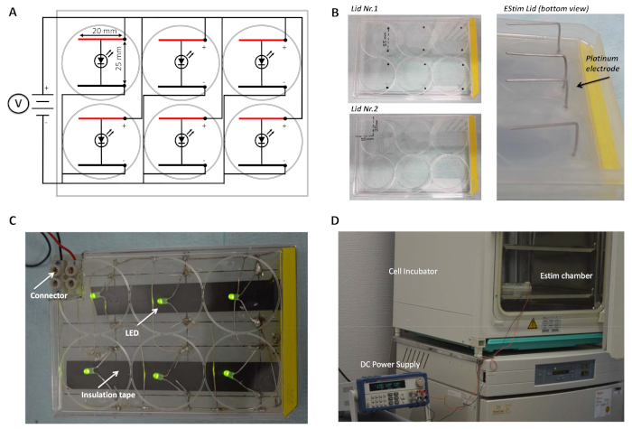
Figure 1: EStim cell culture chamber. (A) Electric circuit diagram of the EStim chamber showing the anodes (black), cathodes (red), and LEDs connected to the DC power supply. (B) Image of the marked 6-well plate lids and L-shaped platinum electrodes (bottom view) incorporated into the 6-well plate lid. (C) Assembled EStim chamber (top view) with wire connector, LEDs, and electrical insulation tape that shields the cells from the LED light (arrows). (D) EStim cell treatment setup with the EStim chamber in the incubator connected to the DC power supply, on the outside. Please click here to view a larger version of this figure.
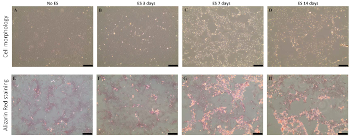
Figure 2: Effect of EStim on MSC morphology and calcium deposition. Cells in osteogenic culture medium, exposed and not exposed (controls) to 100 mV/mm of EStim for 3, 7, and 14 days (1 h/day). (A–D) Morphology and (E–H) calcium deposition (alizarin red staining) on day 14 of culturing. Significant changes in cell morphology and calcium deposits were visible in cells treated with EStim for (C and G) 7 and (D and H) 14 days (10x magnification; the scale bar = 200 µm). This figure was modified from Eischen-Loges et al.9. Please click here to view a larger version of this figure.
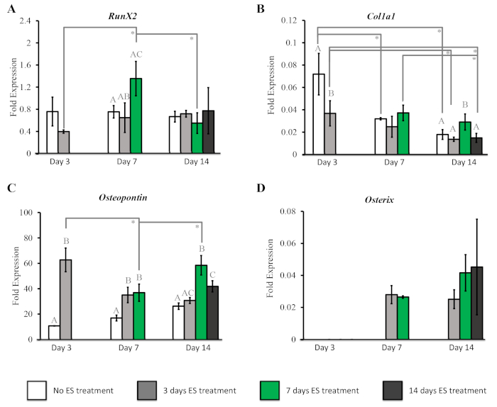
Figure 3: Effect of EStim on MSC osteogenic marker gene expression. The osteogenic marker gene expression (measured with RT-qPCR at days 3, 7, and 14 of culturing) in cells treated with EStim for 3, 7, and 14 days, or nontreated. (A) At day 7 of culturing, the RunX2 expression was significantly higher in cells treated with EStim for 7 days. (B) The ColIa1 expression was significantly higher in cells treated for 7 days, measured at day 14 of culturing. (C) The expression of Osteopontin was significantly increased in EStim-treated cells at days 3, 7, and 14 of culturing. (D) The Osterix expression was absent in control cells at all time points and was seen only at 7 and 14 days of culture in cells exposed to EStim. Different letters on the bars indicate significant (p < 0.05) differences among groups at the same time point. The asterisk indicates significant (p < 0.05) differences between time points within the same group. This figure was modified from Eischen-Loges et al.9. Please click here to view a larger version of this figure.
