This article introduces a comprehensive experimental methodology on two of the latest technologies available to measure the lower limb biomechanics of individuals.
Biomechanical analysis techniques are useful in the study of human movement. The aim of this study was to introduce a technique for the lower limb biomechanical assessment in healthy participants using commercially available systems. Separate protocols were introduced for the gait analysis and muscle strength testing systems. To ensure maximum accuracy for gait assessment, attention should be given to the marker placements and self-paced treadmill acclimatization time. Similarly, participant positioning, a practice trial, and verbal encouragement are three critical stages in muscle strength testing. The current evidence suggests that the methodology outlined in this article may be effective for the assessment of lower limb biomechanics.
The discipline of biomechanics primarily involves the study of stress, strain, loads and motion of biological systems – solid and fluid alike. It also involves the modelling of mechanical effects on the structure, size, shape and movement of the body1. For many years, developments in this field have improved our understanding of normal and pathologic gait, mechanics of neuromuscular control, and mechanics of growth and form2.
The main objective of this article is to present a comprehensive methodology on two of the latest technologies available to measure lower limb biomechanics of individuals. The gait analysis system measures and quantifies gait biomechanics by using a self-paced (SP) treadmill in combination with an augmented reality environment, which integrates a SP algorithm to regulate the treadmill's speed, as described by Sloot et al3. The muscle strength testing equipment is used as an assessment and a treatment tool for upper extremity rehabilitation4. This device can objectively assess a variety of physiological patterns of movement or job simulation tasks in isometric and isotonic modes. It is currently recognized as the gold standard for upper limb strength measurement5 but the evidence related specifically to the lower limb remains unclear. This paper explains the detailed protocol for completing an assessment of gait and isometric strength for the lower extremity.
Within biomechanical analysis, it is useful to combine assessments of functional performance (such as gait analysis) with specific tests of muscular performance. This is because whilst it may be assumed that increased muscle strength improves functional performance, this may not always be apparent6. This understanding is required for the improved future design of rehabilitation protocols and research strategies to assess these approaches.
The method reported was followed in a study that received ethical approval from the Bournemouth University Research Ethics Committee (Reference 15005).
1. Participants
2. Setup and procedures for gait analysis
3. Setup and procedures for muscle strength test
The mean and standard deviation of the spatial-temporal, kinematics, and kinetic gait parameters are given in Table 2. MVIC data for all 30 participants are summarized in Table 3. A typical set of data for the left and right side of one participant showing graphical representation of gait parameters is provided in Figure 4 and Figure 5, respectively.
The data presented are representative of the results obtained across all participants, and are consistent with textbook reference results obtained for gait and isometric strength testing15.
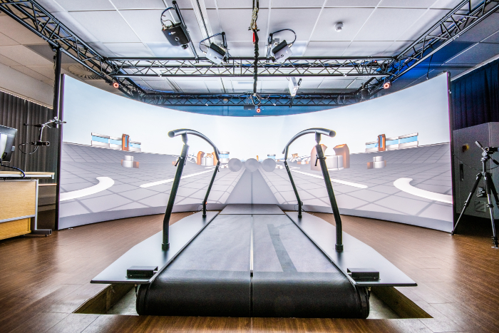
Figure 1: Gait analysis system. The GRAIL system is used to measure gait parameters. This system consists of a split-belt instrumented treadmill, 160° semi-cylindrical projection screen, force sensors, video cameras and optical infrared system. Please click here to view a larger version of this figure.
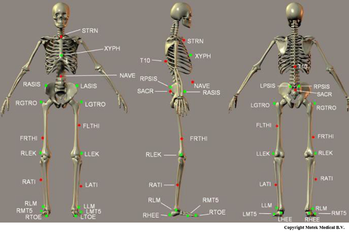
Figure 2: Diagram of markers used in Human Body Model (HBM). This figure shows the exact placements of all markers in the HBM lower body model. Special attention should be paid to the placement of the markers printed in green (bold in Table 1); these are used during initialization to define the biomechanical skeleton. This figure is adapted from the HBM Reference Manual8. Please click here to view a larger version of this figure.
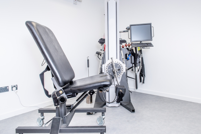
Figure 3: The muscle strength testing equipment (multimodal dynamometer) used to measure participants lower limb muscle strength. This system is used to measure the participants' muscle strength based on Maximum Voluntary Isometric Contraction (MVIC). Please click here to view a larger version of this figure.
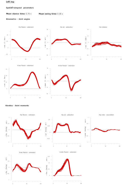
Figure 4: A sample report produced from offline analysis of the gait assessment using the proposed technique. Spatial temporal data and kinematic and kinetic gait cycle for the left side of one participant. Each line represents one gait cycle. The Y-axis represents the joint angles in degrees for the kinematic plots and joint moment in newton meter per kilogram for the kinetic plots. Red lines represent left side gait parameters. Please click here to view a larger version of this figure.
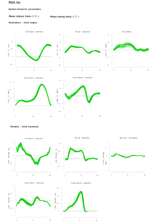
Figure 5: A sample report produced from offline analysis of the gait assessment using the proposed technique. Spatial temporal data and kinematic and kinetic gait cycle for the right side of one participant. Each line represents one gait cycle. The Y-axis represents the joint angles in degrees for the kinematic plots, and joint moment in newton meter per kilogram for the kinetic plots. The Green lines represent right side gait parameters. Please click here to view a larger version of this figure.
Label | Anatomical location | Description |
T10 | T10 | On the 10th thoracic vertebrae |
SACR | Sacrum bone | On the sacral bone |
NAVE | Navel | On the navel |
XYPH | Xiphoid process | Xiphiod procces of the sternum |
STRN | Sternum | On the jugular notch of the sternum |
LASIS | Pelvic bone left front | Left anterior superior iliac spine |
RASIS | Pelvic bone right front | Right anterior superior iliac spine |
LPSIS | Pelvic bone left back | Left posterior superior iliac spine |
RPSIS | Pelvic bone right back | Right posterior superior iliac spine |
LGTRO | Left greater trochanter of the femur | On the center of the left greater trochanter |
FLTHI | Left thigh | On 1/3 on the line between the LGTRO and LLEK |
LLEK | Left lateral epicondyle of the knee | On the lateral side of the joint axis |
LATI | Left anterior of the tibia | On 2/3 on the line between the LLEK and LLM |
LLM | Left lateral malleolus of the ankle | The center of left lateral malleolus |
LHEE | Left heel | Center of the heel at the same height as the toe |
LTOE | Left toe | Tip of big toe |
LMT5 | Left 5th meta tarsal | Caput of the 5th meta tarsal bone, on joint line midfoot/toes |
RGTRO | Right greater trochanter of the femur | On the center of the right greater trochanter |
FRTHI | Right thigh | On 2/3 on the line between the RGTRO and RLEK |
RLEK | Right lateral epicondyle of the knee | On the lateral side of the joint axis |
RATI | Right anterior of tibia | On 1/3 on the line between the RLEK and RLM |
RLM | Right lateral malleolus of the ankle | The center of right lateral malleolus |
RHEE | Right heel | Center of the heel at the same height as toe |
RTOE | Right toe | Tip of big toe |
RMT5 | Right 5th meta tarsal | Caput of the 5th meta tarsal bone, on joint line midfoot/toes |
Table 1: Markers used in the Human Body Model (HBM). This table shows the exact placements of all markers in the HBM lower body model. Special attention should be paid to the placement of the markers written in bold; these are used during initialization to define the biomechanical skeleton. This table is adapted from the HBM Reference Manual8.
Variable name | Side | Mean | Standard Deviation |
Spatial temporal | |||
Walking speed (m/s) | 1.37 | 0.22 | |
Step length (m) | Left | 0.72 | 0.07 |
Right | 0.73 | 0.07 | |
Stride time (s) | Left | 1.07 | 0.10 |
Right | 1.07 | 0.10 | |
Stance time (s) | Left | 0.70 | 0.08 |
Right | 0.70 | 0.08 | |
Swing time (s) | Left | 0.37 | 0.03 |
Right | 0.37 | 0.03 | |
Kinematic | |||
Hip Flex (deg) | Left | 30.05 | 9.08 |
Right | 29.92 | 8.79 | |
Hip Ext (deg) | Left | -13.26 | 7.75 |
Right | -13.36 | 7.68 | |
Hip Abd (deg) | Left | -7.27 | 3.00 |
Right | -7.72 | 3.17 | |
Hip Add (deg) | Left | 8.66 | 4.22 |
Right | 7.81 | 3.72 | |
Hip Int Rot (deg) | Left | 5.38 | 6.95 |
Right | 6.82 | 6.42 | |
Hip Ext Rot (deg) | Left | -9.04 | 7.03 |
Right | -5.77 | 5.97 | |
Knee Flex (deg) | Left | 67.46 | 5.16 |
Right | 68.47 | 4.75 | |
Knee Ext (deg) | Left | -0.43 | 2.26 |
Right | -0.29 | 2.01 | |
Ankle Flex (deg) | Left | -17.20 | 6.94 |
Right | -14.91 | 6.47 | |
Ankle Ext (deg) | Left | 18.13 | 5.92 |
Right | 19.36 | 6.54 | |
Kinetic | |||
Peak Hip Ext (Nm/kg) | Left | 0.82 | 0.21 |
Right | 0.80 | 0.24 | |
Peak Hip Abd (Nm/kg) | Left | 0.91 | 0.15 |
Right | 0.92 | 0.11 | |
Peak Hip Int Rot (Nm/kg) | Left | 0.26 | 0.13 |
Right | 0.26 | 0.14 | |
Peak Knee Ext (Nm/kg) | Left | 0.38 | 0.06 |
Right | 0.39 | 0.06 | |
Peak Ankle Flex (Nm/kg) | Left | 1.85 | 0.21 |
Right | 1.86 | 0.22 |
Table 2: The mean and standard deviation of the spatial-temporal, kinematics, kinetic gait parameters for the 30 participants. Gait parameters are reported for the left and the right side separately.
Variable name | Side | Mean | Standard Deviation |
Knee Ext | Left | 527.17 | 136.42 |
Right | 550.60 | 132.55 | |
Knee Flex | Left | 191.60 | 38.53 |
Right | 203.87 | 47.67 |
Table 3: The mean and standard deviation of the Maximum Voluntary Isometric Contraction (MVIC) for knee joint using the muscle strength testing equipment for the 30 participants.
Supplementary File 1: Matlab coding file. Please click here to view this file (Right click to download).
The contribution of this study is to accurately and comprehensively describe within one protocol the techniques for combined gait analysis and muscle strength testing that have not previously been described together.
In order to achieve accurate results for gait analysis, there are two areas that require maximum attention: 1) marker placements and 2) acclimatization time. The accuracy of the measured data is heavily dependent on the accuracy of the model used. The other key factors that affect accuracy include erroneous marker movement due to superficial skin deformation relative to the underlying skeletal structure, and the resolution of the tracking system16. Figure 2 shows the exact placements of all markers in the HBM lower body model. Special attention should be given to the placement of the markers printed in green; these are used during initialization to define the biomechanical skeleton. Participants were asked to walk for at least 5 min to adapt to SP treadmill walking17,18. The SP mode was chosen in order to allow participants a more natural stride variability3. However, studies have shown that walking speed varies more during SP walking and gait disturbance could occur through acceleration or deceleration of the belt3. In line with other studies13,19, to minimize this effect, we recommend at least five minutes19 should be allowed for acclimatization.
To measure participants' muscle strength using the muscle test equipment, there are three critical stages: 1) alignment of knee joint with the dynamometer axis, 2) practice trial, and 3) verbal encouragement. Inappropriate alignment between the dynamometer and knee joint axis of rotation can introduce a factor confounding accurate isometric assessment20. Throughout the study, all participants were given precise instruction about the system prior to taking part. However, a practice trial and verbal encouragement are two factors that can greatly affect the MVIC14. Many of the individuals who underwent the strength test have very limited or no experience in performing strength testing maneuvers. Strength testing has generally been shown to be reliable21, but it has been shown that strength scores of novice participants are likely to improve on subsequent testing as they become more comfortable and familiar with the test and the system22. Verbal encouragement during exercise testing has been shown to enhance maximal force23, rate of force development23, muscle activation24, muscular endurance25, power26, maximal oxygen consumption27, and time to exhaustion27,28. Therefore, we highly recommend adopting this step.
Overall, the data presented here are representative of textbook reference results for gait and isometric strength testing obtained on other equipment. Therefore, it is proposed that the methodology outlined in this article may be considered effective in the assessment of gait and muscular strength in healthy individuals. Further studies should evaluate the reliability of these systems before they are used in clinical applications.
The authors have nothing to disclose.
We would like to thank Dr. Johnathan Williams for his advice on MATLAB data processing.
701 Small lever | Baltimore Therapeutic Equipment Company (BTE) | Not Available – Online link provided in description | The unique attachment designed for the Primus RS to measure Knee Extension/Flexion – https://store.btetech.com/collections/primus/products/701-small-lever |
D-Flow Software – Vresion 3.26 | Motekforce Link | Not Available – Online link provided in description | Software used to control GRAIL system – https://summitmedsci.co.uk/products/motek-dflow-hbm-software/ |
Gait Offline Analysis (GOAT) – Version 2.3 | Motekforce Link | Not Available – Online link provided in description | Software used for the analysis of the gait parameters – https://www.motekmedical.com/product/grail/ |
Gait Real-time Analysis Interactive Lab (GRAIL) | Motekforce Link | Not Available – Online link provided in description | GRAIL system measures and quantifies gait biomechanics by using a virtual reality based self-paced (SP) treadmill – https://www.motekmedical.com/product/grail/ |
Leg Pad for 701 | Baltimore Therapeutic Equipment Company (BTE) | Not Available – Online link provided in description | The unique attachment designed for the Primus RS to measure Knee Extension/Flexion – https://store.btetech.com/collections/primus/products/701-802-leg-pad |
Positioning Chair | Baltimore Therapeutic Equipment Company (BTE) | Not Available – Online link provided in description | Participant Positioning Chair is designed for assessment and treatment of the lower exteremeties. The chair is designed for multiple positions. https://www.btetech.com/product/primus/ |
Primus RS | Baltimore Therapeutic Equipment Company (BTE) | Not Available – Online link provided in description | Primus RS equipment captures and reports real time objective data in Isotonic, Isometric, and Isokinetic resistance modes – https://www.btetech.com/wp-content/uploads/BTE-Rehabilitation-Equipment-PrimusRS-Brochure-1.pdf |