We describe an intracisternal injection that employs a needle bent at the tip that can be stabilized to the skull, thus eliminating the risk of damage to the underlying parenchyma. The approach can be used for genetic fate mapping and manipulations of leptomeningeal cells and for tracking cerebrospinal fluid movement.
Method Article
Induction of Leptomeningeal Cells Modification Via Intracisternal Injection
In This Article
Summary
Abstract
The protocol outlined here describes how to safely and manually inject solutions through the cisterna magna while eliminating the risk of damage to the underlying parenchyma. Previously published protocols recommend using straight needles that should be lowered to a maximum of 1-2 mm from the dural surface. The sudden drop in resistance once the dural membrane has been punctured makes it difficult to maintain the needle in a steady position. Our method, instead, employs a needle bent at the tip that can be stabilized against the occipital bone of the skull, thus preventing the syringe from penetrating into the tissue after perforation of the dural membrane. The procedure is straightforward, reproducible, and does not cause long-lasting discomfort in the operated animals. We describe the intracisternal injection strategy in the context of genetic fate mapping of vascular leptomeningeal cells. The same technique can, furthermore, be utilized to address a wide range of research questions, such as probing the role of leptomeninges in neurodevelopment and the spreading of bacterial meningitis, through genetic ablation of genes putatively implicated in these phenomena. Additionally, the procedure can be combined with an automatized infusion system for a constant delivery and used for tracking cerebrospinal fluid movement via injection of fluorescently labelled molecules.
Introduction
Leptomeningeal cells are a fibroblast-like population of cells organized in a thin layer overlaying the brain and expressing genes implicated in collagen crosslinking (e.g., Dcn and Lum), and in the establishment of a brain-meningeal barrier (e.g., Cldn11)1,2. Leptomeningeal cells are implicated in a wide range of physiological functions, from strict control over the cerebrospinal fluid drainage3 to guidance of neural progenitors in the developing brain4,5. A recent study has also proposed that leptomeninges in the newborn may harbor radial glia-like cells that migrate into the brain parenchyma and develop into functional cortical neurons6.
Leptomeningeal cells are located in close proximity to surface astrocytes and share with them, as well as other parenchymal astroglia, expression of connexin-30 (Cx30)7. The surgical procedure outlined below allows widespread and specific labelling of these meningeal cells via a one-time delivery of endoxifen into the cisterna magna of transgenic mice conditionally expressing tdTomato in Cx30+ cells (i.e., using a CreER-loxP system for fate mapping). Endoxifen is an active metabolite of Tamoxifen and induces recombination of CreER-expressing cells in the same way as Tamoxifen does. It is, however, the recommended solution for topical application because it dissolves in 5-10% DMSO, instead of high concentrations of ethanol. Additionally, endoxifen does not cross the brain-meningeal barrier, thereby enabling specific recombination of leptomeningeal cells, without labelling of the underlying Cx30+ astroglial population (see Representative Results).
The technique presented here aims at manually and safely injecting the compound in the cerebrospinal fluid, via direct access to the cisterna magna. Unlike other, more invasive procedures requiring craniotomy, this approach allows to infuse compounds without causing damage to the skull or the brain parenchyma. Thus, it is not associated with the induction of inflammatory reactions triggered by activation of parenchymal glia cells. Similar to other injection strategies described before8,9,10, the present approach relies on the surgical exposure of the atlanto-occipital dural membrane covering the cisterna magna, after blunt dissection of the overlaying neck muscles. However, unlike for other procedures, we recommend the use of a needle bent at the tip, which can be stabilized against the occipital bone during administration. This will prevent the risk of the needle penetrating too deep and damaging the underlying cerebellum and medulla.
This surgical procedure is compatible with lineage tracing investigations that aim at mapping changes in cell identities and migration routes through parenchymal layers. It can also be adapted to genetic ablation studies that intend to probe the role of leptomeningeal cells in health and disease, such as their contribution to cortical development5 or the spreading of bacterial meningitis3,11. Finally, it can be utilized to track cerebrospinal fluid movement when combined with delivery of fluorescent tracers in wildtype animals.
Protocol
The surgical procedures hereby presented were approved by Stockholms Norra Djurförsöksetiska Nämnd and carried out in agreement with specifications provided by the research institute (Karolinska Institute, Sweden).
NOTE: Intracisternal injection can be flexibly adapted for multiple research purposes. We present below a procedure developed to efficiently label leptomeningeal cells for fate mapping based on injection of endoxifen in a transgenic mouse line carrying R26R-tdTomato12 and CreER, the latter under the Cx30 promoter13. Labelling of this population of cells may be achieved through injection of viral constructs using the same procedure outlined below. Finally, this approach can be employed for tracking cerebrospinal fluid flow, by infusion of fluorescent tracers.
1. Preparation of the Injection System
NOTE: We recommend carrying out the procedure in a suitable surgical room, and in aseptic conditions. Surgical tools can be sterilized using heat (autoclave, glass bead sterilizer) or sanitized using a high-level chemical disinfectant if they are heat-sensitive. Rinse the instruments before use when employing chemical disinfection or allow them to cool down when sanitized with heat.
- Using forceps, bend the needle of the injection syringe to 30° at 3 mm from the tip.
NOTE: Use Hamilton syringes with a 30 G beveled needle. - Prepare 1 mg/mL of endoxifen solution, diluted in 10% DMSO and backfill the syringe with the bent needle.
NOTE: Administer 5 μL of the compound to ensure widespread exposure of meningeal cells in the adult C57Bl/6j mouse (ca. 25-30 g), although pilot experiments that test different concentrations and injection volumes may be necessary when treating animals of different ages and strains. - Adjust the mouse head holder so that the mouth piece is at approximately 30° from the surface of the surgical table.
NOTE: A stereotactic frame with three-point fixation (i.e., ears and mouth) can also be used for this procedure. In this case, however, the animal will only be fixed with the mouth piece, whereas ear bars can be extended under the animal's forelimbs and be used to support the animal's body during the procedure.
2. Induction of Anesthesia
- For injectable anesthetics, use concentrations and modes of administration recommended by the veterinary unit at the local institution.
- For inhalational anesthetics, such as isoflurane, prepare the administration unit according to the manufacturer's specifications.
- With isoflurane, set concentration of the compound at 4% for induction of anesthesia.
- Deliver air at a rate of 400 mL/min.
- Allow the anesthesia unit to fill the chamber with the anesthetic for a few minutes and place the mouse in the chamber afterwards.
NOTE: For the present experiments, we have used adult (>2 months old and approximately 30 g) male and female transgenic mice carrying Cx30-CreER and R26R-tdTomato, and bred on a C57Bl/6j background. - Monitor the animal during induction of anesthesia. The breathing pattern should slow down when the mouse is under full anesthesia.
- Remove the animal from the chamber and check for suppression of the paw reflex by delicately pinching the hind paws.
NOTE: The reflex may still be present but delayed a couple of seconds. If that is the case, place the animal back in the chamber until complete suppression of the paw reflex has been achieved. - Administer analgesic (e.g., Carprofen, 5 mg/kg through subcutaneous injection) to aid post-operative pain management.
3. Positioning of the Animal for the Procedure
- Fix the animal's head onto the head holder. To improve accessibility to the cisterna magna, position the animal's body at approximately 30° from the surface of the table and the head titled downwards, to establish an angle of 120° with the rest of the body and extend the back of the neck to facilitate access to the cisterna magna (Figure 1A).
- Add paper towels underneath the animal to support its body throughout the procedure.
- Secure anesthesia delivery through the mouth piece and reduce isoflurane concentration to 2.5% and air delivery to 200 ml/min.
- Apply ophthalmic ointment.
4. Exposure of the Cisterna Magna
- Shave the back of the animal's neck and sanitize the area with 70% ethanol and Betadine.
- Using surgical scissors, perform a midline incision (ca. 7 mm in length) starting at the level of the occipital bone and extending it posteriorly.
- Gently separate superficial connective tissue and neck muscles pulling sidewise from the midline with fine tip tweezers. This will expose the dural membrane overlaying the cisterna magna, shaped as an inverted triangle.
- Use sterile absorption spears or cotton swabs to control any resultant bleeding.
- Position a small surgical separator to maintain the neck muscles pulled aside and enable visualization of the cisterna magna throughout the procedure.
5. Intracisternal Injection
- To gain access to the cisterna magna, identify the caudal end of the occipital bone and insert the needle that was previously bent immediately underneath.
NOTE: There will be a sudden drop in resistance as the dural membrane is punctured. However, the tip of the needle will only penetrate slightly underneath the meningeal surface, thanks to its hooked shape. - Once the dura has been perforated, allow the bent tip of the needle to penetrate underneath the dural surface by gently pulling the syringe upwards and parallel to the animal's body, in order to "hook" the needle to the skull (see Figure 1B). This will ensure better stability and will prevent the needle from penetrating deeper, thus avoiding the risk of damaging the underlying cerebellum or medulla.
- Inject the compound slowly to avoid interference with the cerebrospinal fluid's natural flow.
NOTE: Depending on the purpose of the experiment, the infusion rate may vary. If slow and steady infusion rate is required (e.g., when using the procedure to trace cerebrospinal fluid movement), it may be advisable to use an automatized microinfusion system in combination with the syringe. - After the injection, let the needle rest in site for 1 min and carefully remove it. Use fine tip forceps to aid retraction of the needle from its hooked position.
NOTE: The use of a thin needle (e.g., 30 G) does not lead to substantial damage of the meningeal membrane, and consequent outflow of the cerebrospinal fluid. If larger needle sizes are required, we recommend stopping the leaking of fluid by applying pressure at the injection site using a sterile cotton tip.
6. Concluding the Procedure and Post-operative Care
- Remove the separator and allow the muscles to go back to their original position overlaying the dural membrane.
- Close the skin incision using a few drops of cyanoacrylate adhesive.
NOTE: Alternatively, use absorbable sutures (e.g., 5-0 vicryl sutures) and close the skin incision with interrupted stitches. - Apply local anesthetic (e.g., 100 μL of 5% lidocaine) at the incision site.
- Remove the animal from the holder and place in a clean cage positioned on a heating pad. Monitor the animal until it regains consciousness.
NOTE: The procedure is not expected to cause long-lasting pain or distress in the animal. Nevertheless, the mouse should be monitored closely for the first postoperative days and pain relief measures may be undertaken whenever deemed necessary, and in accordance with recommendations provided by the veterinary unit at your institution.
Results
Intracisternal injection of endoxifen in transgenic mice expressing CreER under the Cx30 promoter13 and an inducible fluorescent reporter allows for specific recombination of leptomeningeal cells without labelling the neighboring Cx30-expressing surface and parenchymal astrocytes in the cortex (Figure 1). In order to gain access to the cisterna magna, the anesthetized animal is positioned with its body and its head at an angle of approximately 120°, thus, allowing the back of its neck to be stretched (Figure 1A). The atlanto-occipital portion of dural membrane is then exposed through blunt dissection of the neck muscles, thus gaining access to the underlying cisterna magna. A safe manual injection is performed with a needle bent to approximately 30° at 3 mm from the tip. This allows the syringe to be held against the occipital bone of the cranium, thus improving stability during administration (Figure 1B). Taking advantage of the physiological movement of the cerebrospinal fluid, the endoxifen solution is distributed throughout the subarachnoid space to efficiently recombine leptomeningeal cells overlaying olfactory bulbs, cortex, and cerebellum (Figure 1C). As demonstrated in Figure 1, the solution does not cross the brain-meningeal barrier and does not come in contact with astroglial cells of the parenchyma, as opposed to systemic administration through oral gavage (2 mg/mL per day, on five consecutive days; Figure 1C-E).
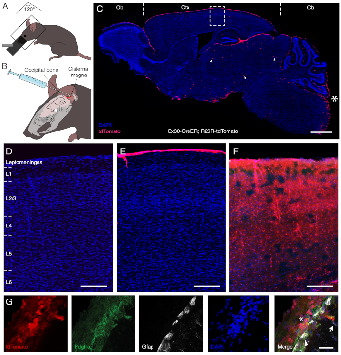
Figure 1: Specific labelling of leptomeningeal cells via intracisternal injection of endoxifen. Panel A and B illustrate the procedure developed for intracisternal administration. In order to gain access to the cisterna magna, the back of the neck should be stretched. The anesthetized animal is, therefore, positioned at an approximate angle of 120° between the body and the head, which is tilted downwards (A). The hooked needle allows to secure the syringe to the skull and to safely proceed with a manual administration of the solution (B). Panel C displays a sagittal section of the brain after intracisternal administration of endoxifen in a transgenic mouse model carrying CreER under the Cx30 promoter and inducible expression of tdTomato fluorescent reporter. The asterisk (*) marks the injection site and demonstrates the absence of operative damage following the procedure. Endoxifen selectively induces genetic recombination and reporter gene expression in cells in the meningeal layer overlaying the olfactory bulb (Ob), cortex (Ctx), and cerebellum (Cb). Only a few astrocytes in the midbrain (arrowhead) become recombined after intracisternal delivery of endoxifen. Panels D-F are magnifications of the boxed area in C in animals that were treated with vehicle solution (D), subjected to intracisternal injection of endoxifen (E), or treated systemically through oral gavage (F). Whereas intracisternal administration specifically labels cells of the leptomeninges, systemic delivery also leads to recombination of Cx30-expressing astrocytes throughout the cortical layers (L1 to L6). Panel G illustrates recombination of leptomeningeal cells (asterisk), identified through Pdgfra reactivity, after intracisternal injection. By contrast, surface (arrowhead) and parenchymal (arrow) astrocytes expressing Gfap remain unlabeled. Scale bars = 1,000 μm (C), 150 μm (D-F), and 40 μm (G). Please click here to view a larger version of this figure.
Discussion
The protocol outlined here presents a straightforward and reproducible procedure to label leptomeningeal cells for fate mapping. We use intracisternal injection of endoxifen, an active metabolite of Tamoxifen, to induce expression of tdTomato fluorescent reporter in Cx30-CreER; R26R-tdTomato mice12,13.
Compared to other protocols used for gaining access to the cerebrospinal fluid through the cisterna magna9, our approach ensures a safe manual administration thanks to the use of a bent needle that can be stabilized to the occipital bone of the skull. Once the dural membrane of the cisterna magna is perforated, there is a sudden drop in resistance. At this point, other protocols recommend lowering the needle to a maximum of 1-2 mm from the dural surface and manually keeping it in a steady position throughout the procedure9. As opposed to a straight needle, the hooked needle is secured to the skull and cannot penetrate deeper in the tissue, thus eliminating the risk of damaging the underlying cerebellum or medulla. Our hooked system allows for a safer administration of solution, particularly when using a slow rate of infusion.
The procedure outlined here is not expected to cause long-lasting discomfort to the operated animal. Care must be taken, however, when administering large volumes of solution. A fast delivery rate may lead to alterations in the intracranial pressure and the development of neurological symptoms in the mouse. We suggest injecting volumes up to 5-10 μL to avoid this risk or assemble the syringe onto a micromanipulator that has control over the delivery rate. This is particularly important when adapting this procedure to the study of the cerebrospinal fluid movement. It is recommended to avoid manual injection and use a slow rate of infusion (e.g., 1 μL/min) to prevent excessive perturbance of the physiological flow. Furthermore, the present protocol is designed to perform a single intracisternal injection, which efficiently labels leptomeningeal cells. We recommend considering ethical specifications, as well as the animal's ability to withstand multiple surgical procedures, should the study require repeated administration of compounds.
In addition to endoxifen administration, the technique outlined here can be combined with delivery of viruses carrying reporter genes under a leptomeningeal cell-specific promoter. Furthermore, the present delivery system can be utilized for acute tracing of the cerebrospinal fluid flow10. For this purpose, fluorescent tracers such as Cell Tracker (ca. 700 Da) or Dextran Fluorescin (ca. 3000 Da) can be delivered through the cisterna magna, and the syringe may be mounted onto a micromanipulator to enable control over the rate of infusion of the compound. This may be important in order to avoiding excessive disturbance of the natural cerebrospinal fluid movement in tracing experiments.
Leptomeningeal cells express claudin-11 and other proteins associated with tight junctions, which contribute to the establishment of a blood-cerebrospinal fluid barrier in the subarachnoid space and to the homeostatic control of fluid and nutrients circulation3. The approach outlined here may be combined with conditional ablation of genes implicated in the junctional control of the barrier to probe their putative role in maintaining strictly regulated cerebrospinal fluid composition. Additionally, cells from the leptomeninges play a role in development, where they provide extrinsic signals that contribute to the generation of cortical neurons5 and the formation of callosal connections4. Our method can also be adapted to gain further insight into the role of the leptomeninges in correct cortical development and axonal pathfinding. Finally, bacteria such as Neisseria meningitidis have been shown to attach to human leptomeningeal cells14, and animal models for the disease have been developed to study bacterial invasion and resulting neurological damage15,16, although surface ligands responsible for the infection are yet to be fully determined. Selective recombination of leptomeningeal cells achieved with our technique could aid identification of the adhesion sites used by bacteria to infect the subarachnoid space. Of note, the protocol hereby presented may require modifications to account for additional ethical and safety requirements necessary to carry out procedures that entail bacterial infections.
In conclusion, the intracisternal injection herein described represents a simple and well-tolerated surgical approach that offers the opportunity to investigate a wide range of leptomeningeal and cerebrospinal fluid functions, when combined with gene-editing approaches or infusion of labelled molecules.
Disclosures
The authors declare no competing interests.
Acknowledgements
This study was supported by grants from the Swedish Research Council, the Swedish Cancer Society, the Swedish Foundation for Strategic Research, Knut och Alice Wallenbergs Stiftelse and the Strategic Research Programme in Stem Cells and Regenerative Medicine at Karolinska Institutet (StratRegen).
Materials
| Name | Company | Catalog Number | Comments |
|---|---|---|---|
| Anesthesia unit | Univentor 410 | 8323102 | Complete of vaporizer, chamber, and tubing that connects to chamber and mouse head holder |
| Anesthesia (Isoflurane) | Baxter Medical AB | 000890 | |
| Betadine | Sigma-Aldrich | PVP1 | |
| Carprofen | Orion Pharma AB | 014920 | Commercial name Rymadil |
| Cyanoacrylate glue | Carl Roth | 0258.1 | Use silk 5-0 sutures, in alternative |
| Medbond Tissue Glue | Stoelting | 50479 | |
| DMSO | Sigma-Aldrich | D2650 | |
| Endoxifen | Sigma-Aldrich | E8284 | |
| Ethanol 70% | Histolab | 01370 | |
| Hamilton syringe (30 G beveled needle) | Hamilton | 80300 | |
| Lidocaine | Aspen Nordic | 520455 | |
| Mouse head holder | Narishige International | SGM-4 | With mouth piece for inhalational anhestetics. Alternatively, use a stereotactic frame |
| Scissors | Fine Science Tools | 15009-08 | |
| Shaver | Aesculap | GT420 | |
| Sterile absorption spears | Fine Science Tools | 18105-01 | Sterile cotton swabs are also a good option |
| Surgical separator | World Precision Instrument | 501897 | |
| Tweezers | Dumont | 11251-35 | |
| Viscotears | Bausch&Lomb Nordic AB | 541760 |
References
- Vanlandewijck, M., et al. A molecular atlas of cell types and zonation in the brain vasculature. Nature. 554 (7693), 475-480 (2018).
- Whish, S., et al. The inner CSF-brain barrier: developmentally controlled access to the brain via intercellular junctions. Frontiers in Neuroscience. 9, 16(2015).
- Weller, R. O., Sharp, M. M., Christodoulides, M., Carare, R. O., Mollgard, K. The meninges as barriers and facilitators for the movement of fluid, cells and pathogens related to the rodent and human CNS. Acta Neuropathologica. 135 (3), 363-385 (2018).
- Choe, Y., Siegenthaler, J. A., Pleasure, S. J. A cascade of morphogenic signaling initiated by the meninges controls corpus callosum formation. Neuron. 73 (4), 698-712 (2012).
- Siegenthaler, J. A., et al. Retinoic acid from the meninges regulates cortical neuron generation. Cell. 139 (3), 597-609 (2009).
- Bifari, F., et al. Neurogenic Radial Glia-like Cells in Meninges Migrate and Differentiate into Functionally Integrated Neurons in the Neonatal Cortex. Cell Stem Cell. 20 (3), 360-373 (2017).
- De Bock, M., et al. A new angle on blood-CNS interfaces: a role for connexins? FEBS Letters. 588 (8), 1259-1270 (2014).
- Ramos, M., et al. Cisterna Magna Injection in Rats to Study Glymphatic Function. Methods in Molecular Biology. 1938, 97-104 (2019).
- Xavier, A. L. R., et al. Cannula Implantation into the Cisterna Magna of Rodents. Journal of Visualized Experiments. (135), (2018).
- Iliff, J. J., et al. A paravascular pathway facilitates CSF flow through the brain parenchyma and the clearance of interstitial solutes, including amyloid beta. Science Translational Medicine. 4 (147), (2012).
- Coureuil, M., Lecuyer, H., Bourdoulous, S., Nassif, X. A journey into the brain: insight into how bacterial pathogens cross blood-brain barriers. Nature Reviews Microbiology. 15 (3), 149-159 (2017).
- Madisen, L., et al. Transgenic mice for intersectional targeting of neural sensors and effectors with high specificity and performance. Neuron. 85 (5), 942-958 (2015).
- Slezak, M., et al. Transgenic mice for conditional gene manipulation in astroglial cells. Glia. 55 (15), 1565-1576 (2007).
- Hardy, S. J., Christodoulides, M., Weller, R. O., Heckels, J. E. Interactions of Neisseria meningitidis with cells of the human meninges. Molecular Microbiology. 36 (4), 817-829 (2000).
- Colicchio, R., et al. The meningococcal ABC-Type L-glutamate transporter GltT is necessary for the development of experimental meningitis in mice. Infection and Immunity. 77 (9), 3578-3587 (2009).
- Ricci, S., et al. Inhibition of matrix metalloproteinases attenuates brain damage in experimental meningococcal meningitis. BMC Infectious Diseases. 14, 726(2014).
Reprints and Permissions
