Here, a protocol for creating a central corneal epithelial abrasion wound in the mouse using a trephine and a blunt golf club spud is described. This corneal wound healing model is highly reproducible and is now being used to evaluate compromised corneal wound healing in the context of diseases.
A subscription to JoVE is required to view this content. Sign in or start your free trial.
Method Article
An Epithelial Abrasion Model for Studying Corneal Wound Healing
In This Article
Summary
Abstract
The cornea is critical for vision, accounting for about two-thirds of the refractive power of the eye. Crucial to the role of the cornea in vision is its transparency. However, due to its external position, the cornea is highly susceptible to a wide variety of injuries that can lead to the loss of corneal transparency and eventual blindness. Efficient corneal wound healing in response to these injuries is pivotal for maintaining corneal homeostasis and preservation of corneal transparency and refractive capabilities. In events of compromised corneal wound healing, the cornea becomes vulnerable to infections, ulcerations, and scarring. Given the fundamental importance of corneal wound healing to the preservation of corneal transparency and vision, a better understanding of the normal corneal wound healing process is a prerequisite to understanding impaired corneal wound healing associated with infection and disease. Toward this goal, murine models of corneal wounding have proven useful in furthering our understanding of the corneal wound healing mechanisms operating under normal physiological conditions. Here, a protocol for creating a central corneal epithelial abrasion in mouse using a trephine and a blunt golf club spud is described. In this model, a 2 mm diameter circular trephine, centered over the cornea, is used to demarcate the wound area. The golf club spud is used with care to debride the epithelium and create a circular wound without damaging the corneal epithelial basement membrane. The resulting inflammatory response proceeds as a well-characterized cascade of cellular and molecular events that are critical for efficient wound healing. This simple corneal wound healing model is highly reproducible and well-published and is now being used to evaluate compromised corneal wound healing in the context of disease.
Introduction
The cornea is the transparent anterior one-third of the eye. The cornea serves several functions including protecting the inner structures of the eye and forming a structural barrier that protects the eye against infections1. More importantly, the cornea is critical for vision, providing about two-thirds of the refractive power of the eye2,3. Crucial to the role of the cornea in vision is its transparency. However, due to its outward position, the cornea is exposed to a wide variety of injuries on a day-to-day basis that can lead to disruption of its barrier function, loss of transparency, and eventual blindness. Loss of corneal transparency is a leading cause of visual impairment worldwide4,5. Corneal abrasions are a common reason for visits to the emergency room (ER), accounting for half of the eye-related cases presented at the ER6. Over 1 million individuals are estimated to suffer from eye-related injuries annually in the United States7. Efficient corneal wound healing in response to these injuries is pivotal for maintaining corneal homeostasis and preservation of its transparency and refractive capabilities. In events of compromised corneal wound healing, the cornea becomes vulnerable to infections, ulcerations, and scarring8,9. Also, the increasing popularity of refractive surgeries places a unique traumatic challenge on the cornea10. Given the fundamental importance of corneal wound healing to the preservation of corneal transparency and vision, a better understanding of the normal corneal wound healing process is a prerequisite to understanding impaired corneal wound healing associated with infection and disease.
To that end, several animal models of corneal wound healing have been developed11,12,13,14,15. Murine models of corneal wound healing have proven useful in furthering our understanding of the corneal wound healing mechanisms operating under normal physiological conditions. Different types of corneal wounds have been employed in studying corneal wound healing, each suitable for investigating different aspects of the wound healing process. The commonest types of wound models used in corneal wound healing studies are the mechanical and chemical wound models. Chemical corneal wounds, mostly involving the creation of alkaline burns on the cornea, are useful for studying corneal ulcers, opacification, and neovascularization13. Mechanical corneal wounds involve debridement (abrasion) wounds and keratectomy wounds14,15,16. An intact or breached corneal epithelial basement membrane defines debridement and keratectomy wounds, respectively. In debridement wounds, the epithelial basement membrane remains intact, while in keratectomy wounds, the basement membrane is breached with penetration mostly into the anterior stroma. Debridement wounds are most useful for studying re-epithelialization, epithelial cell proliferation, immune response, and nerve regeneration following corneal wounding. Keratectomy wounds, on the other hand, are most useful for studying corneal scarring14,15.
Here, a protocol for creating a central corneal epithelial abrasion wound in the mouse using a trephine and a blunt golf club spud is described. This simple corneal wound healing model is highly reproducible and well-published and is now being used to evaluate compromised corneal wound healing in the context of disease17.
Access restricted. Please log in or start a trial to view this content.
Protocol
All animal protocols were approved by the Institutional Animal Care and Use Committees at the University of Houston and Baylor College of Medicine. The guidelines outlined in the Association for Research in Vision and Ophthalmology (ARVO) statement on the use of animals in vision and ophthalmic research were followed in handling and using the mice.
1. Preparation
- Preparation of fluorescein solution
- Prepare 1% fluorescein solution by dissolving 10 mg of sodium fluorescein salt in 1 mL of sterile saline or sterile 1x phosphate buffered saline (PBS).
NOTE: Prepare sodium fluorescein solution on the day of use or a day before to avoid microbial contamination. When prepared a day before use, store fluorescein solution at 4 °C away from light. Wrap tubes with aluminum foil to prevent photobleaching. - Divide the solution into aliquots suitable for use in a single experiment. 1-1.5 µL of fluorescein solution is used per mouse per time point. Use the following formula to calculate the volume of aliquot appropriate for a single experiment:

NOTE: For example, if six mice are studied in a single experiment with wound size monitored at five time points, the volume of aliquot suitable for that single experiment would be:

- Prepare 1% fluorescein solution by dissolving 10 mg of sodium fluorescein salt in 1 mL of sterile saline or sterile 1x phosphate buffered saline (PBS).
- Preparation of ketamine/xylazine cocktail for anesthesia of mice.
- To prepare 10 mL of cocktail, mix 2.0 mL of ketamine (100 mg/mL) with 1.0 mL of xylazine (20 mg/mL), and add 7.0 mL of sterile PBS. Prepare ketamine/xylazine cocktail a day before or on the day of the surgery.
NOTE: All solutions should be used at room temperature unless stated otherwise.
- To prepare 10 mL of cocktail, mix 2.0 mL of ketamine (100 mg/mL) with 1.0 mL of xylazine (20 mg/mL), and add 7.0 mL of sterile PBS. Prepare ketamine/xylazine cocktail a day before or on the day of the surgery.
2. Anesthesia
- Weigh the mouse (8-12 weeks old C57BL/6 wildtype mice) to determine the appropriate amount of anesthetic to administer. Use the two-handed mouse restraint technique to restrain and handle mice for the injection of the anesthetic18. Administer ketamine/xylazine cocktail intraperitoneally (i.p.) at a final concentration of 80 mg/kg of ketamine and 8 mg/kg of xylazine.
- Wait until complete anesthesia is achieved before performing corneal wounding on the mouse. Evaluate the depth of anesthesia by assessing the pedal reflex after toe pinch. When adequate anesthesia is achieved, the mouse should not move upon toe pinch.
NOTE: Because mice quickly lose body heat during anesthesia, it is important to provide a source of warmth for mice to prevent hypothermia. Place mice on a heat source (heating pad) during anesthesia and recovery stages.
3. Creation of corneal wound
- Wound the right or left eye only. Maintain consistency with the eye (i.e., left or right) that is wounded when moving from mouse to mouse.
NOTE: Because abrasions damage corneal nerves and reduce acuity, wounding both eyes can cause significant discomfort and impairment. Since analgesics have the potential to suppress inflammatory responses, their use may be a confounder in certain experiments aimed at understanding corneal inflammation. Avoiding analgesics in corneal wound experiments was approved by our Institutional Animal Care and Use Committee (IACUC). - To create the epithelial corneal wound
- Under a dissecting microscope, use the 2 mm diameter trephine (see Figure 1) to demarcate the center of the cornea, keeping the eye wide open using the thumb and index finger to hold the eyelids. Gently twirl the trephine to make an impression on the corneal epithelium.
NOTE: Care must be taken to not apply excessive pressure when using the trephine as this can result in corneal perforation. Also, care must be taken to position the trephine centrally. The pupils can be used as a landmark to locate the center of the cornea. - Under a dissecting microscope, hold the blunt golf club spud (see Figure 1) at approximately 45° from the surface of the cornea within the area demarcated with the trephine. Carefully and continuously scrape the epithelium within the demarcated area with the spud to debride the epithelium.
NOTE: Do not apply excessive force in debridement as that can also cause corneal perforation. The eyes of mice can dry up during the wounding process, making debridement difficult. In such a case, apply sterile PBS to the ocular surface to maintain optimal hydration.
- Under a dissecting microscope, use the 2 mm diameter trephine (see Figure 1) to demarcate the center of the cornea, keeping the eye wide open using the thumb and index finger to hold the eyelids. Gently twirl the trephine to make an impression on the corneal epithelium.
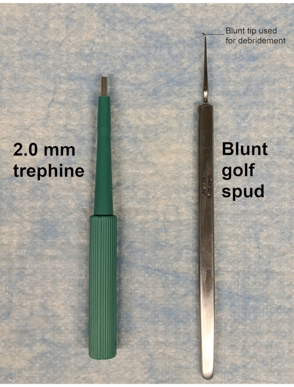
Figure 1: 2 mm trephine and blunt golf club spud. The trephine is used to demarcate a circular region at the cornea center, and the golf club spud is used to remove the epithelium within the demarcated region. Please click here to view a larger version of this figure.
4. Monitoring of wound closure and re-epithelialization
- Pipette 1-1.5 µL of 1% fluorescein solution onto the wounded surface and image cornea using a digital microscope with a blue light source.
- Acquire images within the first minute of fluorescein solution addition to avoid spreading to surrounding epithelium leading to overestimation of wound size. Wounded corneas are imaged at specific times (i.e., 0 h, 12 h, 18 h, 24 h, and 30 h) after wounding.
NOTE: At all time points, imaging is performed under anesthesia; ketamine/xylazine is used at the time of wounding while isoflurane is used at subsequent timepoints. Mice are housed individually in separate cages for the duration of wound closure monitoring. When housed together, mice tend to lick littermate's eyes, a behavior which has been shown to affect wound healing in different tissues19,20. - Analyze captured images, tracing wound area using an image analysis software. The wound area for each time point is expressed as a percentage of the original wound area at 0 h.
5. Immunofluorescence imaging and analysis
- Euthanize mice by an IACUC-approved method (in this case, carbon dioxide overdose followed by cervical dislocation) at desired time points after wounding.
- Harvest eyeballs by gently pressing on the lateral canthus to displace the eyeball using iris curved scissor. Guide the scissors behind the eyeball to firmly grasp the optic nerve, and then cut the nerve, which allows the eyeball to be removed.
- Fix each eyeball in 1 mL of 1x PBS containing 2% paraformaldehyde for 1 h at room temperature, and then wash three times in 1 mL of 1x PBS for 5 min each.
- Under a dissecting microscope, use a surgical blade to make an incision in the sclera, about 500 µm distal to the limbus, and then cut through the globe. Using forceps, gently remove the iris matter from the cornea, and then carefully trim the scleral tissue away, making sure to leave the limbus intact.
- Make four partial radial cuts, each approximately 1 mm in length, which extend from the peripheral cornea and stop short of the center to allow the cornea to flatten.
- Permeabilize and block corneas in 1 mL of 2% bovine serum albumin (BSA) and 0.01% TritonX -100 in 1x PBS for 15 min followed by blocking in 2% BSA in 1x PBS for an additional 45 min at room temperature.
- Incubate corneas overnight at 4 °C in a cocktail of directly labeled unique fluorochrome conjugated antibodies prepared in 1x PBS containing 2% BSA.
NOTE: Antibodies are targeted to label specific cells and tissues of interest. For example, endothelium, neutrophils, and platelets are labeled with anti-CD3121,22, anti-Ly-6G23,24, and anti-CD4125,26 antibodies, respectively. 4',6-diamidino-2-phenylindole (DAPI) is added to the antibody cocktail to visualize nuclei. - After incubation, wash corneas three times in 1x PBS for 15 min each.
- Mount corneas on a microscope slide in a drop of anti-fade fluorescence mounting medium, cover with a coverslip, and image with the desired magnification (4x to 100x objective) using a fluorescence light microscope. Take full-thickness images of the cornea across different regions as illustrated in Figure 2A.
NOTE: The pattern of microscopic analysis illustrated in Figure 2A is used for analyzing specific regional changes in inflammation and cell division. Both DAPI and Ly-6G staining are used to identify the extravascular neutrophils. With DAPI staining, spherical neutrophils have a distinct horseshoe or donut shaped nucleus (Figure 2B).
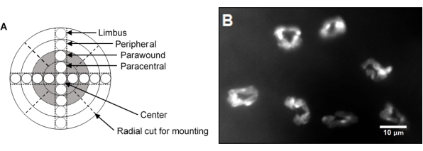
Figure 2: Corneal imaging strategy and neutrophil infiltration after central abrasion. (A) Schematic representation of a cornea wholemount showing nine microscopic fields across the diameter of the cornea. The gray area represents the original wound area. The width of each region is 500 µm. (B) Note the distinct horseshoe or donut shape of neutrophil nuclei with DAPI staining. Please click here to view a larger version of this figure.
Access restricted. Please log in or start a trial to view this content.
Results
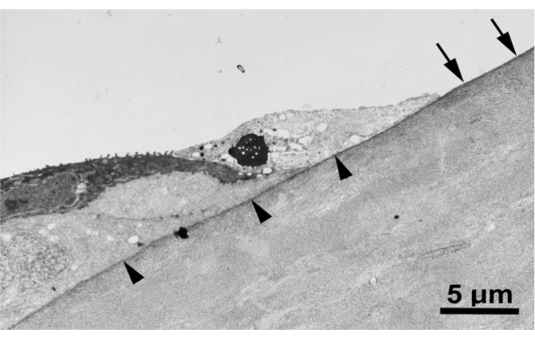
Figure 3 shows a transmission electron micrograph of a corneal wound created with the blunt golf club spud, demonstrating that the epithelial basement membrane is indeed intact after injury.
Access restricted. Please log in or start a trial to view this content.
Discussion
The purpose of this methods paper was to describe a protocol for creating a central corneal epithelial abrasion wound in the mouse using a trephine and a blunt golf club spud. This murine model has been used to study corneal inflammation and its contribution to wound healing. This type of model can be used to study corneal wound healing mechanisms under normal physiological conditions and in pathologies17,28,29,
Access restricted. Please log in or start a trial to view this content.
Disclosures
The authors have nothing to disclose.
Acknowledgements
Funding: Supported by: NIH EY018239 (A.R.B., C.W.S., and R.E.R.), P30EY007551 (A.R.B.), and Sigma Xi Grant in Aid of Research (P.K.A.). The content is solely the responsibility of the authors and does not represent the official views of the National Institutes of Health, or Sigma Xi.
Access restricted. Please log in or start a trial to view this content.
Materials
| Name | Company | Catalog Number | Comments |
|---|---|---|---|
| Anti-CD31 antibody | BD Bioscience, Pharmingen | 550274 | |
| Anti-CD41 antibody | BD Bioscience, Pharmingen | 553847 | |
| Anti-Ly6G antibody | BD Bioscience, Pharmingen | 551459 | |
| Bovine serum albumin (BSA) | ThermoFisher scientific | B14 | |
| C57BL/6 mice | Jackson Laboratories | 664 | |
| DAPI | Sigma Aldrich | D8417 | |
| DeltaVision wide-field deconvolution fluorescence microscope | GE Life Sciences | ||
| Dissecting microscope | Leica microsystems | ||
| Electronic Toploading Balances (Weighing scale) | Fisher Scientific | ||
| Ethanol | ThermoFisher scientific | T038181000CS | |
| Golf-club spud | Stephens instruments | S2-1135 | |
| Iris curve scissors | Fisher Scientific | 31212 | |
| Isoflurane | Patterson veterinary | 07-893-1389 | |
| Ketamine | Patterson veterinary | 07-890-8598 | |
| Phospate buffered saline (PBS) | ThermoFisher scientific | AM9624 | |
| Sodium fluorescein salt | Sigma Aldrich | 46970 | |
| Surgical blade (scapel blade) | Fine Science tools | 10022-00 | |
| Trephine | Integra Miltex | 33-31 | |
| TritonX -100 | Fisher Scientific | 50-295-34 | |
| Forcep | Fine Science tools | 11923-13 | |
| Xylazine | Patterson veterinary | 07-808-1947 |
References
- DelMonte, D. W., Kim, T. Anatomy and physiology of the cornea. Journal of Cataract and Refractive Surgery. 37 (3), 588-598 (2011).
- Meek, K. M., Knupp, C. Corneal structure and transparency. Progress in Retinal and Eye Research. 49, 1-16 (2015).
- Sridhar, M. S. Anatomy of cornea and ocular surface. Indian Journal of Ophthalmology. 66 (2), 190-194 (2018).
- Flaxman, S. R., et al. Global causes of blindness and distance vision impairment 1990-2020: a systematic review and meta-analysis. The Lancet Global Health. 5 (12), 1221-1234 (2017).
- Robaei, D., Watson, S. Corneal blindness: A global problem. Clinical & Experimental Ophthalmology. 42 (3), 213-214 (2014).
- McGwin, G., Owsley, C. Incidence of emergency department-treated eye injury in the United States. Archives of Ophthalmology. 123 (5), 662-666 (2005).
- Ljubimov, A. V., Saghizadeh, M. Progress in corneal wound healing. Progress in Retinal and Eye Research. 49, 17-45 (2015).
- Wilson, S. L., Haj, A. J. E., Yang, Y. Control of scar tissue formation in the cornea: Strategies in clinical and corneal tissue engineering. Journal of Functional Biomaterials. 3 (3), 642(2012).
- Vaidyanathan, U., et al. Persistent corneal epithelial defects: A review article. Medical Hypothesis, Discovery and Innovation in Ophthalmology. 8 (3), 163-176 (2019).
- Netto, M., et al. Wound healing in the cornea: a review of refractive surgery complications and new prospects for therapy. Cornea. 24 (5), 509-522 (2005).
- Friedenwald, J. S., Buschke, W. Some factors concerned in the mitotic and wound-healing activities of the corneal epithelium. Transactions of the American Ophthalmological Society. 42, 371-383 (1944).
- Xu, K., Yu, F. -S. X. Impaired epithelial wound healing and EGFR signaling pathways in the corneas of diabetic rats. Investigative Ophthalmology & Visual Science. 52 (6), 3301-3308 (2011).
- Bai, J. Q., Qin, H. F., Zhao, S. H. Research on mouse model of grade II corneal alkali burn. International Journal of Ophthalmology. 9 (4), 487-490 (2016).
- Blanco-Mezquita, J. T., Hutcheon, A. E. K., Stepp, M. A., Zieske, J. D. αVβ6 integrin promotes corneal wound healing. Investigative Ophthalmology and Visual Science. 52 (11), 8505-8513 (2011).
- Blanco-Mezquita, J. T., Hutcheon, A. E. K., Zieske, J. D. Role of thrombospondin-1 in repair of penetrating corneal wounds. Investigative Ophthalmology and Visual Science. 54 (9), 6262-6268 (2013).
- Stepp, M. A., et al. Wounding the cornea to learn how it heals. Experimental Eye Research. 121, 178-193 (2014).
- Hargrave, A., et al. Corneal dysfunction precedes the onset of hyperglycemia in a mouse model of diet-induced obesity. PLoS ONE. 15, 0238750(2020).
- Machholz, E., Mulder, G., Ruiz, C., Corning, B. F., Pritchett-Corning, K. R. Manual restraint and common compound administration routes in mice and rats. Journal of Visualized Experiments: JoVE. (67), e2771(2012).
- Bodner, L., Dayan, D. Effect of parotid submandibular and sublingual saliva on wound healing in rats. Comparative Biochemistry and Physiology. Part A, Physiology. 100 (4), 887-890 (1991).
- Abbasian, B., Azizi, S., Esmaeili, A. Effects of rat's licking behavior on cutaneous wound healing. Iranian Journal of Basic Medical Sciences. 13 (1), 242-247 (2010).
- DeLisser, H. M., et al. Involvement of endothelial PECAM-1/CD31 in angiogenesis. The American Journal of Pathology. 151 (3), 671-677 (1997).
- Piali, L., et al. CD31/PECAM-1 is a ligand for alpha v beta 3 integrin involved in adhesion of leukocytes to endothelium. The Journal of Cell Biology. 130 (2), 451-460 (1995).
- Fleming, T. J., Fleming, M. L., Malek, T. R. Selective expression of Ly-6G on myeloid lineage cells in mouse bone marrow. RB6-8C5 mAb to granulocyte-differentiation antigen (Gr-1) detects members of the Ly-6 family. The Journal of Immunology. 151 (5), 2399-2408 (1993).
- Fleming, T. J., Malek, T. R. Multiple glycosylphosphatidylinositol-anchored Ly-6 molecules and transmembrane Ly-6E mediate inhibition of IL-2 production. The Journal of Immunology. 153 (5), 1955-1962 (1994).
- Phillips, D. R., Charo, I. F., Scarborough, R. M. GPIIb-IIIa: the responsive integrin. Cell. 65 (3), 359-362 (1991).
- Nieswandt, B., et al. Acute systemic reaction and lung alterations induced by an antiplatelet integrin gpIIb/IIIa antibody in mice. Blood. 94 (2), 684-693 (1999).
- Li, Z., Burns, A. R., Rumbaut, R. E., Smith, C. W. γδ T cells are necessary for platelet and neutrophil accumulation in limbal vessels and efficient epithelial repair after corneal abrasion. American Journal of Pathology. 171 (3), 838-845 (2007).
- Liu, Q., Smith, C. W., Zhang, W., Burns, A. R., Li, Z. NK cells modulate the inflammatory response to corneal epithelial abrasion and thereby support wound healing. American Journal of Pathology. 181 (2), 452-462 (2012).
- Gao, Y., et al. NK cells are necessary for recovery of corneal CD11c+ dendritic cells after epithelial abrasion injury. Journal of Leukocyte Biology. 94 (2), 343-351 (2013).
- Xiao, C., et al. Acute tobacco smoke exposure exacerbates the inflammatory response to corneal wounds in mice via the sympathetic nervous system. Communications Biology. 2, 33(2019).
- Wang, H., et al. Epothilone B speeds corneal nerve regrowth and functional recovery through microtubule stabilization and increased nerve beading. Scientific Reports. 8 (1), 2647(2018).
- Li, Z., Burns, A. R., Smith, C. W. Lymphocyte function-associated Antigen-1-dependent inhibition of corneal wound healing. Cell Injury. 169, 1590-1600 (2006).
- Wu, M., et al. The neuroregenerative effects of topical decorin on the injured mouse cornea. Journal of Neuroinflammation. 17 (1), 1-14 (2020).
- Rodrigues, M., Kosaric, N., Bonham, C. A., Gurtner, G. C. Wound healing: A cellular perspective. Physiological Reviews. 99 (1), 665-706 (2019).
- Rennard, S. I. Inflammation and repair processes in chronic obstructive pulmonary disease. American Journal of Respiratory and Critical Care Medicine. 160 (5), Pt 2 12-16 (1999).
- Landén, N. X., Li, D., Ståhle, M. Transition from inflammation to proliferation: a critical step during wound healing. Cellular and Molecular Life Sciences. 73 (20), 3861-3885 (2016).
- Li, Z., Rumbaut, R. E., Burns, A. R., Smith, C. W. Platelet response to corneal abrasion is necessary for acute inflammation and efficient re-epithelialteation. Investigative Ophthalmology and Visual Science. 47, 4794-4802 (2006).
- Lam, F. W., Burns, A. R., Smith, C. W., Rumbaut, R. E. Platelets enhance neutrophil transendothelial migration via P-selectin glycoprotein ligand-1. American Journal of Physiology - Heart and Circulatory Physiology. 300 (2), 468-475 (2011).
- La Cruz, A. D., et al. Platelet and erythrocyte extravasation across inflamed corneal venules depend on CD18, neutrophils, and mast cell degranulation. International Journal of Molecular Sciences. 22 (14), 7360(2021).
- Li, Z., Burns, A. R., Smith, C. W. Two waves of neutrophil emigration in response to corneal epithelial abrasion: Distinct adhesion molecule requirements. Investigative Ophthalmology and Visual Science. 47 (5), 1947-1955 (2006).
- Li, Z., Burns, A. R., Han, L., Rumbaut, R. E., Smith, C. W. IL-17 and VEGF Are Necessary for Efficient Corneal Nerve Regeneration. The American Journal of Pathology. 178 (3), 1106-1116 (2011).
- Xue, Y., et al. Modulation of circadian rhythms affects corneal epithelium renewal and repair in mice. Investigative Ophthalmology and Visual Science. 58 (3), 1865-1874 (2017).
- Zhang, W., Magadi, S., Li, Z., Smith, C. W., Burns, A. R. IL-20 promotes epithelial healing of the injured mouse cornea. Experimental Eye Research. 154, 22-29 (2017).
- Li, Z., Burns, A. R., Miller, S. B., Smith, C. W. CCL20, γδ T cells, and IL-22 in corneal epithelial healing. FASEB Journal. 25 (8), 2659-2668 (2011).
- Li, Z., Burns, A. R., Han, L., Rumbaut, R. E., Smith, C. W. IL-17 and VEGF are necessary for efficient corneal nerve regeneration. American Journal of Pathology. 178 (3), 1106-1116 (2011).
- Reins, R. Y., Hanlon, S. D., Magadi, S., McDermott, A. M. Effects of topically applied Vitamin D during corneal wound healing. PLoS ONE. 11 (4), 0152889(2016).
- Gagen, D., et al. ICAM-1 mediates surface contact between neutrophils and keratocytes following corneal epithelial abrasion in the mouse. Experimental Eye Research. 91 (5), 676-684 (2010).
- Li, Z., Rivera, C. A., Burns, A. R., Smith, C. W. Hindlimb unloading depresses corneal epithelial wound healing in mice. Journal of Applied Physiology. 97 (2), 641-647 (2004).
- Byeseda, S. E., et al. ICAM-1 is necessary for epithelial recruitment of γδ T cells and efficient corneal wound healing. American Journal of Pathology. 175 (2), 571-579 (2009).
- Li, Z., Burns, A. R., Rumbaut, R. E., Smith, C. W. γδ T cells are necessary for platelet and neutrophil accumulation in limbal vessels and efficient epithelial repair after corneal abrasion. American Journal of Pathology. 171 (3), 838-845 (2007).
- Petrescu, M. S., et al. Neutrophil interactions with keratocytes during corneal epithelial wound healing: A role for CD18 integrins. Investigative Ophthalmology and Visual Science. 48 (11), 5023-5029 (2007).
- Pal-Ghosh, S., Pajoohesh-Ganji, A., Tadvalkar, G., Stepp, M. A. Removal of the basement membrane enhances corneal wound healing. Experimental Eye Research. 93 (6), 927-936 (2011).
- Pal-Ghosh, S., et al. Cytokine deposition alters leukocyte morphology and initial recruitment of monocytes and γδT cells after corneal injury. Investigative Ophthalmology & Visual Science. 55 (4), 2757-2765 (2014).
- Pal-Ghosh, S., Tadvalkar, G., Jurjus, R. A., Zieske, J. D., Stepp, M. A. BALB/c and C57BL6 mouse strains vary in their ability to heal corneal epithelial debridement wounds. Experimental Eye Research. 87 (5), 478-486 (2008).
- Kato, T., Chang, J. H., Azar, D. T. Expression of type XVIII collagen during healing of corneal incisions and keratectomy wounds. Investigative Ophthalmology and Visual Science. 44 (1), 78-85 (2003).
- Kure, T., et al. Corneal neovascularization after excimer keratectomy wounds in matrilysin-deficient mice. Investigative Ophthalmology and Visual Science. 44 (1), 137-144 (2003).
- Lin, A., et al. Bacterial keratitis preferred practice pattern. Ophthalmology. 126 (1), 1-55 (2019).
- Cable, E. J., Onishi, K. G., Prendergast, B. J. Circadian rhythms accelerate wound healing in female Siberian hamsters. Physiology and Behavior. 171, 165-174 (2017).
- Lyons, A. B., Moy, L., Moy, R., Tung, R. Circadian rhythm and the skin: A review of the literature. Journal of Clinical and Aesthetic Dermatology. 12 (9), 42-45 (2019).
- Westman, J., Grinstein, S., Marques, P. E. Phagocytosis of Necrotic Debris at Sites of Injury and Inflammation. Frontiers in Immunology. 10, 3030(2020).
- Gaudry, M., et al. Intracellular pool of vascular endothelial growth factor in human neutrophils. Blood. 90 (10), 4153-4161 (1997).
- Pan, Z., et al. Vascular endothelial growth factor promotes anatomical and functional recovery of injured peripheral nerves in the avascular cornea. FASEB Journal. 7, 2756-2767 (2013).
- Di, G., et al. VEGF-B promotes recovery of corneal innervations and trophic functions in diabetic mice. Scientific Reports. 7 (1), 1-13 (2017).
- Thomas, M. R., Storey, R. F. The role of platelets in inflammation. Thrombosis and Haemostasis. 114 (3), 449-458 (2015).
- Margraf, A., Zarbock, A. Platelets in inflammation and resolution. The Journal of Immunology. 203 (9), 2357-2367 (2019).
Access restricted. Please log in or start a trial to view this content.
Reprints and Permissions
