ラマン散乱現象は、1928年にC. V. Raman1によって初めて観測された。入射光子がサンプルと相互作用しているとき、非弾性散乱事象が自発的に起こり得、光子のエネルギー変化は分析された化学種の振動遷移と一致する。このプロセスでは、化学タグを使用する必要がないため、サンプルの摂動を最小限に抑えながら、化学分析のための汎用性の高いラベルフリーツールになります。その利点にもかかわらず、自発的ラマン散乱は、低散乱断面(典型的には赤外線[IR]吸収断面よりも1011 低い)に悩まされ、分析2のために長い取得時間を必要とする。したがって、ラマン散乱プロセスの感度を高めるための探求は、ラマン技術をリアルタイムイメージングに推進する上で不可欠です。
ラマン散乱の感度を大幅に高める効果的な方法の1つは、コヒーレントラマン散乱(CRS)プロセスによるものであり、そのために2つのレーザーパルスが分子振動遷移を励起するために典型的に使用される3,4。2つのレーザー間の光子エネルギー差がサンプル分子の振動モードと一致すると、強いラマン信号が生成されます。イメージングに最も一般的に使用される2つのCRSプロセスは、コヒーレント抗ストークスラマン散乱(CARS)と刺激ラマン散乱(SRS)5です。過去20年間、技術開発により、CARSおよびSRS顕微鏡技術が進歩し、生体サンプル中の標識のない定量化および化学変化の解明のための強力なツールになりました。
CARS顕微鏡による化学イメージングは、Duncanら6によって実証されたように、レーザースキャニングがCARS画像を取得するために最初に適用された1982年にさかのぼることができます。CARS顕微鏡の近代化は、レーザー走査型多光子蛍光顕微鏡7の幅広い応用の後、大幅に加速されました。高繰り返し速度レーザーを使用したXieグループの初期の研究は、CARSを生物学的サンプル中の分子の特性評価のための高速、ラベルフリーの化学イメージングプラットフォームに移行しました8,9,10。CASイメージングの主な問題の1つは、非共振背景の存在であり、これは画像のコントラストを低下させ、ラマンスペクトルを歪める。非共鳴バックグラウンド11、12、13、14、15を減少させるか、またはCARSスペクトル16、17から共鳴ラマン信号を抽出するために多くの努力がなされている。この分野を大幅に進歩させたもう1つの進歩は、ハイパースペクトルCARイメージングであり、これは改善された化学的選択性18、19、20、21で各画像ピクセルでのスペクトルマッピングを可能にする。
刺激ラマン散乱(SRS)は、CARSよりも若いイメージング技術ですが、22年初めに発見されました。2007年に、低繰り返し率レーザー光源23を用いたSRS顕微鏡法が報告された。まもなく、いくつかのグループは、高繰り返し速度レーザー24、25、26を用いた高速SRSイメージングを実証した。CARSに対するSRS顕微鏡法の主な利点の1つは、非共振バックグラウンド27がないことであるが、クロスフェーズ変調(XPM)、過渡吸収(TA)、2光子吸収(TPA)、および光熱(PT)効果などの他のバックグラウンドがSRS28で起こり得る。さらに、SRS信号およびサンプル濃度は、二次信号濃度依存性を有するCARSとは異なり、線形関係を有する29。これにより、化学定量とスペクトルアンミキシングが簡素化されます。マルチカラーおよびハイパースペクトルSRSは、異なる形態30、31、32、33、34、35、36で進化しており、スペクトル集束は化学イメージングのための最も一般的なアプローチの1つである37,38。
CARSとSRSの両方は、信号励起のための分子の振動遷移に一致させるために、ポンプとストークスレーザービームをサンプルに集束させる必要があります。CARS顕微鏡とSRS顕微鏡にも多くの共通点があります。しかし、これら2つのプロセスの根底にある物理学と、これらの顕微鏡技術に関わる信号検出には、3,39の格差があります。CARSは、正味の光子-分子エネルギー結合3を持たないパラメトリックプロセスである。しかし、SRSはノンパラメトリックプロセスであり、光子と分子系27との間のエネルギー移動に寄与する。CARSでは、アンチストークス周波数で新しい信号が生成されますが、SRSはポンプとストークスレーザービーム間のエネルギー移動として現れます。
CARS 信号は Eq (1)28 を満たします。
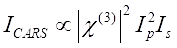 (1)
(1)
一方、SRS信号はEq(2)28と書くことができる。
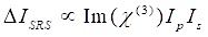 (2)
(2)
ここで、Ip、Is、I CARSおよびΔISRSは、それぞれポンプビーム、ストークスビーム、CARS信号、およびSRS信号の強度である。χ(3) はサンプルの 3 次非線形光学感受性であり、実数部と虚数部で構成される複素数値です。
これらの方程式は、CARSとSRSのスペクトルプロファイルと信号濃度依存性を表します。物理学の違いにより、これら2つの顕微鏡技術の検出スキームが異なります。CARSにおける信号検出は、通常、新たに生成された光子のスペクトル分離と、光電子増倍管(PMT)または電荷結合素子(CCD)を用いた検出を伴う。SRSの場合、ポンプビームとストークスビーム間のエネルギー交換は、通常、光変調器を使用した高速強度変調と、ロックインアンプと対になったフォトダイオード(PD)を使用した復調によって測定されます。
近年、CARとSRSの両方の分野で多くの技術開発と応用が発表されていますが、特にハイパースペクトルCARSとSRS顕微鏡では、2つのCRS技術の体系的な比較は同じプラットフォーム上で行われていません。感度、空間分解能、スペクトル分解能、および化学分離能力を直接比較することで、生物学者は化学定量に最適なモダリティを選択できます。このプロトコルでは、フェムト秒レーザーシステムとスペクトル集束に基づくハイパースペクトルCARモダリティとSRSモダリティの両方を備えたマルチモーダルイメージングプラットフォームを構築するための詳細な手順が提供されます。2つの技術は、スペクトル分解能、検出感度、空間分解能、および細胞のイメージングコントラストについて順方向で比較されてきた。
