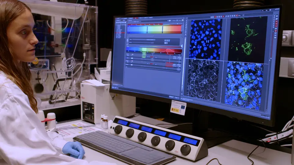
Classically, the endocardium of the mouse embryonic valve primordium has been analyzed using transversal, coronal, or sagittal sections. Our novel approach for en face, two-dimensional imaging of the endocardium at valvulogenic regions allows planar polarity and cell rearrangement analysis of the endocardium during valve development.
A subscription to JoVE is required to view this content. Sign in or start your free trial.
Method Article
En Face Endocardial Cushion Preparation for Planar Morphogenesis Analysis in Mouse Embryos
In This Article
Summary
Abstract
The study of the cellular and molecular mechanisms underlying the development of the mammalian heart is essential to address human congenital heart disease. The development of the primitive cardiac valves involves the epithelial-to-mesenchymal transition (EMT) of endocardial cells from the atrioventricular canal (AVC) and outflow tract (OFT) regions of the heart in response to local inductive myocardial and endocardial signals. Once the cells delaminate and invade the extracellular matrix (cardiac jelly) located between the endocardium and the myocardium, the primitive endocardial cushions (EC) are formed. This process implies that the endocardium has to fill the gaps left by the delaminated cells and has to reorganize itself to converge (narrow) or extend (lengthen) along an axis. Current research has implicated the planar cell polarity (PCP) pathway in regulating the subcellular localization of the factors involved in this process. Classically, the initial phases of cardiac valve development have been studied in cross-sections of embryonic hearts or in ex vivo AVC or OFT explants cultured on collagen gels. These approaches allow the analysis of apico-basal polarity but do not allow the analysis of cell behavior within the plane of the epithelium or of the morphological changes of migrating cells. Here, we show an experimental approach that allows the visualization of the endocardium at valvulogenic regions as a planar field of cells. This experimental approach provides the opportunity to study PCP, planar topology, and intercellular communication within the endocardium of the OFT and AVC during valve development. Deciphering new cellular mechanisms involved in cardiac valve morphogenesis may contribute to understanding congenital heart disease associated with endocardial cushion defects.
Introduction
The heart is the first functional organ of a mammalian embryo. Around embryonic day (E) 7.5 in mice, bilateral precardiac mesoderm cells form the cardiac crescent in the ventral side1.The cardiac crescent contains two populations of precardiac cells that include progenitors of the myocardium and the endocardium2. Around E8.0, the cardiac precursors fuse in the midline, forming the primitive heart tube consisting of two epithelial tissues, the outer myocardium, and the inner endocardium, which is a specialized endothelium separated by an extracellular matrix named cardiac jelly. Later, at E8.5, the heart tube undergoes ri....
Access restricted. Please log in or start a trial to view this content.
Protocol
Animal studies were approved by the Centro Nacional de Investigaciones Cardiovasculares (CNIC) Animal Experimentation Ethics Committee and by the Community of Madrid (ref. PROEX 155.7/20). All animal procedures conformed to EU Directive 2010/63EU and Recommendation 2007/526/EC regarding the protection of animals used for experimental and other scientific purposes, enacted in Spanish law under Real Decreto 1201/2005.
1. Obtention of AVC and/or OFTs (adapted from Xiao et al.25)
- Set up crosses in the afternoon between female mTmG and male VE-Cadherin-Cre-ERT/+
Access restricted. Please log in or start a trial to view this content.
Results
The data generated using this protocol show that it is possible to perform en face imaging of the endocardium of the AVC. The first objective was to analyze the cell shape of the endocardium during the formation of the valves at a cellular resolution (Figure 1). In order to highlight individual endocardial cells at E9.5, we used two transgenic mouse strains. (1) ROSAmT/mG is a two-color fluorescent Cre reporter allele (tdTomato/mT and EGFP/mG) w.......
Access restricted. Please log in or start a trial to view this content.
Discussion
The endocardium is an epithelial monolayer that covers the entire inner surface of the embryonic heart tube. During valve development, endocardial cells at the prospective valvular regions undergo EMT, thus endocardial cells transform and rearrange their cytoskeleton to delaminate from the endocardium toward the cardiac jelly. We and others have obtained relevant data about valve development in mouse embryos by analyzing transversal sections of E8.5 and E9.5 embryonic hearts, where the endocardium is shown as a row of ce.......
Access restricted. Please log in or start a trial to view this content.
Disclosures
The authors have nothing to disclose.
Acknowledgements
This study was supported by grants PID2019-104776RB-I00 and CB16/11/00399 (CIBER CV) from MCIN/AEI/10.13039/501100011033 to J. L. P. J.G.-B. was funded by Programa de Atracción de Talento from Comunidad de Madrid (2020-5ª/BMD-19729). T.G.-C. was funded by Ayudas para la Formación de Profesorado Universitario (FPU18/01054). We thank the CNIC Unit of Microscopy and Dynamic Imaging, CNIC, ICTS-ReDib, co-financed by MCIN/AEI /10.13039/501100011033 and FEDER "A way to make Europe" (#ICTS-2018-04-CNIC-16). We also thank A. Galicia and L. Méndez for mouse husbandry. The cost of this publication was supported in part by funds from the European Regi....
Access restricted. Please log in or start a trial to view this content.
Materials
| Name | Company | Catalog Number | Comments |
|---|---|---|---|
| 4-OH-Tamoxifen | Sigma Aldrich | H-6278 | |
| 16 % Paraformaldheyde | Electron Microscopy Sciences | 157-10 | Dilute to 4% in water |
| anti-GFP | Aves Labs | FGP-1010 | |
| anti-VECadherin | BD Biosciences | 555289 | |
| Goat anti-Chicken, Alexa Fluor 488 | Thermo Fisher Scientific | A-11039 | |
| Goat Anti-Mouse Alexa Fluor 647 | Jackson ImmunoResearch | 115-605-174 | |
| DAPI | AppliChem | A4099,0005 | |
| Slides Superfrost PLUS | VWR | 631-0108 | 25 mm x 75 mm x 1.0 mm |
| Triton X-100 | Sigma Aldrich | X100-100ML | |
| Tween 20 | A4974,0500 | AppliChem | |
| Vectashield Mounting Medium | Vector Laboratories | H-1000-10 |
References
- Buckingham, M., Meilhac, S., Zaffran, S. Building the mammalian heart from two sources of myocardial cells. Nature Reviews Genetics. 6 (11), 826-835 (2005).
- Kelly, R. G., Buckingham, M. E., Moorman, A. F. Heart fields and cardiac morphogenesis. Cold Spring Harbou....
Access restricted. Please log in or start a trial to view this content.
Reprints and Permissions