Presented here is a reproducible, affordable, and robust method for the isolation and expansion of primary bronchial epithelial cells for long-term biobanking and the generation of differentiated epithelial cells by culture at the air-liquid interface.
Method Article
Isolating Bronchial Epithelial Cells from Resected Lung Tissue for Biobanking and Establishing Well-Differentiated Air-Liquid Interface Cultures
Opens in a new tab
In This Article
Summary
Loading...
$$\rightleftharpoonup{xx}$$
$$\longleftharp{xx}$$,
$$\longrightharp{xx}$$,
Abstract
Loading...
$$\rightleftharpoonup{xx}$$
$$\longleftharp{xx}$$,
$$\longrightharp{xx}$$,
The airway epithelial cell layer forms the first barrier between lung tissue and the outside environment and is thereby constantly exposed to inhaled substances, including infectious agents and air pollutants. The airway epithelial layer plays a central role in a large variety of acute and chronic lung diseases, and various treatments targeting this epithelium are administered by inhalation. Understanding the role of epithelium in pathogenesis and how it can be targeted for therapy requires robust and representative models. In vitro epithelial culture models are increasingly being used and offer the advantage of performing experiments in a controlled environment, exposing the cells to different kinds of stimuli, toxicants, or infectious agents. The use of primary cells instead of immortalized or tumor cell lines has the advantage that these cells differentiate in culture to a pseudostratified polarized epithelial cell layer with a better representation of the epithelium compared to cell lines.
Presented here is a robust protocol, that has been optimized over the past decades, for the isolation and culture of airway epithelial cells from lung tissue. This procedure allows successful isolation, expansion, culture, and mucociliary differentiation of primary bronchial epithelial cells (PBECs) by culturing at the air-liquid interface (ALI) and includes a protocol for biobanking. Furthermore, the characterization of these cultures using cell-specific marker genes is described. These ALI-PBEC cultures can be used for a range of applications, including exposure to whole cigarette smoke or inflammatory mediators, and co-culture/infection with viruses or bacteria.
The protocol provided in this manuscript, illustrating the procedure in a step-by-step manner, is expected to provide a basis and/or reference for those interested in implementing or adapting such culture systems in their laboratory.
Introduction
Loading...
$$\rightleftharpoonup{xx}$$
$$\longleftharp{xx}$$,
$$\longrightharp{xx}$$,
The role of the airway epithelium in a variety of acute and chronic lung diseases has been described in various reviews1,2,3,4,5,6,7. Well-differentiated cultures of airway epithelial cells are an important tool to unravel the role of the airway epithelium. Air-liquid interface (ALI) airway epithelial cell culture is broadly applied to promote the differentiation of airway basal epithelial cells and thereby study the airway epithelium reliably in vitro8,9. In the past years, the use of such models has increased even further as a result of new research initiatives related to the COVID-19 pandemic and a worldwide transition into animal-free research. Therefore, the increased use of this model cell line emphasizes the need for sharing procedures and experiences to obtain robust results. This will also allow the comparison of results between research groups. The robustness of the procedure is the key characteristic and therefore needs to be subjected to quality control. Several laboratories have invested in developing protocols for culturing primary airway epithelial cells at the ALI. The time, effort, and required budget can be reduced when these procedures are shared in detail. These details include, for example, the choice of cell culture plastics and media provided by various manufacturers, since this was found to influence the characteristics of the cultures obtained10,11,12. This stresses the importance of sharing experiences and details of culture procedures, since in the absence of such insights, outcomes may be affected and/or validation efforts across various laboratories may be hampered.
The human lung epithelium comprises various cell types, including major types such as basal cells, ciliated cells, goblet cells, and club cells. In order to reliably mimic the epithelial cell layer in the airways in vitro, these cell types need to be represented in the culture models, and their polarization and function maintained13,14,15,16. The realization that donor characteristics (including disease state) and the anatomical origin of the cells (i.e., nasal, tracheal, large, and small airways) may affect the cellular composition and functional responses of the cell culture is equally important. Relevant expertise and practice are a prerequisite to successfully culture primary airway epithelial cells and assess the quality of the culture both intuitively (by visual inspection during culture) and quantifiably. The aim of this contribution is to provide a cost- and time-effective method for the isolation and culture of primary human bronchial epithelial cells (PBECs) that can also be applied to the culture of tracheal and small airway epithelial cells. In addition to describing a method for isolating such cells from resected lung tissue, a method for expansion and biobanking, and finally for the establishment and characterization of a well-differentiated ALI-culture within a reasonable cost and time period is presented and discussed.
Protocol
Loading...
$$\rightleftharpoonup{xx}$$
$$\longleftharp{xx}$$,
$$\longrightharp{xx}$$,
Cells were isolated from macroscopically normal lung tissue obtained from patients undergoing resection surgery for lung cancer at the Leiden University Medical Center, the Netherlands. Patients from which this lung tissue was derived were enrolled in the biobank via a no-objection system for coded anonymous further use of such tissue (www.coreon.org). However, since 01-09-2022, patients have been enrolled in the biobank using active informed consent in accordance with local regulations from the LUMC biobank with approval by the institutional medical ethical committee (B20.042/Ab/ab and B20.042/Kb/kb).
NOTE: All procedures are performed in a biological safety cabinet, according to local biological safety rules, and under sterile working conditions while wearing surgical gloves and a lab coat, unless stated otherwise. For all the media, reagents, and other solutions used in the protocol, please see the Table of Materials and Supplementary Table 1. Please see Figure 1 for detailed protocol steps
1. Isolation of bronchial epithelial cells from human lung tissue
NOTE: To obtain an optimal success rate for bronchial epithelial cell isolation, the excised bronchial ring should be kept submerged at 4 °C for a maximum of 24 h in phosphate-buffered saline (PBS) with Primocin added.
- Preparations before the start of the procedure
- Prepare a coating solution in PBS, as described in Supplementary Table 1, and coat an appropriate number of 6-well plates with 1.5 mL of coating solution per well. Incubate for 2 h in a cell culture incubator at 37 °C and 5% CO2.
NOTE: The number of wells to be coated is dependent on the size of the excised tissue. As a rough guideline, four 6-well plates are coated when the excised bronchial ring is 10 mm in diameter and 4 mm in width. - Prepare complete keratinocyte serum-free medium (c-KSFM), as described in Supplementary Table 1; use 2 mL of the medium per well of a 6-well plate.
NOTE: This c-KSFM can be stored at 4 °C for 7 days. c-KSFM is a low-calcium medium used for the expansion of airway epithelial cells while inhibiting the growth of contaminating fibroblasts.
- Prepare a coating solution in PBS, as described in Supplementary Table 1, and coat an appropriate number of 6-well plates with 1.5 mL of coating solution per well. Incubate for 2 h in a cell culture incubator at 37 °C and 5% CO2.
- Clean the bronchial ring by gently rinsing the ring in 10 mL of sterile PBS in a 10 cm Petri dish. Use tweezers to carefully hold the ring (only touch outside), and small scissors to remove any excess connective tissue and blood remnants. For further processing, cut the ring in two.
NOTE: All tools used in the process should be sterilized before use. - Submerge the two halves of the bronchial ring in 10 mL of a prewarmed solution of protease XIV (1.8 mg/mL) in Hank's balanced salt solution (HBSS), including Primocin in a closed sterile container, and incubate for exactly 2 h at 37 °C in a cell culture incubator.
NOTE: HBSS is used as a diluent for protease XIV to detach the cells from the tissue during a 2 h incubation period. HBSS is a balanced isotonic solution that enables the maintenance of cell viability during short-term incubations. - After the incubation, transfer the pieces of tissue to a Petri dish with 10 mL of warm PBS and scrape the inside of the ring using bent tweezers to obtain a cell solution.
NOTE: The tissue appears softer and somewhat expanded. - Discard the ring, transfer the cell solution to a 50 mL tube, and add warm PBS to obtain a final volume of 50 mL. Centrifuge for 7 min at 230 x g and at room temperature (RT).
- Aspirate the supernatant and resuspend the pellet in 10 mL of warm PBS. Further, make up the volume up to 50 mL with warm PBS. Centrifuge for 7 min at 230 x g at RT.
- Aspirate the supernatant and resuspend the cell pellet in an appropriate amount of warm c-KSFM containing Primocin.
NOTE: The Primocin is used for a minimum of 7 days to eliminate any bacteria, fungi, or (importantly) mycoplasma that may have been present in the tissue; after 7 days, addition of only penicillin/streptomycin to the medium is sufficient. - Aspirate the coating solution from the 6-well plates and add 2 mL of cell suspension per well.
- Allow the cells to grow until 80% to 90% confluency is reached and change the medium three times per week (e.g., every Monday, Wednesday, and Friday). The desired degree of confluency is usually reached between 7 and 14 days; if the time required for reaching the desired confluency exceeds 14 days, discard the cells.
NOTE: In the first few days, only a small number of cells start proliferating; groups of cells are noticeable after a few days.
2. Cryopreservation of human primary bronchial epithelial cells (PBECs)
NOTE: When working with temperatures of -80 °C and -196 °C, cryo-gloves are used for protection, and tweezers are used to transfer frozen vials. When working with liquid nitrogen, cryo-gloves and a face-shield are used for personal protection.
- Aspirate the medium and wash the wells once with 2 mL of warm PBS per well.
- Trypsinize the cells by adding 0.5 mL of soft trypsin per well (see Supplementary Table 1 for composition of the soft trypsin solution). Incubate the cells for 5 to a maximum of 10 min at 37 °C. Swirl the trypsin solution in the plate and release the cells by gently tapping the plate.
- Transfer the detached cells to a 50 mL centrifuge tube containing 1.1 mg/mL soybean trypsin inhibitor (SBTI; to inhibit trypsin activity) dissolved in KSFM with penicillin/streptomycin. The volume of SBTI must be double the total volume of soft trypsin (i.e., 1 mL per well).
NOTE: Do not add SBTI directly to the wells, as the cells will reattach within minutes. - Centrifuge the tube for 7 min at 230 x g at RT.
- Discard the supernatant and resuspend the pelleted cells in 10 mL of RT KSFM containing penicillin/streptomycin but no other additives. Count the cells using a hemocytometer or automated cell counter. Perform a live/dead cell count by adding trypan blue in a 1:1 ratio, or use an alternative live/dead cell counting procedure.
- Cryopreserve the cells at a concentration of 400,000 cells per mL freezing medium (refer to Supplementary Table 1 for composition) and add 1 mL of this suspension per cryovial. Transfer the cryovials to a coolcell container and place it at -80 °C. After 24 h, transfer the vials to -196 °C liquid nitrogen for long-term storage.
NOTE: Two options are possible for transferring the cells to freezing medium, both of which work well: 1) Pellet the cells again by centrifugation and resuspend in cold freezing medium at the required cell concentration; or 2) add cold freezing medium and adjust the concentration of the cryopreservant (dimethyl sulfoxide [DMSO]) based on the volume of KSFM in which the cells are present.
3. Thawing cryopreserved PBECs and growing them for culture on inserts
- Coat a T75 cell culture flask overnight with 10 mL of coating solution in PBS with the lids tightly closed. Incubate the flask in a cell culture incubator at 37 °C and 5% CO2.
- Before thawing the cryopreserved PBECs, remove the coating solution from the flask and fill it with 10 mL of c-KSFM. Let it warm to 37 °C in a cell culture incubator, with slightly opened lids to let incubator air in.
- Thaw the cells quickly in a 37 °C water- or bead-bath.
NOTE: A bead-bath is preferred over a water-bath because of the lower risk of contamination and lower energy consumption. - Add the complete content of the cryovial to the pre-warmed T75 flask with medium (step 3.2) and distribute the cells evenly.
NOTE: Do not centrifuge the cells at this stage, since they will not survive the centrifugation step. - After approximately 4 h, ensure that the cells are sufficiently attached. Replace the medium with 10 mL of fresh, warm c-KSFM.
NOTE: This way, the DMSO from the freezing medium is removed. This step should take place between 4 h and 24 h after seeding the cells in the flask. - Grow the cells until 80% to 90% confluency is reached, changing the medium every Monday, Wednesday, and Friday.
4. Establishing an air-liquid interface culture with primary bronchial epithelial cells (ALI-PBEC)
NOTE: The following procedure is for the culture of PBECs on 11.9 mm inner diameter inserts.
- Coat an appropriate number of cell culture inserts with 0.4 mL of coating solution per insert. Incubate overnight at 37 °C in a cell culture incubator.
- Prepare complete BD medium (cBD medium), as described in Supplementary Table 1.
NOTE: cBD medium is a composite medium (see Supplementary Table 1) formulated to support the growth of bronchial epithelial cells for longer periods of time, while allowing their differentiation following an increase in retinoic acid (RA) (or an analog of RA as used in the present protocol) concentration and culture at the ALI, as outlined in step 4.10. - Trypsinize the PBECs in the T75 flask, using 2 mL of soft trypsin per flask. Incubate the cells for 5 to 10 min to allow the cells to detach (based on visual inspection). After 5 min of incubation, facilitate detachment of the cells by swirling the trypsin in the flask and gently tapping the flask (repeat if needed).
- Add 4 mL of SBTI to the flask and directly transfer the cell suspension to a 25 mL centrifuge tube.
NOTE: The cells reattach within minutes. Therefore, when working with more than one flask, the cell suspension obtained in step 4.4 must be directly transferred to a centrifuge tube before adding SBTI to a second flask. In the procedure, a maximum of five flasks are processed simultaneously. - Centrifuge the tubes for 7 min at 230 x g at RT.
- Resuspend the cells in 6 mL of cBD medium and count the cells using a hemocytometer or automated cell counter. Perform a live/dead cell count by, for example, adding trypan blue in a 1:1 ratio, or using another live/dead cell counting procedure.
- Remove the coating solution from the cell culture inserts.
- Dilute the cell suspension, generated in step 4.6, with cBD medium supplemented with 1 nM EC 23 to a concentration of 80,000 cells per mL, and add 0.5 mL onto the top of the membrane in the insert. Add 1.5 mL of cBD medium supplemented with 1 nM EC 23 to the well underneath the insert.
- Change the medium with cBD medium supplemented with 1 nM EC 23 three times a week until the cultures are ready for air exposure (i.e., 2 days after 100% confluency is reached). Every time, 0.5 mL of the medium is added inside the insert (on the cells) and 1.5 mL is added to the bottom compartment (the well).
NOTE: In general, the cell layer reaches 100% confluency approximately 5 days after seeding the PBECs on the inserts. Based on the visual inspection of the confluency of the cells, the decision is made to transfer the cells to the ALI stage 2 days later. - When the cells are ready for transfer to the ALI (i.e., 2 days after reaching 100% confluency), remove the medium from the inserts and the well, do not add new medium inside the insert, and add new medium (1 mL of cBD medium supplemented with 50 nM EC 23) only to the well. Change the medium in the wells three times a week.
- To remove excess mucus and cellular debris, gently add 200 µL of warm PBS on the apical side of the cell layer inside the insert (preferably via the side of the insert and not by directly pipetting on the cells) and incubate for 10 min in a cell culture incubator at 37 °C. Then, aspirate the PBS to remove excess mucus and cellular debris.
NOTE: From this point onward (start of ALI culture), before changing the medium of the lower compartment, wash the apical side of the cells with PBS every time. - Culture the cells at the ALI for a minimum of 2 weeks to make sure all the major cell types are represented.
5. Establishing an ALI-PBEC culture from a mixed donor population
- Use cells from up to five individual donors to start PBEC cultures from a mixed population.
- Mix an equal number of cells per donor using the cells generated in step 4.7 to reach a total of 150,000 cells per insert (i.e., 30,000 cells per donor when using five donors). This will make sure that proliferation in the insert is kept to a minimum and equal numbers of cells from the individual donors are present in the culture.
- Continue the ALI-PBEC culture as described in steps 4.9-4.12.
6. Quality control of the ALI-PBEC culture
- Monitoring trans epithelial electrical resistance (TEER) during cell culture
NOTE: The electrical resistance measurement, based on using a voltohmmeter, can be performed at any given time during ALI-PBEC culture and is performed every time according to the same protocol under the same conditions, since the electrical resistance measurement is influenced by various variables, including the electrode position, temperature, medium, and handling. TEER can be calculated using the measured electrical resistance by applying the following formula based on Ohm's law: , wherein Rm is the measured electrical resistance, Rb is the baseline electrical resistance of an insert without coating and cells, and SA is the surface area of the membrane of the insert17.
- Gently add 200 µL of warm PBS to the apical side of the cell layer to remove the mucus and debris on the cells. Incubate the insert for 10 min in a cell culture incubator at 37 °C and remove the PBS again.
- Gently add 700 µL of warm PBS to the apical side of the cell layer and incubate the insert for 10 min at RT to allow stabilization of the temperature for measurements.
- Calibrate the voltohmmeter using the 1,000 Ω test resistor, set the voltohmmeter to measure Ohms, and use a screw-driver to adjust the "R ADJ" calibration screw until set to 1,000 Ω.
- Rinse the electrode by moving it up and down a few times in sterile water (RT) and then in sterile PBS (RT).
- Measure the electrical resistance of the cell layer in the inserts. To this end, place the electrode in a vertical position in the well with the long arm of the electrode touching the bottom of the plate. This way, the short arm is above the cell layer inside the insert. Read the value displayed on the voltohmmeter.
NOTE: The displayed value will not fully stabilize; read the value the moment the value is intermittent. - Between measurements, clean the electrode by moving it up and down a few times in sterile PBS (RT).
- Clean the electrode when the measurements are finished by moving it up and down a few times in sterile water (RT), sterile PBS (RT), and 70% ethanol (RT). Store the electrode dry.
- Perform a baseline measurement of an insert (without coating) and cells by adding 700 µL of warm PBS inside the insert and 1 mL of warm PBS to the well and measuring the electrical resistance alongside the other inserts.
- Evaluating the cellular composition of the ALI-PBEC culture
- During the ALI stage, check the differentiation by visually assessing beating cilia. These can be observed via standard brightfield microscopy as early as 9 days after air exposure on the apical side of the cells.
NOTE: Beating cilia are best visible directly after washing the apical surface. Goblet cells produce mucus, and therefore the presence of mucus, as observed during washing of the apical surface, is a sign that goblet cells are formed. However, the level of goblet cells and the amount of mucus produced is highly donor-dependent. The presence of mucus can be seen when aspirating the PBS after washing the apical surface of the cell layer in the insert; in this case, the aspirated PBS is more viscous, and mucus threads may be observed while aspirating. - Evaluation of the cellular composition using immunostainings and fluorescent microscopy or fluorescence-activated cell sorting (FACS), or gene expression analysis by real time-quantitative polymerase chain reaction (RT-qPCR) provides important information on the differentiation state of the culture, but its description is beyond the scope of this contribution.
- During the ALI stage, check the differentiation by visually assessing beating cilia. These can be observed via standard brightfield microscopy as early as 9 days after air exposure on the apical side of the cells.
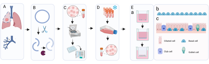
Figure 1: Schematic overview of the isolation, expansion, and culture procedure of primary bronchial epithelial cells. (A) Lung tissue is obtained during cancer resection surgery and the pathologist excises bronchial ring tissue that is macroscopically normal and tumor-free. (B) The bronchial ring is cleaned and exposed to enzymatic treatment to detach and dissociate the cell layer. (C) The retrieved cell suspension is washed and the cells are distributed in wells of a 6-well plate for expansion. (D) Upon sufficient expansion of the isolated cells in c-KSFM with Primocin, cell layers are dissociated by trypsinization and cells are resuspended in freezing medium for cryopreservation. When needed, cryopreserved cells are thawed and again expanded using c-KSFM with penicillin/streptomycin in cell culture flasks. After expansion, they are seeded in cBD medium on cell culture inserts; (Ea) ALI-PBEC culture takes place in two main stages: the submerged stage in cBD medium supplemented with 1 nM EC 23 until the cells reach full confluence, followed by removal of apical medium and culture at the ALI to allow differentiation; in this ALI phase, the cells are cultured in cBD medium supplemented with 50 nM EC 23. (Eb) Graphical representation of basal cells that cover the insert during submerged culture. (Ec) Graphical representation of the differentiated epithelial cell layer obtained following culture at the ALI in the presence of increased concentrations of EC 23. Please click here to view a larger version of this figure.
Results
Loading...
$$\rightleftharpoonup{xx}$$
$$\longleftharp{xx}$$,
$$\longrightharp{xx}$$,
Expansion using submerged culture
Using the method presented here, an average of eight cryovials with 400,000 cells/cryovial can be obtained from one 6-well plate for long-term storage in liquid nitrogen (Figure 2A). To achieve this, isolated PBECs are cultured in 6-well plates for a minimum of 7 days and a maximum of 14 days (Figure 2B) in the presence of Primocin to exclude microbial (especially mycoplasma) contamination. Figure 2A,B provides insight into the cell numbers obtained and required culture time among different isolations from various donors. Before harvesting the cells by trypsinization for storage in liquid nitrogen, the confluency must be over 80%. If this is not achieved within 14 days, the cells should not be cryopreserved. Importantly, when harvesting for storage and passaging, the confluency of the cell layer should not exceed ~95% (Figure 2C). After storage in liquid nitrogen, the cells can be thawed and cultured for expansion until sufficient cell numbers are obtained for ALI cultures. The medium used for expansion of the cells at this stage is c-KSFM, similar as during the initial culture following harvest from the bronchial ring18. There is however no need for Primocin at this stage, because the risk for additional microbial contamination originating from the lung tissue is absent, and therefore Primocin can be changed for penicillin/streptomycin. This medium favors epithelial cells over fibroblasts and therefore prevents possible overgrowth of the culture by faster proliferating fibroblasts19,20,21. Using c-KSFM medium, the cells are spread out in the flask and do not connect to each other, which is markedly different from the morphology of cultured cells submerged at this stage in cBD medium (Figure 2D,E). After 5 or 6 days of culturing the thawed cells in a T75 flask, the cell layer should be 80%-95% confluent, which translates to approximately 3 x 10⁶ cells in total (Figure 2F). From this, approximately 75 inserts (12-well plate size) can be generated for ALI culture.
The method for isolation and culture described in this contribution can also be adapted for use with bronchial biopsies or bronchial brushes as starting material.
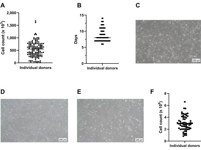
Figure 2: Basal cell expansion before and after cryopreservation. Cells were isolated according to the described protocol and cultured using c-KSFM. The number of cells generated per donor was monitored, the live cell counts were performed using an automated cell counter (A) when harvesting the passage 0 (P0) cells from the 6-well plates (step 2 of the protocol), n = 123 donors, the cell count was presented as the number of cells harvested per well; each dot represents one donor and the median is indicated by a horizontal bar. (B) As part of quality control, the time the P0 cells required to reach 80% to 90% confluency in the 6-well plates was monitored and shown as days after starting the submerged culture in c-KSFM (n = 127 different donors). Each individual donor is indicated by a dot, all dots belonging to 1 day merge into a line; the wider the line the more donors it stands for; the median number of days the cells are in culture is indicated by a thinner horizontal bar. (C) Representative brightfield image of P0 cells grown submerged in c-KSFM at the moment the cells were harvested for long-term cryopreservation. (D) Representative brightfield image of P1 cells grown submerged in c-KSFM at the moment the cells were harvested and transferred to inserts and (E) P1 cells grown submerged in cBD medium. (F) The number of live cells generated per donor was monitored using an automated cell counter when harvesting P1 cells from the T75 flask (section 4 of the protocol), n = 63 different donors; the cell count is presented as the number of cells per T75 flask, each donor is indicated by a dot, and the median cell count is indicated by a horizontal bar. Please click here to view a larger version of this figure.
Air-liquid interface (ALI) culture
At 7 days after starting the ALI culture, the electrical resistance of the cell layer is measured and should be more than 300 Ω (Figure 3A); if this is not achieved, the culture is regarded as failed because of a possible lack of tight junction formation. It is recommended to exclude the possibility of getting low TEER values caused by damage to the epithelial layer in individual inserts resulting from, for instance ,damage to the cell layer during washing and aspiration. This can be checked by visual microscopic inspection of the culture inserts. In our experience, the inter-donor variability in electrical resistance can be significant (Figure 3B), which is also reported in the literature14, and as observed is also markedly influenced by the origin of Dulbecco's modified eagle medium (DMEM) used (Figure 3C).
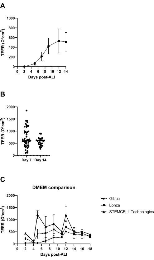
Figure 3: Trans-epithelial electrical resistance as a quality control of ALI-PBEC cultures. PBECs were isolated and expanded, and well-differentiated ALI-PBEC cultures were established. At several time points during culture, electrical resistance was measured, and subsequently the TEER was calculated (Ω·cm2). (A) Electrical resistance was measured over the course of 14 days post-ALI. n = 4 different donors. The data is depicted as the mean value ± standard deviation (SD). (B) As part of ALI-PBEC cell culture quality control, electrical resistance was measured at day 7 (n = 50) and day 14 post-ALI (n = 25); each dot represents one donor and the median TEER (Ω·cm2) is indicated by a horizontal bar. Data were tested for significance using a nonparametric Mann-Whitney test, and no significant difference was found. (C) Media from three different DMEM suppliers were tested to assess the influence on TEER formation. n = 4 different donors; mean values are depicted ± SD. Please click here to view a larger version of this figure.
While establishing a well-differentiated ALI-PBEC culture, from the start of air exposure, the concentration of RA increases22. This way, cells switch from proliferation to mucociliary differentiation, which is visible as early as 9 days (donor-dependent) after air exposure using brightfield microscopy. Movement of the first cilia is visible at this point, and somewhat earlier when based on gene expression of markers of differentiated luminal cells23 (Figure 4).
As described in the protocol, mucus production can also be observed when rinsing the apical surface during the medium change. RA is very sensitive to light, which leads to highly variable activity at the same stock concentration. For this reason, RA is replaced by the synthetic RA analogue EC 23 and used in the same concentration, with similar results as determined experimentally. For this reason and to avoid changing the procedure, the selected EC 23 concentration was kept equal to the RA concentration (i.e., 50 nM) previously used24,25 (Figure 5). Figure 5A depicts the TEER values achieved when using different concentrations of EC 23, showing a maximal TEER at 50 nM within this range of concentrations tested. The results shown in Figure 5B confirm that gene expression of markers for ciliated and goblet cells are similar when using 50 nM EC 23 or RA. EC 23 is also required during culture at the submerged stage (although at a much lower concentration), since omitting it at this submerged stage and only adding it at the ALI stage results in a culture that never reaches full confluency. The time needed to generate a well-differentiated ALI-PBEC culture with visible ciliary beating activity and mucus production is around 14 days, and most of the experiments are therefore initiated between 14-21 day ALI cultures (Figure 4). All the major different cell types (basal, ciliated, goblet, and club cells) are observed upon 14 days of ALI culture, although levels of expression are highly donor-dependent. This is demonstrated by assessing gene expression of TP63, FOXJ1, MUC5AC, and SCGB1A1 by RT-qPCR, or protein expression using antibodies directed against p63, α-tubulin, Muc5AC, and CC-16 by immune fluorescence (IF) staining, to detect markers for basal, ciliated, goblet, and club cells, respectively25,26. However, whereas 14-21 days may be regarded as a rule of thumb for most experiments, for select experiments, a longer duration of differentiation may be considered, as found for xenobiotic metabolism, SARS-CoV-2 infection, and the assessment of mucociliary clearance27,28,29.
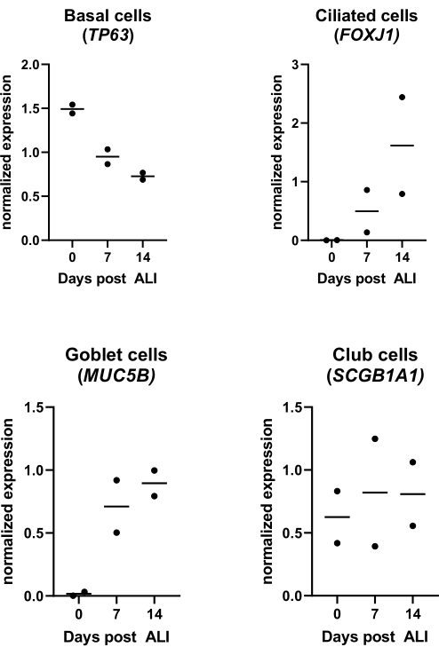
Figure 4: Air-liquid interface (ALI) culture. PBECs were isolated and expanded, and well-differentiated ALI-PBEC cultures were established. ALI-PBEC cultures were monitored over the course of 14 days post-ALI. Cell cultures were lysed for the isolation of RNA on days 0, 7, and 14 post-ALI. Data from two different donors are shown, each dot represents one individual donor monitored over time, and the median is indicated by a horizontal bar. Gene expression of basal, ciliated, goblet, and club cell markers (TP63, FOXJ1, MUC5B, and SCGB1A1, respectively) was measured by qPCR and normalized for RPL13A and ATP5B gene expression (see reference 23 for details). Please click here to view a larger version of this figure.
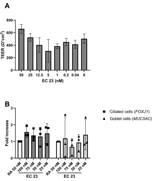
Figure 5: Comparison of retinoic acid (RA) and its synthetic analogue EC 23. PBECs were isolated and expanded, and well-differentiated ALI-PBEC cultures were established. Upon start of air exposure of the PBEC cultures, RA (50 nM) was replaced by various concentrations of EC 23. (A) Electrical resistance was measured at day 14 post-ALI and subsequently the TEER was calculated (Ω·cm2), n = 2 donors, the bars depict the mean value ± SD. (B) At day 14 post-ALI, cell cultures were lysed for RNA isolation and subsequent gene expression analysis of cell markers for respectively ciliated and goblet cells (FOXJ1, MUC5AC) using qPCR, and normalized for RPL13A (n = 3 donors). A fold increase is showed against ALI-PBEC cultured with 50 nM RA and depicted as the mean value ± SD. (See reference 23 for details) Please click here to view a larger version of this figure.
Over the past years, the performance of alternative products in the culture system has been examined, such as the media and culture plastics. There were various reasons for such evaluations, including changes in medium composition by the manufacturers, new media introduced, as well as a shortage of products during the COVID-19 pandemic (2020-2022). The observation was made that similar products from different suppliers result in differentiated epithelial cell cultures based on the assessment of markers of epithelial cell types, although the final cellular composition may vary substantially (Figure 6A), whereas differences in TEER were less pronounced (Figure 6B). On the other hand, medium from different suppliers did result in substantial differences in cellular composition; when using inserts from different brands, such differences were limited (Figure 6C). In particular, when using the airway epithelial cell culture medium PneumaCult from STEMCELL Technologies, a different morphology and more rapid formation of visible ciliary activity was observed. Besides these observations, a difference in TEER values and a difference in cellular composition of the ALI-PBEC compared to cBD medium was also noted (Figure 6D).
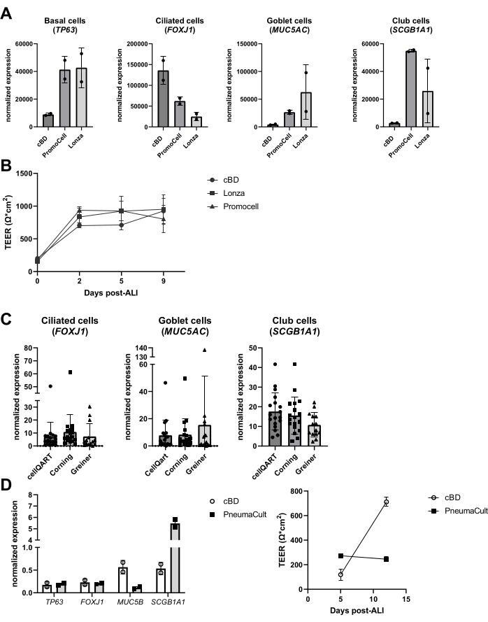
Figure 6: Comparing different suppliers of epithelial cell medium and cell culture inserts. PBEC were isolated, expanded and well differentiated ALI-PBEC cultures were established. (A) ALI-PBECs were cultured for 14 days, and then the cell layers were lysed for RNA isolation. Gene expression of basal, ciliated, goblet, and club cell markers (TP63, FOXJ1, MUC5AC, and SCGB1A1, respectively) was measured by qPCR and normalized for RPL13A and ATP5B. n = 2 donors; the bars depict the mean value ± SD. (B) Over the course of 9 days post-ALI, electrical resistance was measured and subsequently the TEER was calculated (Ω·cm2). n = 3 different donors; mean values are depicted ± SD. (C) ALI-PBECs were cultured for 14 days using cell culture inserts purchased from three different suppliers, and then the cell layers were lysed for RNA isolation. Gene expression of ciliated, goblet, and club cell markers (FOXJ1, MUC5AC, and SCGB1A1, respectively) was measured by qPCR and normalized for RPL13A. n = 18 different donors, the bars depict the mean value ± SD. Data were tested for significance using a one-way ANOVA nonparametric Kruskal-Wallis test and no significant difference was found. (D) ALI-PBECs were cultured either in cBD medium or PneumaCult medium (STEMMCELL technologies) for 14 days post-ALI, and then the cell layers were lysed for RNA isolation. Gene expression of basal, ciliated, goblet, and club cell markers (TP63, FOXJ1, MUC5B, and SCGB1A1, respectively) was measured by qPCR and normalized for RPL13A (the bars depict the mean value ± SD), and electrical resistance was measured at days 5 and 12 post-ALI and used to calculate the TEER (Ω·cm2). n = 2; the mean value is depicted ± SD (see reference 23 for details). Please click here to view a larger version of this figure.
Supplementary Table 1: Composition of the solutions and media used in the protocol. Please click here to download this File.
Discussion
Loading...
$$\rightleftharpoonup{xx}$$
$$\longleftharp{xx}$$,
$$\longrightharp{xx}$$,
The protocol presented here describes the isolation of human bronchial epithelial cells from resected lung tissue, a method for the optimal expansion of cells without loss of differentiation potential, a cryopreservation procedure, and a procedure for generating well-differentiated ALI-PBEC cultures. Furthermore, a description of quality control is provided, as well as instructions for monitoring and evaluation of the differentiated ALI-PBECs.
The protocol described starts with a macroscopically normal, tumor-free bronchial ring that is resected from a lung lobe from patients undergoing surgery related to their lung cancer diagnosis. It therefore needs to be noted that these rings strictly cannot be regarded as healthy tissue, which may therefore affect cell culture characteristics. Alternative sources for obtaining bronchial epithelial cells include using bronchial biopsies, bronchial brushings, or tissue from a transplant donor or recipient lungs. Regardless of the source, when using lung tissue, a risk of microbial contamination should be considered, and therefore antibiotics are used in the different culture media to reduce the risk of microbial contamination of the cell culture. In particular, mycoplasma is a high and common risk in cell culture, because of its wide variety of effects on cell culture, resistance to antibiotics commonly used in cell culture, and the fact that mycoplasma contamination can only be confirmed by mycoplasma detection assays. Therefore, in the initial stage of cell culture following the isolation of cells from lung tissue, the broad-spectrum antimicrobial formulation Primocin is used, and during the culture process, randomly selected samples are tested for the presence of mycoplasma.
The isolation procedure starting with a bronchial ring provides sufficient starting material to allow the degree of expansion of these primary cells needed to start cultures at the ALI without compromising differentiation capacity. However, starting the expansion of the isolated epithelial cells with a limited number of cells may pose issues with obtaining a sufficient number of inserts with enough cells that can be seeded for ALI culture. Extended culture and repeated passaging of primary cells may result in replicative senescence. Various solutions have been proposed to overcome this limitation. Horani et al. showed that the Rho kinase inhibitor (ROCK) Y-27632 increased the proliferation of basal cells30, Mou et al. used dual Smad inhibition to expand basal stem cells while maintaining the characteristics of the differentiated epithelial cell layer31, and Sachs et al. have developed an airway organoid system that can be used to expand airway epithelial cells and maintain their differentiation potential over the course of multiple passages32. The latter method was also used to expand cells from sources with very low cell numbers, such as tracheal aspirates (TAs) from preterm infants (<28 week gestation age) and bronchoalveolar lavage (BAL) fluid, before transfer to the ALI culture as described here33. It was found that cells isolated from BAL and TAs showed a differentiation capacity that was similar to cells generated from bronchial tissue, although differences were observed when the differentiation was skewed toward more ciliated or more goblet cell-containing cultures using Notch signalling inhibition or the Th2 cytokine IL-1333. It is therefore recommended that if ALI-PBECs are cultured from a starting material with low epithelial cell numbers using similar approaches, to always check the cultures for the basic quality criteria, as discussed in section 6 of the protocol. Importantly, the use of feeder cells may also help in obtaining larger cell numbers, which is essential in a setting for transplantable scaffold engineering where time and cell number are essential. This is illustrated by a study in which autologous epithelial cells were cultured from biopsies derived from a patient with tracheal disease and cells were rapidly expanded in the presence of a murine embryonic feeder layer (mitotically inactivated 3T3-J2 fibroblasts) and the above-mentioned inhibitor of the Rho/ROCK pathway (Y-27632)34. The resulting cell culture was found to be useful for repopulation of tracheal scaffolds, and thus this could be viewed as a suitable protocol for a transplant model.
When using the protocol described in this contribution, but also when using other culture protocols, inevitably a selection bias is introduced. It is important to realize that differences in protocol details, such as the origin of cells used to initiate cultures, medium composition, and other protocol details, can lead to changes in cellular composition of the cultures and thereby changes in the response of the ALI culture33,35. In addition, differences in cell properties have also been observed when comparing different media for differentiating the airway cells10,11. When comparing PneumaCult and cBD medium, differences were observed in goblet cell and club cell mRNA markers, TEER values, and cell layer thickness. Based on these observations, despite the lack of statistical underpinning, due to the low number of donors used, the medium composition is unknown to customers, and higher costs of the PneumaCult medium, the decision was made in our laboratory to use cBD medium.
As discussed, cells can be initially expanded using organoid culture and subsequently transferred to the 2D ALI insert system. This is important, since airway epithelial organoids are not suitable for exposure to airborne substances, whereas use of the ALI 2D system allows evaluation of the impact of airborne substances such as cigarette smoke23,36 on cultured airway epithelial cells. A different approach for establishing ALI airway epithelial cell cultures is to generate airway epithelial cells by the differentiation of human pluripotent stem cells (hiPSCs)37. In such protocols, at the final stage of the differentiation protocol after differentiation to proximal airway progenitors, cells can be differentiated by culture to the ALI using procedures similar to the ones described here.
In the current protocol, cBD medium is used for culture at the ALI. cBD medium is a serum-free medium that is prepared by adding a mix of different supplements, inspired by Fulcher et al.38 as well as other studies. The supplement solution contains 52 µg/mL bovine pituitary extract (BPE), 0.5 µg/mL hydrocortisone, 0.5 ng/mL human EGF, 0.5 µg/mL epinephrine, 10 µg/mL transferrin, 5 µg/mL insulin, 6.5 ng/mL triiodothyronine, and 0.1 ng/mL RA39. Since BPE is a tissue extract and is subjected to batch wise variation, the medium cannot be considered as a fully defined medium, nor is it animal-free. Cell culture medium that is fully defined is preferred to minimize batch to batch differences. In view of the transition into animal-free research, it is important that efforts are made for producing defined media that do not contain animal products and that are affordable for the scientific community.
Various experimental setups can be used based on the ALI model, depending on the research question. For instance, to investigate the impact of compounds that may influence the differentiation process, this can be addressed by adding the compounds to the culture during the different stages of submerged culture, during differentiation, or at the well-differentiated stage. The cellular composition of the ALI-PBEC culture can be influenced by adding specific compounds; for instance, differentiating ALI-PBECs in the presence of IL-13 generates a culture with more goblet cells and fewer ciliated cells, while treatment with the γ-secretase inhibitor DAPT (used to block Notch signaling) during differentiation results in a culture with more ciliated cells at the expense of goblet cells23,40,41,42.
Furthermore, agents to stimulate cells or block certain processes can either be applied to the basal compartment or (in a very small volume) to the apical compartment of the culture. Cells can also be exposed to airborne substances from the apical side. Such exposure designs have been used to study the effect of diesel exhaust or whole cigarette smoke on PBECs23,43,44. The medium can be harvested every time the medium is changed to monitor secreted proteins at the basal side; the same applies for the apical side of the cells that is washed with PBS while refreshing the basal medium. The so-called apical wash is harvested and optional dithioerythritol (DTE) is added to dissociate the mucus that is produced by the goblet cells more efficiently. Cell lysates can be obtained for isolation of total protein, RNA, and chromosomal and mitochondrial DNA. The cells can be further studied using antibodies for specific markers, by cutting the polyethylene terephthalate (PET) membrane from the plastic insert and further cutting this membrane into smaller pieces for multiple immunofluorescence stainings45. Furthermore, flow cytometry or FACS can also be used following trypsinization of the cells in the inserts. During the ALI stage, development of the cellular barrier can be monitored by measuring the electrical resistance and subsequently calculating the TEER, where the electrical resistance is inversely proportional to the surface area of the membrane insert. The calculation is based on Ohm's law using the following formula: , wherein Rm is the measured electrical resistance, Rb is the baseline electrical resistance of an insert without coating and cells, and SA is the surface area of the membrane of the insert. Measuring the electrical resistance using EVOM2 and STX/chopstick electrodes is straightforward but highly dependent on handling procedures when introducing it into the well. Also, the shape of the electrode has been suggested to affect the measurement of the barrier function of the relatively large surface area17.
Further improvement in the ALI cell culture system, aimed at increasing accurate tissue representation, includes coculture of additional cell type,s such as leukocytes, fibroblasts or endothelial cells46,47,48. It has been observed that that co-culture of ALI-PBEC with granulocyte-macrophage colony-stimulating factor (GM-CSF) or M-CSF-differentiated macrophages markedly affects innate epithelial responses and repair48. It is important to note that in such coculture models, medium compatibility can be an issue. Since the medium used for the airway epithelial cell culture is developed specifically for PBECs and may not be optimally suited for other cell types, optimization is necessary. Another type of advancement seen in the field of airway biology for which isolated PBECs can be used is the use of Organs-on-Chips (OoC) technology49,50. Using this technology, the influence of the mechanical forces of breathing and blood flow, such as stretch, air, and medium flow, can be studied 29.
Inter-donor variability can be significant when using PBECs from various donors, and therefore it is important to consider using cells from several donors to account for this variability in epithelial cell culture studies. Since the culture of ALI-PBECs is time-consuming and associated with considerable costs, the option to establish ALI-PBEC cultures by mixing cells from different donors in one cell culture insert is examined. This way, pilot experiments can be readily performed using primary cells, before analyzing the responses of cultures derived from various individual donors . In addition, donors with different characteristics (e.g., different age category or gender) can be grouped for explorative studies. When using donor mixes, it is important to make sure that equal cell numbers of different donors are present, to prevent the possibility that one donor dominates the outcomes as a result of a higher proliferation rate. Therefore, cells from individual donors are expanded separately and seeded at a higher density in the insert compared to seeding cells from an individual donor, to minimize proliferation in the insert before transition to the ALI. Responses of donor mixes and corresponding individual donors were compared by studying the infection kinetics of SARS-CoV-2. Using RT-qPCR and immunofluorescence staining, it was observed that the donor mix provided a good representation of the various individual donors, by showing similar numbers of virus particles produced and similar numbers of infected cells28.
To become an acceptable alternative for animal models, gene editing of cultured bronchial epithelial cells should be feasible51. RNA interference technology by using small interfering RNAs (siRNAs) in ALI-PBECs is examined, however since the cells need to be transfected with siRNA during the submerged phase of the culture, knockdown is not sufficiently maintained during ALI culture because of the long culture duration, unless siRNA transfection is frequently repeated during culture52. Nevertheless, siRNAs can be successfully used for modifying gene expression in submerged basal cells. Others have successfully used CRISPR/Cas9 technology to achieve gene editing in primary ALI airway epithelial cell cultures with ribonucleoprotein (RNP) delivery 53. When using such techniques, it is essential that the cells maintain their full differentiation capacity. Because primary airway cell cultures cannot be passaged indefinitely, clonal expansion of the gene edited cells is not easy and the addition of medium to select transfected cells is cumbersome. Therefore, it is difficult to achieve the desirable knockdown in all the cultured cells. An alternative to generate knockout clones is the use of knock-out strategies in hiPSCs54 and the use of these cells to generate airway epithelial cells. Another, albeit suboptimal, alternative is establishing an immortalized PBEC line in order to clonally expand gene-edited cells55.
The protocol presented here is one way of generating a well-differentiated pseudostratified ALI-PBEC, but other protocols have also been found to establish such a culture, with smaller and bigger differences in comparison to the presented protocol. In our opinion, across laboratory validation of culture methods and stringent quality control are essential for the ALI-PBEC system and similar culture systems of airway epithelial cells, to become a valid alternative for animal experiments.
Disclosures
Loading...
$$\rightleftharpoonup{xx}$$
$$\longleftharp{xx}$$,
$$\longrightharp{xx}$$,
The authors declare that they have no relevant conflicts of interest.
Acknowledgements
Loading...
$$\rightleftharpoonup{xx}$$
$$\longleftharp{xx}$$,
$$\longrightharp{xx}$$,
The studies using the model described in this contribution have been supported by a variety of fundings organisations, including the Lung Foundation Netherlands, the Netherlands Organization for Health Research and Development (ZonMw, COVID-19 MKMD grant), the Dutch Society for the Replacement of Animal Testing (Stichting Proefdiervrij, grant #114025007), as well as research grants from companies like Boehringer Ingelheim and Galapagos. Figure 1 was created with BioRender.com.
Materials
| Name | Company | Catalog Number | Comments |
|---|---|---|---|
| 1,000 ohm test resistor | World Precision Instruments | N/A | Used to calibrate the EVOM2 Epithelial Voltohmmeter |
| 4-[2-(5,6,7,8-Tetrahydro-5,5,8,8-tetramethyl-2-naphthalenyl)ethynyl)-benzoic acid (EC 23) | Tocris | 4011 | Used in cBD medium |
| 6-well Clear TC-treated Multiple Well Plates | Corning | 3506 | Used in the first step to grow the cells isolated form the bronchial ring |
| Airway Epithelial Cell Growth Medium Kit | PromoCell | C-21160 | Used to compare to cBD medium |
| Bead Bath 20 Liter | Lab Armor | 74220-720 | Used to pre-warm cell culture solutions |
| BEGM Bronchial Epithelial Cell Growth Medium BulletKit | LONZA | CC-3170 | Used to compare to cBD medium |
| Bovine albumin fraction V (BSA) | Thermo Fisher Scientific | 15260037 | Used in coating solution |
| Bovine pituitary extract (BPE) | Thermo Fisher Scientific | 37000-015 | Used in c-KSFM |
| Bronchial epithelial cell growth supplement (BEpiCGS) | ScienCell Research Laboratories | 3262 | Used in cBD medium |
| Bronchial epithelial cell medium-basal (BEpiCM-b) | ScienCell Research Laboratories | SCC3211-b | Used in cBD medium |
| Cell culture inserts; 12 mm Transwell with 0.4 µm pore polyester membrane insert | Corning | 3460 | Cell culture inserts used in the protocol |
| Cell culture inserts; 12-well inserts, 0.4 µm PET clear | CellQART made by SABEU | 9310412 | Cell culture inserts used to compare with Corning cell culture inserts |
| Cell culture inserts; 12-well ThinCert Tissue culture Inserts | Greiner Bio-One | 82050-032 | Cell culture inserts used to compare with Corning cell culture inserts |
| CELLSTAR flask, TC, PS, 250 ml, 75 cm2 | Greiner Bio-One | 658170 | Used to expand the number cells |
| CFX Maestro 1.0 | Bio-Rad | N/A | Software program for analyzing qPCR data generated with the CFX384 System |
| CFX384 Touch Real-Time PCR Detection System | Bio-Rad | 1855484 | qPCR detection system |
| Chopstick electrode set | World Precision Instruments | STX2 | Used to measure electrical resistance in ALI-PBEC |
| CO2-Incubator | PHCbi | MCO-170AICUV-PE | Cell culture incubator used for mycplasma free cell cultures |
| CO2-Incubator | Hereaus | Heracell 150 | Cell culture incubator used for possibly mycplasma infected cell cultures |
| Coolcell Container | Corning | 432006 | Used to cryopreserve cells at -80 °C before transfer to liquid N2 |
| Countess 3 Automated cell counter | Thermo Fisher Scientific | AMQAX2000 | Used to count cells and determine the cell concentration |
| Cryovials | Nalgene | 479-3224 | Used to cryopreserve cells in |
| D-Glucose | Avantor VWR BDH CHEMICALS | 101174Y | Used in soft trypsin |
| Dimethyl sulfoxide (DMSO) | Avantor VWR | 0231 | Used in cell freeze medium |
| dNTP (10 mM) | Promega | U1515 | Used in the synthesis of cDNA |
| Dulbecco's Modified Eagle's Medium (DMEM) + 4500 mg/l D-Glucose | STEMCELL Technologies | 36250 | Used in cBD medium |
| Dulbecco's Modified Eagle's Medium (DMEM) 4.5 g/l glucose with l-glutamine | LONZA | LOBE12-604F | Used in cBD medium to compare with DMEM from other manufacturers |
| Dulbecco's Modified Eagle's Medium (DMEM), high glucose, pyruvate | Thermo Fisher Scientific | 41966029 | Used in cBD medium to compare with DMEM from other manufacturers |
| Epidermal growth factor (EGF) | Thermo Fisher Scientific | 37000-015 | Used in c-KSFM |
| Ethylenediaminetetraacetic acid (EDTA) | Avantor VWR BDH CHEMICALS | 443885J | Used in soft trypsin |
| EVOM2 Epithelial Voltohmmeter | World Precision Instruments | 91799 | Used with the chopstick electrode set to measure electrical resistance in ALI-PBEC |
| Fibronectin solution, Human | PromoCell | C-43060 | Used in coating solution |
| Glutamax | Thermo Fisher Scientific | 35050038 | Used in cBD medium |
| Hanks balanced salt solution (HBSS) | ScienCell Research Laboratories | SCC0313 | Used to dissolve protease XIV |
| IQ SYBR Green Super mix | Bio-Rad | 170887 | qPCR reagent |
| Isoproterenol hydrochloride, (-)- | Sigma-Aldrich | I-6504 | Used in c-KSFM |
| Keratinocyte-SFM (KSFM) | Thermo Fisher Scientific | 17005-034 | Used in c-KSFM |
| Maxwell RSC Instrument | Promega | AS4500 | Automated RNA isolation system |
| Maxwell RSC simplyRNA Tissue Kit | Promega | AS1340 | Used to isolate total RNA with the Maxwell RSC Instrument |
| M-MLV Reverse transcriptase | Promega | M5301 | Used in the synthesis of cDNA |
| M-MLV Reverse transcriptase 5X reaction buffer | Promega | M531A | Used in the synthesis of cDNA |
| MycoStrip | InvivoGen | rep-mys-10 | Used to detect the presence of mycoplasma in cell culture samples |
| N-2-hydroxyethylpiperazine-N-2-ethane sulfonic acid (HEPES) | Thermo Fisher Scientific | 15630056 | Used in cBD medium |
| Oligo(dT)15 | Qiagen | 79237 | Used in the synthesis of cDNA |
| Penicillin/Streptomycin solution (Pen/Strep) | ScienCell Research Laboratories | SCC0513 | Used as antibiotic in c-KSFM and cBD medium |
| Phosphate buffered saline (PBS) | LUMC pharmacy | N/A | Used in different steps of the protocol |
| Pneumacult-ALI Medium | STEMCELL Technologies | 05002 | Used to grow cells in the differentiation stage to compare to cBD medium |
| Pneumacult-Ex Plus Medium | STEMCELL Technologies | 05040 | Used to grow cells in the submerged stage to compare to cBD medium |
| Primer, ATP5B, forward | Integrated DNA Technologies | N/A | Nucleotide sequence: TCACCCAGGCTGGTTCAGA |
| Primer, ATP5B, reverse | Integrated DNA Technologies | N/A | Nucleotide sequence: AGTGGCCAGGGTAGGCTGAT |
| Primer, FOXJ1, forward | Integrated DNA Technologies | N/A | Nucleotide sequence: GGAGGGGACGTAAATCCCTA |
| Primer, FOXJ1, reverse | Integrated DNA Technologies | N/A | Nucleotide sequence: TTGGTCCCAGTAGTTCCAGC |
| Primer, MUC5AC, forward | Integrated DNA Technologies | N/A | Nucleotide sequence: CCTTCGACGGACAGAGCTAC |
| Primer, MUC5AC, reverse | Integrated DNA Technologies | N/A | Nucleotide sequence: TCTCGGTGACAACACGAAAG |
| Primer, MUC5B, forward | Integrated DNA Technologies | N/A | Nucleotide sequence: GGGCTTTGACAAGAGAGT |
| Primer, MUC5B, reverse | Integrated DNA Technologies | N/A | Nucleotide sequence: AGGATGGTCGTGTTGATGCG |
| Primer, RPL13A, forward | Integrated DNA Technologies | N/A | Nucleotide sequence: AAGGTGGTGGTCGTACGCTGTG |
| Primer, RPL13A, reverse | Integrated DNA Technologies | N/A | Nucleotide sequence: CGGGAAGGGTTGGTGTTCATCC |
| Primer, SCGB1A1, forward | Integrated DNA Technologies | N/A | Nucleotide sequence: ACATGAGGGAGGCAGGGGCTC |
| Primer, SCGB1A1, reverse | Integrated DNA Technologies | N/A | Nucleotide sequence: ACTCAAAGCATGGCAGCGGCA |
| Primer, TP63, forward | Integrated DNA Technologies | N/A | Nucleotide sequence: CCACCTGGACGTATTCCACTG |
| Primer, TP63, reverse | Integrated DNA Technologies | N/A | Nucleotide sequence: TCGAATCAAATGACTAGGAGGGG |
| Primocin | InvivoGen | ant-pm-2 | Used as antimicrobial agent against bacteria, mycoplasma, and fungi in c-KSFM medium |
| Protease XIV | Sigma-Aldrich | P5147 | Used for the enzymatic treatment of the bronchial ring |
| RNAsin Recombinant Ribonuclease inhibitor | Promega | N2515 | Used in the synthesis of cDNA |
| Soybean trypsin inhibitor (SBTI) | Sigma-Aldrich | T9128 | Used to inhibit the action of soft trypsin |
| T100 Thermal Cycler | Bio-Rad | 1861096 | Used in the synthesis of cDNA |
| TissueSAFE plus | MILESTONE MEDICAL | N/A | Vacuum transfer system for biological specimens |
| Trypan blue solution | Thermo Fisher Scientific | 15250061 | Used to count live- and dead cells |
| Trypsin 1:250 | Thermo Fisher Scientific | 27250-018 | Used in soft trypsin |
| Type I collagen solution (PureCol) | Advanced BioMatrix | 5005-B | Used in coating solution |
| Universal container, PP, with PE screw cap | Avantor VWR | 216-2053 | Used in the protocol for the Protease XIV treatment of the bronchial ring |
References
Loading...
$$\rightleftharpoonup{xx}$$
$$\longleftharp{xx}$$,
$$\longrightharp{xx}$$,
- Aghapour, M., et al. Role of air pollutants in airway epithelial barrier dysfunction in asthma and COPD. European Respiratory Review. 31 (163), 210112(2022).
- de Waal, A. M., Hiemstra, P. S., Ottenhoff, T. H., Joosten, S. A., vander Does, A. M. Lung epithelial cells interact with immune cells and bacteria to shape the microenvironment in tuberculosis. Thorax. 77 (4), 408-416 (2022).
- Duchesne, M., Okoye, I., Lacy, P. Epithelial cell alarmin cytokines: Frontline mediators of the asthma inflammatory response. Frontiers in Immunology. 13, 975914(2022).
- Hewitt, R. J., Lloyd, C. M. Regulation of immune responses by the airway epithelial cell landscape. Nature Reviews Immunology. 21 (6), 347-362 (2021).
- Ruysseveldt, E., Martens, K., Steelant, B. Airway basal cells, protectors of epithelial walls in health and respiratory diseases. Frontiers in Allergy. 2, 787128(2021).
- Alysandratos, K. D., Herriges, M. J., Kotton, D. N. Epithelial stem and progenitor cells in lung repair and regeneration. Annual Review of Physiology. 83, 529-550 (2021).
- Hammad, H., Lambrecht, B. N. Barrier epithelial cells and the control of type 2 immunity. Immunity. 43 (1), 29-40 (2015).
- Hynds, R. E., Bonfanti, P., Janes, S. M. Regenerating human epithelia with cultured stem cells: feeder cells, organoids and beyond. EMBO Molecular Medicine. 10 (2), 139-150 (2018).
- Hiemstra, P. S., Tetley, T. D., Janes, S. M. Airway and alveolar epithelial cells in culture. The European Respiratory Journal. 54 (5), 1900742(2019).
- Saint-Criq, V., et al. Choice of differentiation media significantly impacts cell lineage and response to CFTR modulators in fully differentiated primary cultures of cystic fibrosis human airway epithelial cells. Cells. 9 (9), 2137(2020).
- Leung, C., Wadsworth, S. J., Yang, S. J., Dorscheid, D. R. Structural and functional variations in human bronchial epithelial cells cultured in air-liquid interface using different growth media. American Journal of Physiology. Lung Cellular and Molecular Physiology. 318 (5), L1063-L1073 (2020).
- Morgan, R., et al. A medium composition containing normal resting glucose that supports differentiation of primary human airway cells. Scientific Reports. 12 (1), 1540(2022).
- Ghosh, B., et al. Strong correlation between air-liquid interface cultures and in vivo transcriptomics of nasal brush biopsy. American Journal of Physiology. Lung Cellular and Molecular Physiology. 318 (5), L1056-L1062 (2020).
- Pezzulo, A. A., et al. The air-liquid interface and use of primary cell cultures are important to recapitulate the transcriptional profile of in vivo airway epithelia. American Journal of Physiology. Lung Cellular and Molecular Physiology. 300 (1), L25-L31 (2011).
- Dvorak, A., Tilley, A. E., Shaykhiev, R., Wang, R., Crystal, R. G. Do airway epithelium air-liquid cultures represent the in vivo airway epithelium transcriptome. American Journal of Respiratory Cell and Molecular Biology. 44 (4), 465-473 (2011).
- Legebeke, J., et al. Temporal whole-transcriptomic analysis of characterized in vitro and ex vivo primary nasal epithelia. Frontiers in Cell and Developmental Biology. 10, 907511(2022).
- Srinivasan, B., et al. TEER measurement techniques for in vitro barrier model systems. Journal of Laboratory Automation. 20 (2), 107-126 (2015).
- van Wetering, S., et al. Regulation of secretory leukocyte proteinase inhibitor (SLPI) production by human bronchial epithelial cells: increase of cell-associated SLPI by neutrophil elastase. Journal of Investigative Medicine. 48 (5), 359-366 (2000).
- Balk, S. D. Calcium as a regulator of the proliferation of normal, but not of transformed, chicken fibroblasts in a plasma-containing medium. Proceedings of the National Academy of Sciences. 68 (2), 271-275 (1971).
- Gail, M. H., Boone, C. W., Thompson, C. S. A calcium requirement for fibroblast motility and prolifertion. Experimental Cell Research. 79 (2), 386-390 (1973).
- Dulbecco, R., Elkington, J. Induction of growth in resting fibroblastic cell cultures by Ca. Proceedings of the National Academy of Sciences. 72 (4), 1584-1588 (1975).
- van Wetering, S., et al. Epithelial differentiation is a determinant in the production of eotaxin-2 and -3 by bronchial epithelial cells in response to IL-4 and IL-13. Molecular Immunology. 44 (5), 803-811 (2007).
- Amatngalim, G. D., et al. Aberrant epithelial differentiation by cigarette smoke dysregulates respiratory host defence. The European Respiratory Journal. 51 (4), 1701009(2018).
- Christie, V. B., et al. Retinoid supplementation of differentiating human neural progenitors and embryonic stem cells leads to enhanced neurogenesis in vitro. Journal of Neuroscience Methods. 193 (2), 239-245 (2010).
- Schrumpf, J. A., Ninaber, D. K., vander Does, A. M., Hiemstra, P. S. TGF-β1 impairs vitamin D-induced and constitutive airway epithelial host defense mechanisms. Journal of Innate Immunity. 12 (1), 74-89 (2020).
- Schrumpf, J. A., et al. Proinflammatory cytokines impair vitamin D-induced host defense in cultured airway epithelial cells. American Journal of Respiratory Cell and Molecular Biology. 56 (6), 749-761 (2017).
- Boei, J. J. W. A., et al. Xenobiotic metabolism in differentiated human bronchial epithelial cells. Archives of Toxicology. 91 (5), 2093-2105 (2017).
- Wang, Y., et al. Impact of human airway epithelial cellular composition on SARS-CoV-2 infection biology. bioRxiv. , (2021).
- Nawroth, J. C., et al. Breathing on Chip: Dynamic flow and stretch tune cellular composition and accelerate mucociliary maturation of airway epithelium in vitro. bioRxiv. , (2022).
- Horani, A., Nath, A., Wasserman, M. G., Huang, T., Brody, S. L. Rho-associated protein kinase inhibition enhances airway epithelial Basal-cell proliferation and lentivirus transduction. American Journal of Respiratory Cell and Molecular Biology. 49 (3), 341-347 (2013).
- Mou, H., et al. Dual SMAD signaling inhibition enables long-term expansion of diverse epithelial basal cells. Cell Stem Cell. 19 (2), 217-231 (2016).
- Sachs, N., et al. Long-term expanding human airway organoids for disease modeling. The EMBO Journal. 38 (4), 100300(2019).
- Eenjes, E., et al. Disease modeling following organoid-based expansion of airway epithelial cells. American Journal of Physiology. Lung Cellular and Molecular Physiology. 321 (4), L775-L786 (2021).
- Butler, C. R., et al. Rapid expansion of human epithelial stem cells suitable for airway tissue engineering. American Journal of Respiratory and Critical Care Medicine. 194 (2), 156-168 (2016).
- Amatngalim, G. D., et al. Antibacterial defense of human airway epithelial cells from chronic obstructive pulmonary disease patients induced by acute exposure to nontypeable Haemophilus influenzae: modulation by cigarette smoke. Journal of Innate Immunity. 9 (4), 359-374 (2017).
- Plebani, R., et al. 3D lung tissue models for studies on SARS-CoV-2 pathophysiology and therapeutics. International Journal of Molecular Sciences. 23 (17), 10071(2022).
- Wong, A. P., et al. Directed differentiation of human pluripotent stem cells into mature airway epithelia expressing functional CFTR protein. Nature Biotechnology. 30 (9), 876-882 (2012).
- Fulcher, M. L., Gabriel, S., Burns, K. A., Yankaskas, J. R., Randell, S. H. Well-differentiated human airway epithelial cell cultures. Methods in Molecular Biology. 107, 183-206 (2005).
- Cao, J., Wong, C. K., Yin, Y., Lam, C. W. K. Activation of human bronchial epithelial cells by inflammatory cytokines IL-27 and TNF-alpha: implications for immunopathophysiology of airway inflammation. The Journal of Cellular Physiology. 223 (3), 788-797 (2010).
- Tsao, P. N., et al. Notch signaling controls the balance of ciliated and secretory cell fates in developing airways. Development. 136 (13), 2297-2307 (2009).
- Laoukili, J., et al. IL-13 alters mucociliary differentiation and ciliary beating of human respiratory epithelial cells. The Journal of Clinical Investigation. 108 (12), 1817-1824 (2001).
- Mertens, T. C. J., et al. Cigarette smoke differentially affects IL-13-induced gene expression in human airway epithelial cells. Physiological Reports. 5 (13), e13347(2017).
- Zarcone, M. C., et al. Effect of diesel exhaust generated by a city bus engine on stress responses and innate immunity in primary bronchial epithelial cell cultures. Toxicology in Vitro. 48, 221-231 (2018).
- vander Does, A. M., et al. Early transcriptional responses of bronchial epithelial cells to whole cigarette smoke mirror those of in-vivo exposed human bronchial mucosa. Respiratory Research. 23 (1), 227(2022).
- Wang, Y., Ninaber, D. K., van Schadewijk, A., Hiemstra, P. S. Tiotropium and fluticasone inhibit rhinovirus-induced mucin production via multiple mechanisms in differentiated airway epithelial cells. Frontiers in Cellular and Infection Microbiology. 10, 278(2020).
- Ronaghan, N. J., et al. M1-like, but not M0- or M2-like, macrophages, reduce RSV infection of primary bronchial epithelial cells in a media-dependent fashion. PLoS One. 17 (10), 0276013(2022).
- Gindele, J. A., et al. Opposing effects of in vitro differentiated macrophages sub-type on epithelial wound healing. PLoS One. 12 (9), e0184386(2017).
- van Riet, S., et al. Modulation of airway epithelial innate immunity and wound repair by M(GM-CSF) and M(M-CSF) macrophages. Journal of Innate Immunity. 12 (5), 410-421 (2020).
- Huh, D., et al. Reconstituting organ-level lung functions on a chip. Science. 328 (5986), 1662-1668 (2010).
- Stucki, A. O., et al. A lung-on-a-chip array with an integrated bio-inspired respiration mechanism. Lab on a Chip. 15 (5), 1302-1310 (2015).
- Peters-Hall, J. R., et al. Long-term culture and cloning of primary human bronchial basal cells that maintain multipotent differentiation capacity and CFTR channel function. American Journal of Physiology. Lung Cellular and Molecular Physiology. 315 (2), L313-L327 (2018).
- Bartman, C. M., Stelzig, K. E., Linden, D. R., Prakash, Y. S., Chiarella, S. E. Passive siRNA transfection method for gene knockdown in air-liquid interface airway epithelial cell cultures. American Journal of Physiology. Lung Cellular and Molecular Physiology. 321 (1), L280-L286 (2021).
- Koh, K. D., et al. Efficient RNP-directed human gene targeting reveals SPDEF is required for IL-13-induced mucostasis. American Journal of Respiratory Cell and Molecular Biology. 62 (3), 373-381 (2020).
- Bhargava, N., et al. Development of an efficient single-cell cloning and expansion strategy for genome edited induced pluripotent stem cells. Molecular Biology Reports. 49 (8), 7887-7898 (2022).
- Angelopoulou, A., Papaspyropoulos, A., Papantonis, A., Gorgoulis, V. G. CRISPR-Cas9-mediated induction of large chromosomal inversions in human bronchial epithelial cells. STAR Protocols. 3 (2), 101257(2022).
Reprints and Permissions
Request permission to reuse the text or figures of this JoVE article
Request Permission