The in vivo experiments were done in accordance with the guidelines and protocol approved by the Institutional Animal Care and Use Committee of SingHealth (IACUC) and the Association for Research in Vision and Ophthalmology (ARVO) Statement for the use of animals in Ophthalmic and Vision Research. The pups were immunosuppressed from P17 (pre-transplantation) to P30 (post-transplantation) by feeding them drinking water containing cyclosporine (260 g/L).
1. Preparation of Day 32 hESC-derived photoreceptor progenitors after cryopreservation
2. Sub-retinal delivery of the hESCs in rd10 mice
The 10 µL glass syringe was assembled according to the manufacturer's instructions (Figure 1), and the blunt needle used to deliver the cell suspension/media is shown in Figure 1B. Different approaches for sub-retinal injection are illustrated in Figure 2. We describe the pars plana approach in this protocol (Figure 2C). The blunt needle mounted on a glass syringe was inserted through a sclerotomy wound and accessed the sub-retinal space across the globe. As shown in Figure 3A, the trajectory of the needle, penetration through the retina, and delivery of the cells were monitored directly under the microscope when performing the injection. Successful delivery of the cells/media was confirmed by observing a bleb at the injection site (Figure 3B). A successful bleb can be identified as a light whitish color resembling a water balloon. A failed delivery is observed by leakage of the cells/media into the vitreous space at the injection site and failure to form a bleb. The OCT scan was performed on the injected area, and the scan showed floating individualized hESCs in the cell-treated eye (Figure 4A), while the media-treated eye showed clear fluid without cells in the sub-retinal space (Figure 4B). The individualized hESCs are identified as hyperreflective materials distributed in the sub-retinal space (Figure 4A). The success rate of the injection was computed by noting the number of eyes with successful formation of the bleb. We included the applications that adopted this approach in our laboratory and the success rate of the injections (Table 1).
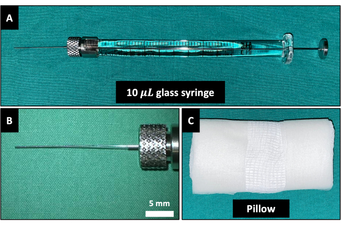
Figure 1: Instruments used during the injection. (A) The 10 µL glass syringe is mounted with a 33G blunt needle. (B) Zoom-in picture of the 33G blunt needle. (C) Pillow for the animal's head to rest on. Please click here to view a larger version of this figure.
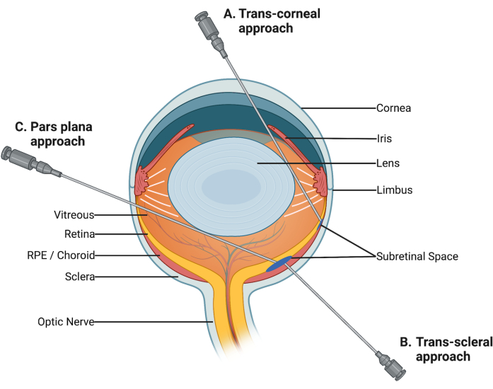
Figure 2: Different routes of sub-retinal injection. (A) Trans-corneal route: The injection needle passes through the cornea and the pupil to enter the sub-retinal space. (B) Trans-scleral route: The sub-retinal space is directly accessed through the sclera. (C) Pars plana route: The injection needle is inserted into the vitreous space via an incision at the limbus. The needle reaches the sub-retinal space by penetrating the retina. Please click here to view a larger version of this figure.
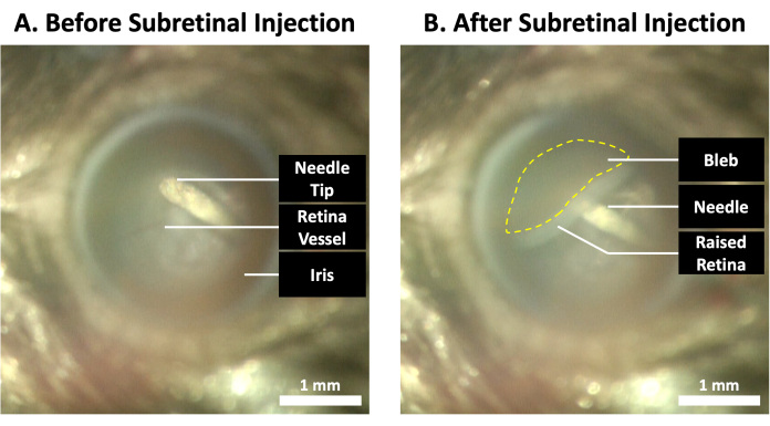
Figure 3: Fundus images of the eye during the sub-retinal injection. (A) Before the sub-retinal injection was performed, the tip of the needle could be seen in the vitreous space touching the retina, avoiding the major retinal blood vessels. (B) After the sub-retinal injection, a visible bleb was formed at the injection site (yellow dotted line). Please click here to view a larger version of this figure.
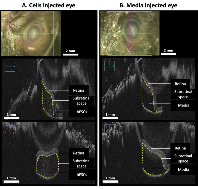
Figure 4: Intraoperative OCT scans of the injected eyes. The scans were done immediately after the injection. (A) hESCs treated eye: the top panel showed the location of the OCT scan (cyan and pink cross-sectional lines) on the eye; hESCs were observed in the treated eye in the sub-retinal space (yellow dotted line, middle and bottom panels). (B) Media-treated eye: the top panel showed the location of the OCT scan (cyan and pink cross-sectional lines) on the eye; clear fluid without cells was observed in the sub-retinal space in the media-injected eye (yellow dotted line, middle and bottom panels). Please click here to view a larger version of this figure.
Applications | Recipient Strain | Success Rate |
AAV | Rpe65rd12/J | 80% |
AAV | C57BL/6 | 95% |
hESC derived progenitor cells | Rd10-/- | 95% |
Table 1: The success rate of sub-retinal injection in different applications.
0.3% Tobramycin | Novartis | NDC 0078-0813-01 | Tobrex (3.5 g) |
0.3% Tobramycin and 0.1% Dexamethasone | Novartis | NDC 0078-0876-01 | Tobradex (3.5 g) |
0.5% Proparacaine hydrochloride | Alcon | NDC 0998-0016-15 | 0.5% Alcaine (15 mL) |
1 mL Tuberculin syringe | Turemo | SS01T2713 | |
1% Tropicamide | Alcon | NDC 0998-0355-15 | 1% Mydriacyl (15 mL) |
2.5% Phenylephrine hydrochloride | Alcon | NDC 0998-0342-05 | 2.5% Mydfrin (5 mL) |
24-well tissue culture plate | Costar | 3526 | |
30 G Disposable needle | Becton Dickinson (BD) | 305128 | |
33 G, 20 mm length blunt needles | Hamilton | 7803-05 | |
Automated Cell Counter | NanoEnTek | Model: Eve | |
B27 without Vitamin A | Life Technologies | 12587001 | 2%36 |
Buprenorphine | Ceva | Vetergesic vet (0.3 mg/mL) | |
CKI-7 | Sigma | C0742 | 5 µM36 |
Cyclosporine | Novartis | 260 g/L in drinking water | |
Day 32 hESC-derived photoreceptor progenitor cells | DUKE-NUS Medical School | Human embryonic stem cells are differentiated for 32 days. See protocol in Ref 36. | |
Gauze | Winner Industries Co. Ltd. | 1SNW475-4 | |
Glasgow Minimum Essential Medium | Gibco | 11710–035 | |
hESC cell line H1 | WiCell Research Institute | WA01 | |
Human brain-derived neurotrophic factor (BDNF) | Peprotech | 450-02-50 | 10 ng/mL36 |
Human ciliary neurotrophic factor (CNTF) | Prospec-Tany Technogene | CYT-272 | 10 ng/mL36 |
Ketamine hydrochloride (100 mg/mL) | Ceva Santé Animale | KETALAB03 | |
LN-521 | Biolamina | LN521-02 | 1 µg36 |
mFreSR | STEMCELL Technologies | 5854 | |
Microlitre glass syringe (10 mL) | Hamilton | 7653-01 | |
N-[N-(3,5-difluorophenacetyl-L-alanyl)]-S-phenylglycine t-butyl ester (DAPT) | Selleckchem | S2215 | 10 µM36 |
N-2 supplement | Life Technologies | A13707-01 | 1%36 |
Non-essential amino acids (NEAA) | Gibco | 11140–050 | 1x36 |
NutriStem XF Media | Satorius | 05-100-1A | |
Operating microscope | Zeiss | OPMI LUMERA 700 | With Built-in iOCT function |
PRDM (Photoreceptor differentiation medium, 50ml) | DUKE-NUS Medical School | See media composition36. Basal Medium, 10 µM DAPT, 10 ng/mL BDNF, 10 ng/mL CNTF, 0.5 µM Retinoic acid, 2% B27 and 1% N2. Basal Medium: 1x GMEM, 1 mM sodium pyruvate, 0.1 mM B-mercaptoethanol, 1x Non-essential amino acids (NEAA). | |
Pyruvate | Gibco | 11360–070 | 1 mM36 |
Rd10 mice | Jackson Laboratory | B6.CXB1-Pde6brd10/J mice | Gender: male/female, Age: P20 (injection), Weight: 3-6 g |
Retinoic acid | Tocris Bioscience | 0695/50 | 0.5 µM36 |
Round Cover Slip (12 mm) | Fisher Scientific | 12-545-80 | |
SB431542 | Sigma | S4317 | 0.5 µM36 |
Vidisic Gel (10 g) | Dr. Gerhard Mann | ||
Xylazine hydrochloride (20 mg/mL) | Troy Laboratories | LI0605 | |
β-mercaptoethanol | Life Technologies | 21985–023 | 0.1 mM36 |
Regeneration of photoreceptor cells using human pluripotent stem cells is a promising therapy for the treatment of both hereditary and aging retinal diseases at advanced stages. We have shown human recombinant retina-specific laminin isoform matrix is able to support the differentiation of human embryonic stem cells (hESCs) to photoreceptor progenitors. In addition, sub-retinal injection of these cells has also shown partial restoration in the rd10 rodent and rabbit models. Sub-retinal injection is known to be an established method that has been used to deliver pharmaceutical compounds to the photoreceptor cells and retinal pigmented epithelial (RPE) layer of the eye due to its proximity to the target space. It has also been used to deliver adeno-associated viral vectors into the sub-retinal space to treat retinal diseases. The sub-retinal delivery of pharmaceutical compounds and cells in the murine model is challenging due to the constraint in the size of the murine eyeball. This protocol describes the detailed procedure for the preparation of hESC-derived photoreceptor progenitor cells for injection and the sub-retinal delivery technique of these cells in genetic retinitis pigmentosa mutant, rd10 mice. This approach allows cell therapy to the targeted area, in particular the outer nuclear layer of the retina, where diseases leading to photoreceptor degeneration occur.