The identification of RNA and protein biomarkers from tears in mouse models holds great promise for early diagnostics in various diseases. This manuscript provides a comprehensive protocol for optimizing the efficacy and efficiency of mRNA and protein isolation from mouse tears.
The tear film is a highly dynamic biofluid capable of reflecting pathology-associated molecular changes, not only in the ocular surface but also in other tissues and organs. Molecular analysis of this biofluid offers a non-invasive way to diagnose or monitor diseases, assess medical treatment efficacy, and identify possible biomarkers. Due to the limited sample volume, collecting tear samples requires specific skills and appropriate tools to ensure high quality and maximum efficiency. Various tear sampling methodologies have been described in human studies. In this article, a comprehensive description of an optimized protocol is presented, specifically tailored for extracting tear-related protein information from experimental animal models, especially mice. This method includes the pharmacological stimulation of tear production in 2-month-old mice, followed by sample collection using Schirmer strips and the evaluation of the efficacy and efficiency of the protocol through standard procedures, SDS-PAGE, qPCR, and digital PCR (dPCR). This protocol can be easily adapted for the investigation of the tear protein signature in a variety of experimental paradigms. By establishing an affordable, standardized, and optimized tear sampling protocol for animal models, the aim was to bridge the gap between human and animal research, facilitating translational studies and accelerating advancements in the field of ocular and systemic disease research.
Tears are considered a plasma ultrafiltrate and have also been described as an intermediate fluid between plasma serum and cerebrospinal fluid due to a significant overlap in the biomolecules they share1. It has been reported that human tears contain proteins, tear lipids, metabolites, and electrolytes2. Recently, other biomolecules such as mRNAs, miRNAs, and extracellular vesicles have also been identified3,4,5,6,7.
In humans, basal tears are located in the tear film, which consists of three layers: the outer lipid layer, which maintains the tear surface smooth to allow us to see through it and prevent tear evaporation; the middle aqueous layer, which keeps the eye hydrated, repels bacteria, protects the cornea, and constitutes 90% of the tear film; and finally, the mucin layer, a family of high molecular weight proteins that are in contact with the cornea and allow the tear to adhere to the eye8. Tear distribution across the ocular surface begins with secretion from the lacrimal gland. This fluid is then guided through tear ducts to pass over the eye's surface and flow into drainage channels. Each blink allows tears to disperse evenly over the entire eye, keeping it moist9.
The tear film is a highly dynamic biofluid capable of reflecting molecular changes that occur not only on the ocular surface but also in other tissues and organs. The analysis of differential expression in this biofluid represents a promising approach for the discovery of biomarkers in human diseases10,11. The utilization of tear film as a source of biomarkers for early diagnosis in various pathologies has been greatly facilitated by the presence of non-invasive collection methods. The most common method for collecting tears in human and veterinary clinics involves membrane-based support (Schirmer's strip), which operates on the principle of capillary action, allowing the water in tears to travel along the length of a paper test strip or capillary tubes placed in the subject's lower conjunctival sac12,13,14. Despite the inherent limitation of obtaining a small sample volume through this method, the biochemical analysis of tear composition using various sensitive techniques has facilitated the identification of potential biomarker molecules11,15. Protocols for optimizing and evaluating the elution of tear proteins from Schirmer strips and capillaries in human patients are well documented16,17. However, complete descriptions of optimized protocols specifically designed to extract molecular information related to tears from experimental animal models are available but scarce. Existing methods, such as tear induction through direct stimulation of the lacrimal gland18, while allowing for the collection of larger volumes, are invasive and can cause discomfort to the animals. Non-invasive methods, such as collecting tears from the ocular surface, have been described as a way to isolate DNA and miRNAs19,21.
This protocol aims to establish a cost-effective and optimized method for collecting and processing tears in mice. The method prioritizes non-invasiveness while obtaining sufficient tear volumes suitable for molecular analysis through techniques like SDS-PAGE, qPCR, and dPCR. The extracted protein and mRNA content information can then be utilized to identify potential biomarkers in existing experimental models of disease.
All procedures described here were approved by the Animal Ethics Committee of Cinvestav (CICUAL, # 0354/23). The laboratory animals were treated and handled in strict accordance with the journal's animal use guidelines and according to the Association for Research in Vision and Ophthalmology (ARVO) Statement for the Use of Animals in Ophthalmic and Vision Research. Ocular health before and after the procedures was evaluated by assessing ocular discharges, swollen eyelids, ocular abnormalities, and behavior changes.
1. Animals and reagent preparation
2. Tear production stimulation and collection
3. Protein isolation
4. SDS-PAGE analysis
5. mRNA isolation for qPCR and digital PCR
The protocol described in this paper provides an easy and affordable method for obtaining molecular information from tear fluid using techniques commonly available in most molecular biology laboratories. Furthermore, the protocol can be scaled up by employing highly sensitive techniques such as ELISA for enzymatic activity detection.
After these procedures, the total protein yield was approximately 3-4 µg/µL. Coomassie-stained SDS page analysis of total protein extracts reveals patterns of protein expression in total retina and tears (Figure 2A).
Isolated RNA had a 260/280 UV absorption ratio between 1.9 and 2.0. The average yield was 134.8 ng/µL. qPCR was performed using primer sets for a housekeeping gene (glyceraldehyde 3-phosphate dehydrogenase, GAPDH)3 and a putative biomarker gene (DNA-methyl transferase 3a, DNMT3a)26. An undiluted cDNA sample 1:1 (150 ng) was used.
A melting curve analysis was performed, and Ct rates were calculated in the qPCR software. All the experiments were performed in duplicate. The analysis revealed Ct values between 20 and 24, which is a suitable range of starting amount of target mRNA in the sample (Figure 2B). However, the melt curve analysis in Figure 2C suggests the low presence of target mRNA. Nonetheless, the 2% agarose gel confirmed the presence of amplicons at the expected size (Figure 2D).
dPCR was performed using the same pair of primer sets for DNMT3a. The dilution of cDNA samples used were 1:1 (150 ng), 1:2 (75 ng), 1:4 (37.5 ng), 1:8 (18.75 ng). All experiments were performed in duplicate. dPCR was able to detect from 1.33 copies/µL in the undiluted sample, to 0.45 copies/µL in the 8-fold diluted sample (Figure 3A,3B). Thus, dPCR showed an increased sensitivity to the target mRNA present in tears compared to qPCR.
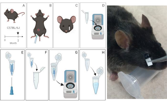
Figure 1: Schematic of the tear extraction protocol from mice. (A) Tear Collection. Three 2-month-old C57BL/6J mice were used. (B) Tear production was pharmacologically induced by intraperitoneal administration of pilocarpine. Photograph illustrating the placement of the Schirmer strip. (C) Tear Processing. Tears were then collected using Schirmer strips for 10 min and transferred into microcentrifuge tubes. (D) Tears collected in the Schirmer strips were centrifuged with sterile water and protease inhibitors or just sterile water. (E, F) These tubes were perforated at the bottom and subsequently transferred to a new sterilized 1.5 mL tube to facilitate fluid elusion. (G, H) The tears are collected in the 1.5 tube mL. (I) Representative photograph of the procedure to collect tears. Please click here to view a larger version of this figure.
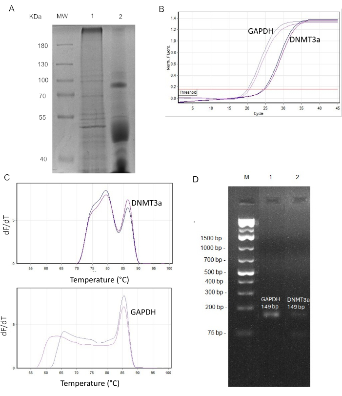
Figure 2: Downstream analysis of tear film for molecular applications. (A) SDS-PAGE of mouse tears and a retinal protein extract. MW: Molecular weight marker.; lane 1: Protein profiles of retina (30 µg); lane 2: protein profiles of tears (30 µg). (B) qPCR for GAPDH and DNMT3a in mice tears. Average CT and ΔCT are shown in Table 2. (C) Melting curve analysis for DNMT3a and GAPDH PCR products. The y-axis represents the rate of change of fluorescence in the amplification reaction (dF/dT), and the x-axis represents temperature in °C (D) Analysis of the PCR amplicon in 2% agarose gel electrophoresis containing 0.5 µg/mL ethidium bromide. Lane M: 1 kb Plus DNA Ladder. Lane 1,2: GAPDH and DNMT3a PCR amplicon, respectively. Please click here to view a larger version of this figure.
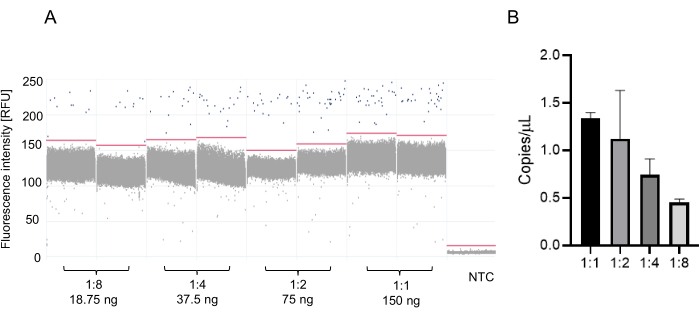
Figure 3: Digital PCR (dPCR) for DNMT3a expression in mice tears. (A) dPCR results are displayed as a 1D scatter plot showing in blue dots the DNMT3a transcript copies. Black dots are partitions without the DNMT3a transcript copies present. (B) A dilution series of cDNA was generated to determine the optimal concentration for quantifying DNMT3a transcript copies. Error bars represent standard deviation. Please click here to view a larger version of this figure.
Name of Material | Description | ||||
10% SDS Gel | For one separating gel: 3.8 mL distilled H2O; 2.6 mL Tris; pH 8.8; 1.5 M 3.4 mL acrylamide; 100 µL 10% SDS; 100 µL 10% APS; 4 µL TEMED. | ||||
For one collecting gel: 2.72 mL distilled H2O, 0.52 mL Tris; pH 6.8; 1.5 M 0.68 mL acrylamide, 40 µL 10% SDS, 40 µL 10% APS 4 µL TEMED. | |||||
Electrophoresis Buffer | 250 mM Tris base, 2 M Glycine, 1% SDS | ||||
Staining Solution | Dissolve the following reagents in 43 mL of water (store at room temperature in an amber bottle): Coomassie 0.25 g; acetic acid 7 mL; methanol 50 mL | ||||
Destaining solution | Dissolve the following reagents in 63 mL of water (store at room temperature): acetic acid 7 mL; ethanol 30 mL |
Table 1. Set up for SDS-Page.
Gene name | CT1 | CT2 | Average with SD | ΔCT |
GAPDH | 20.14 | 20.73 | 20.44±0.17 | |
DNMT3a | 24.59 | 24.97 | 24.78±0.07 | 4.35 |
Table 2. Ct values of DNMT3a and GAPDH in tears of mice.
Tear fluid is easily accessible, and the determination of biomarkers in tears can be employed as a successful complementary technique for the early diagnosis of various human diseases27. While the biochemical analysis of tear composition in experimental animal models complements this approach and promises significant progress in understanding the molecular basis of diseases, there is a scarcity of available data and protocols, which led us to develop one. The method described in this report is technically simple to perform and allows for the preliminary identification of the differential expression of different disease-associated candidate molecules.
Tear collection can be achieved through various methods such as capillary tubes and Schirmer strips16,17, swabs28and sponges29. The Schirmer strip method was preferred due to its ease of access, quantitative capabilities, adaptability during sample collection, cost, and simplicity in extracting tear components for laboratory processing. Additionally, this method is cost-effective, minimally invasive, and poses no risk of injury or damage to the cornea, thanks to the composition of the paper filter strip16,17. Critical steps of this protocol include the need for pharmacological stimulation of tear production using pilocarpine and the selection of an appropriate elution buffer.
Pilocarpine is a cholinergic agonist that binds to M3 muscarinic receptors and can cause pharmacological stimulation of exocrine glands, enhancing tear production, salivary secretion, and urination. Although studies in humans demonstrated that tear stimulation does not change the osmolarity of the collected fluid30, it clearly influences protein profiles31. The use of pilocarpine as a cholinergic pharmacological stimulator of exocrine glands and tear secretion enhancer has been successfully reported in rabbits and mice19,32,33, but the effect of stimulation on protein content has not been evaluated. To avoid this limitation and allow accurate comparison of tear profiles between experimental conditions, it is crucial to consider the specific collection settings. Similarly, the introduction of a rinsing step prior to tear collection could be beneficial to avoid prior contamination of the eye surface. In this protocol, a concentration of 30 mg/kg of pilocarpine was employed13. As the mice remained immobile and calm after the injection of this drug, the tears were collected without any anesthesia, making sample collection easy.
The choice of an appropriate elution buffer is closely related to the selected method for analyzing the biochemical composition of a tear sample and the recovery rate of biological material from the strip. For example, in a comparative study focusing on protein retention from Schirmer's strips in patients, it was established that a buffer comprising 100 mM ammonium bicarbonate along with 0.25% Nonidet P40 (NP40) yielded superior results for proteomics or multiplex ELISA analysis applications17. In our hands, the use of sterile water proved to be sufficient to visualize total protein extracts in SDS-PAGE and to detect the mRNA of the genes of interest by PCR.
To assess the presence of DNMT3a and GAPDH mRNA in mice tears, we compared the performance of qPCR and dPCR. The presence of primer dimers observed in the qPCR melting curve is attributed to the low abundance of the DNMT3a transcript. As demonstrated, dPCR has significantly greater sensitivity and efficacy in detecting the target mRNA in these samples in undiluted and diluted samples. This precise detection capability makes dPCR the preferred method for tear analysis in mice, offering researchers and clinicians a reliable tool for future studies and potential clinical applications.
The development of a standardized protocol for tear sample collection in animal models represents a significant advancement in research. This protocol not only facilitates the systematic and precise collection of tear samples but also opens a wide range of possibilities for their application in various areas of study. The ability to obtain tear samples from animal models allows for the investigation of a broad spectrum of diseases and conditions, as well as the exploration of new biomarkers and potential therapies. Furthermore, by establishing a standardized protocol, the reproducibility of results across different studies and laboratories is promoted, thereby contributing to the validity and reliability of research. Ultimately, this advancement in tear sample collection methodology in animal models has the potential to drive important discoveries and improve the understanding of the pathophysiology of various diseases, opening new avenues for diagnosis, treatment, and prevention.
The authors have nothing to disclose.
This work was supported by VELUX STIFTUNG [project 1852] to M.L. and postgraduate fellowship grants from CONAHCYT to M.B. (836810), E.J.M.C. (802436) and A.M.F (CVU 1317418). Sincere gratitude to all the members of the laboratory, Centro de Investigación sobre el Envejecimiento, and Departamento de Farmacobiología (Cinvestav) for their contributions to the stimulating discussions.
2-mercaptoethanol | Gibco | 1985023 | |
2x Laemmli buffer | Bio-Rad | 16-0737 | |
Acetic Acid | Quimica Meyer | 64-19-7 | |
Acrylamide | Sigma-Aldrich | A4058 | |
Bradford Reagent | Sigma-Aldrich | B6916 | |
Chloroform | Sigma-Aldrich | 1003045143 | |
Coomassie Blue R 250 | US Biological | 6104-59-2 | |
Ethanol | Quimica Rique | 64-17-5 | |
GeneRuler 1kb plus DNA Ladder | Thermofisher scientific | SM1331 | |
Glycerol | US Biological | G8145 | |
Glycine | SANTA CRUZ | SC- 29096 | |
Glycogen | Roche | 10901393001 | |
HCl | Quimica Rique | 7647-01-0 | |
Isopropyl alcohol | Quimica Rique | 67-63-0 | |
Methanol | Quimica Meyer | 67-56-1 | |
Micro tubes 1.5 ml | Axygen | MCT-150-C | |
Micro tubes 600 µl | Axygen | MCT-060-C | |
NaCl | Sigma-Aldrich | S3014 | |
PCR tubes & strips | Novasbio | PCR 0104 | |
Pilocarpine | Sigma-Aldrich | P6503-10g | |
Protease inhibitor | Roche | 11873580001 | |
QIAcuity EvaGreen PCR Kit (5mL) | Qiagen | 250112 | |
QIAcuity Nanoplate 26k 24-well (10) | Qiagen | 250001 | |
Real qPlus 2x Master Mix Green | Ampliqon | A323402 | |
RevertAid First Strad cDNA Synthesis Kit | Thermofisher scientific | K1622 | |
Schirmer's test strips | Laboratorio Santgar | SANT1553 | |
SDS | Sigma-Aldrich | L3771 | |
TEMED | Sigma aldrich | 102560430 | |
TRI reagent | Sigma-Aldrich | T9424-200ML | monophasic solution of phenol and guanidinium isothiocyanate |
Tris | US Biological | T8650 | |
Tris base | Chem Cruz | sc-3715A |
.