Note: this protocol suggests the use of potentially hazardous materials (lead and chloretone). Acquire, read, and follow MSDS for all potentially hazardous materials.
1. Animal Culture, Selection, and Preparation
- For animal culture and handling use planarian water (1X Montjuïc salts9) and plastic transfer pipettes.
- Sexual biotype Schmidtea mediterranea can be used when raised in the laboratory under normal culture conditions10. To produce asexual specimens of requisite size, S. mediterranea raised at room temperature under normal conditions9 should be fed at double to triple normal frequency (2-3 times per week) for one to two months prior to use. Alternatively, asexual animals fed at normal frequency may be kept at 10 degrees Celsius indefinitely in order to increase their average size.
- Select animals that are between 1 to 2 cm in length and wider than 2 mm then starve animals 3-7 days prior to use.
- If performing any pharmacological or radiological treatments on intended hosts, donors, or partially irradiated animals, perform treatment(s) at this point.
- If pharmacological treatments were performed that required feeding the animals, starve animals an additional 3 to 7 days prior to use.
2. Preparation of Solutions and Materials
- Prepare chloretone solution, a mild local anesthetic, by dissolving 0.1-0.2% w/v chloretone in planarian water and chilling the solution on ice.
- If performing partial irradiation only, proceed to step 2.7. For tissue transplantation continue on to step 2.3.
- Using a Bunsen burner, bend 0.75 mm interior diameter, used for cutting the graft tissue, and 0.7 mm exterior diameter, used for creating a hole in the host which will receive the graft, capillary tubes to a 90° angle at 1-2 cm from the end of each tube. To save materials, bend both ends of each capillary tube and break them in half to produce two tools. Take care not to flame the very ends of the tubes.
- Cut the following papers to the indicated sizes:
- Black filter paper (cut into rectangles approx. 2.5 cm x 1.5 cm)
- Whatman #3 filter paper (cut into rectangles approx. 2 cm x 0.5 cm)
- Kimwipe (folded and cut into wads approx. 3 cm x 0.5 cm x 4 ply)
- Cigarette rolling paper (remove gum strip and cut into rectangles approx. 3 cm x 2 cm)
- Prepare modified Holtfreter’s solution (3.5 g/L NaCl, 0.2 g/L NaHCO3, 0.05 g/L KCl, 0.2 g/L MgSO4, 0.1 g/L CaCl2, pH 7.0-7.5) and casein saturated Holtfreter’s solution and chill both to 4 °C.
- Attach a folded Kimwipe to a square of Parafilm and place on Peltier cooler plate or other cooling device situated under a dissecting microscope. Saturate the Kimwipe with chilled Holtfreter’s solution and place two black filter paper rectangles on the Kimwipe.
- Line Petri dishes with Whatman #2 filter paper. Moisten the filter paper with Holtfreter’s solution and chill the dishes on ice. For partial irradiation a larger dish and filter paper liner may be used.
3. Anesthetization and Immobilization
- Fill a Petri dish with chilled chloretone solution and pipette worms into the dish. For transplantation only anesthetize one host and one donor at a time. For partial irradiation many (n > 10) animals can be anesthetized at once.
- Allow worms to soak in the chloretone solution until they become motionless (5-10 min).
- Rinse worms by pipetting them into a dish filled with chilled Holtfreter’s solution.
- Immobilize animals by pipetting them onto black filter paper saturated with chilled Holtfreter’s solution and orient them ventral side down with forceps. If animals are able to locomote, soak up excess Holtfreter’s solution, slightly decrease the temperature of your Peltier or cold plate, or increase the length of chloretone treatment.
4. Partial Irradiation
Note: Follow these steps to prepare animals for partial irradiation. If performing transplantation instead, proceed to section 5.
- Place a chilled Petri dish from step 2.7 on ice in an ice bucket that will fit inside a top source X-ray irradiator.
- Arrange anesthetized animals in a Petri dish by moving the black filter paper on which they are immobilized. Using forceps to move anesthetized worms directly may injure them.
- Transport arranged animals to a top-source X-ray irradiator and situate the ice bucket such that the distance from the cathode tube to the animals is minimized, thus maximizing the effective dose rate.
- Position lead shield(s) (Figure 1) between the animals and the cathode tube as desired. Shields should be 4.5 to 6 mm thick to allow for 97 to 99% attenuation of a 325kV X-ray beam11.
- Deliver X-ray dose. If complete stem cell ablation from non-shielded regions is desired, deliver 30 Gy or greater using an X-ray irradiator. For reference, 30 Gy is equivalent to 3.6 minutes at 320 kilovolts and 10 milliamps in a Precision X-Ray Inc. XRAD320 with a field-to-source distance of 30 centimeters.
- Immediately after dosing is complete, handling worms by the black filter paper, transfer animals into chilled planarian water. Allow the planarian water to warm to room temperature and the animals to dislodge themselves from the black filter paper. The partial irradiation procedure is now complete.
5. Tissue Transplantation
- Using a transfer pipette and forceps, arrange anesthetized host and donor worms on separate rectangles of black filter paper on the Kimwipe which has been cooled on the Peltier or cooler plate under the dissecting microscope.
- Using a 0.75 mm inner diameter capillary tube cut out the graft plug from the donor and, using forceps, place it on an out of the way portion of the host. If graft material gets stuck in the capillary tube, dislodge with forceps.
- Using a 0.7 mm outer diameter capillary tube remove a plug from the host and using forceps position the graft into the hole that is left behind.
- Transfer the transplanted host on its black filter paper rectangle into the Petri dish prepared in step 2.7.
- Wet a piece of rolling paper with casein saturated Holtfreter’s solution and place it on top of the transplanted host as diagramed in Figure 2A.
- Soak four pieces of filter paper in casein saturated Holtfreter’s solution and encase the transplanted host as diagramed in Figure 2B.
- Soak four wads of cut Kimwipe in casein saturated Holtfreter’s solution and lay them over the filter paper from step 5.6 (Figure 2B). Replace the lid and put the Petri dish on ice.
- Transfer the donor worm into planarian water to recover, heal, and regenerate.
- When all transplants are completed, place the transplanted worms into a 10 °C incubator overnight.
- The following morning, taking care not to disturb the graft, uncover the worm and transfer it (on its black filter paper) to a Petri dish filled with planaria water.
- Either allow the worm to dislodge itself from the filter paper or gently remove it with forceps.
- Change the planarian water once every 2-3 days.
6. Representative Results
Immediately following partial irradiation planaria will appear normal and unaffected. Depending on the delivered dose and the geometry of the shield used, irradiated tissue may regress and even disintegrate7. Shielded tissue should remain intact. Following tissue regression and a loss of tissue integrity, a blastema will form and missing structures will be regenerated (Figure 3A). If an amputation is made in the partially irradiated region, the irradiated tissue will be rescued (i.e. prevented from regressing or disintegrating) (Figure 3B). In both the uninjured and the amputated case regeneration will be delayed as compared to an amputated non-irradiated planaria (Figure 3C). If an X-ray dose of 30 Gy was delivered and the partially irradiated animal was uninjured, successful stem cell ablation in a pattern corresponding with the lead shield used can be confirmed 2 to 3 days following partial irradiation by in situ hybridization for the stem cell marker Smed-piwi-112 (a.k.a. smedwi-1) (Figure 4).
The morning following transplantation, a successful graft of the transplanted tissue should be obvious within the host tissue, having adhered to both the dorsal and ventral surfaces of the host (Figure 5A). Occasionally, the graft will adhere to only the ventral or dorsal surface. If the transplantation was completely unsuccessful, no sign of the graft will be visible from either the dorsal or ventral surface of the host (Figure 5B). Shortly following a successful graft of non-irradiated tissue into a host that had been ablated of stem cells by lethal irradiation13,14 , in situ hybridization for Smed-piwi-1 will reveal that stem cells are present primarily within the graft (Figure 5C). Additionally, successful grafts of non-irradiated tissue into lethally irradiated hosts will result in rescue of host tissue and long-term survival of the host15.
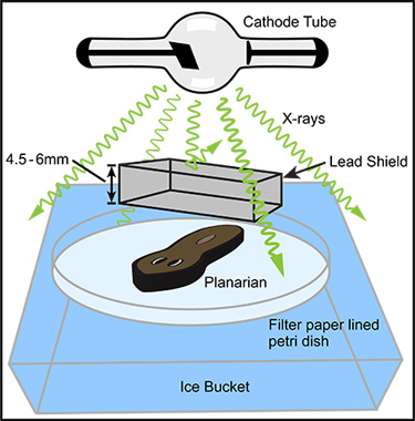
Figure 1. General arrangement of basic components for partial irradiation. In an X-ray irradiator with a top positioned X-ray source (cathode tube) the anesthetized planarian is positioned directly below the X-ray source within the irradiation field. In order to maximize the X-ray dose rate, the distance between the planarian and the X-ray source should be minimized. A lead shield should be positioned between the cathode tube and the anesthetized worm, as close to the worm as practical. The lead shield should be designed, manufactured, and positioned so that it shields the desired tissue but completely exposes the rest of the worm. Many commercial manufactures will produce custom lead shields from your simple design diagram; we have successfully used Alpha Systems Corp. (Bluffdale, UT). The lead should be sufficiently thick to allow for the desired amount of shielding. For example, if near complete shielding of a 320 kV X-ray beam is desired, the lead should be 4.5 to 6 mm thick. The planarian and shield are positioned on a Holtfreter’s soaked filter paper lined Petri dish which rests in an ice bucket. Given a sufficiently large X-ray irradiation field and a number of identical lead shields, many specimens may be partially irradiated at one time (not pictured).
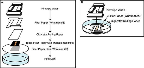
Figure 2. Building the recovery chamber. (A) An exploded view of the tissue transplantation recovery chamber, showing all of the components that are layered on top of one another after being soaked in casein saturated Holtfreter’s Solution. (B) A nearly completed recovery chamber, illustrating the interlocking placement of the Whatman #3 filter paper rectangles which snugly encase the anesthetized planarian, preventing movement during healing. The careful construction of the recovery chamber prevents animal movement and desiccation, promoting rapid healing and greater efficacy of tissue transplantation.
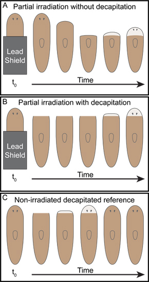
Figure 3. Representative outcomes of simple posterior partial irradiation. As Dubois described7, (A) when the posterior half of planarians were shielded with lead and then exposed to x-ray irradiation the anterior tissue was observed to regress back to the boundary between the irradiated and shielded tissue at which point unpigmented blastemas formed and the animals began to regenerate. (B) On the other hand, when animals were decapitated following the same partial irradiation performed in (A), tissue regression was not observed and the remaining irradiated anterior tissue was rescued. The decapitated partially irradiated animals regenerated heads (B), however, regeneration was significantly delayed as compared to unirradiated decapitated controls (C).
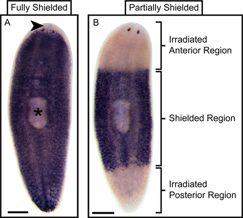
Figure 4. Representative outcome of partial stem cell ablation following partial irradiation. Whole-mount In situ hybridization (WISH) for the stem cell marker Smed-piwi-1 reveals that wild type planaria have stem cells distributed throughout their bodies with the exception of the tissue anterior to the photoreceptors (arrowhead) and the pharynx proper (asterisk)14. (A) WISH for Smed-piwi-1 in irradiated but fully shielded control planaria fixed three days following irradiation shows a stem cell distribution that is indistinguishable from that of wild type planaria. (B) On the other hand, WISH for Smed-piwi-1 in animals that were only partially shielded, leaving the anterior and posterior exposed, but were also fixed three days following irradiation shows that stem cells are ablated from the non-shielded regions. Scale bars are 500 microns.
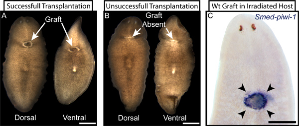
Figure 5. Examples of successful and unsuccessful tissue transplantation. (A) Live images of dorsal and ventral views of a successfully transplanted planarian three days following transplantation. The graft (indicated) is clearly visible on both dorsal and ventral surfaces and is surrounded with characteristic unpigmented tissue at the graft-host interface. (B) Correspondingly, unsuccessful transplantations display no visible graft tissue at the transplantation site (indicated) and instead show a healed, unpigmented, lateral wound from the failed transplantation. A graft that adheres to only the dorsal or the ventral surface can resemble a successful transplantation (A) when viewed from one side and an unsuccessful transplantation (B) when viewed from the other. (C) When wild type (wt) tissue is grafted into an irradiated host that has been ablated of resident stem cells and the transplanted stem cells are later revealed by WISH for Smed-piwi-1 two days following transplantation, the success of the transplantation is clearly displayed by the specific presence of stem cells only in or around the graft location (arrowheads). Scale bars are 500 microns.