1. Maintenance and Propagation of C2C12 Myoblasts
2. C2C12 Cell Transfection
3. Assessment of Cell Survivability/MTT Assay
4. Preparation of Myoblasts for Transplant
5. Cell Implantation
6. Injection of Fluc Substrate, D-luciferin, into Mdx Mouse
7. BLI to Target Luc-expressing MPCs Following Implant into Mouse Models of DMD
Upon 50-60% confluency, C2C12 myoblasts were transiently transfected with the above-mentioned fusion reporter gene construct composed of firefly luciferase [fluc], monomeric red fluorescent protein [mrfp] and sr39 thymidine kinase [sr39tk](Figure 1A). Transfection efficiency was calculated via fluorescence microscopy (Figures 1B,C), making use of the mrfp sequence in our reporter construct. Cell survivability was not affected by labeling with the BLI substrate, D-luciferin (Figure 1D). Following transfection, approximately 100,000 mrfp-expressing myoblasts were implanted intramuscularly into the gastrocnemius muscle of mdx mice (determined previously, manuscript submitted); 100,000 untransfected cells were similarly implanted into the contralateral hind limb as a control. Immediately following cell implantation, mice were injected intraperitoneally (IP) with 150 mg/kg D-luciferin. Following an uptake period of ~20 min, mice were imaged on a small animal optical scanner (GE ExplorOptix black box that is equipped for live animal bioluminescence and fluorescence). As previously demonstrated, both in vitro and post-implantation (manuscript submitted) uptake of D-luciferin was specific to fluc-expressing myoblasts, with no detectable bioluminescence in untransfected cells (Figure 2). Immunohistochemistry confirmed intramuscular transplantation of myoblasts (Figure 3)
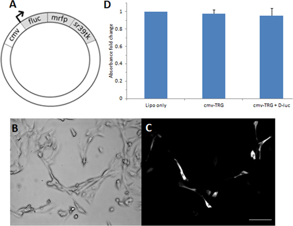
Figure 1. Schematic of CMV-trifusion reporter construct (A); brightfield/fluorescence images of C2C12 myoblasts transfected with the trifusion reporter plasmid (B,C); MTT assay to assess C2C12 cell survivability following labeling with BLI substrate, D-luciferin (D).
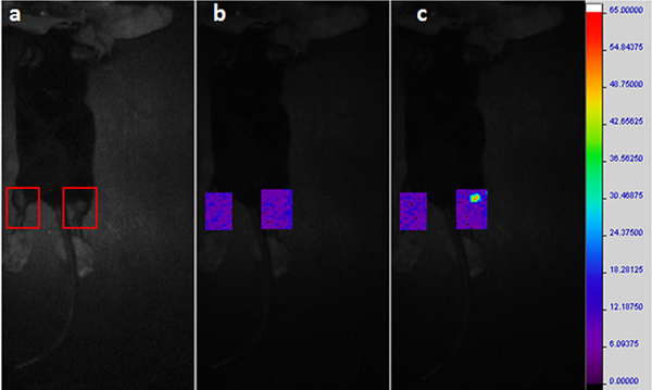
Figure 2. Bioluminescence imaging (BLI). A region of interest (ROI) is drawn to enclose the plucked hind limb area where myoblasts are injected (A). Bioluminescence is not detected during a background scan (B). At 23 min after injection of D-luciferin, a clear signal is detected from the right hind limb where luciferase-expressing myoblasts are injected. No bioluminescence is detected in the contralateral hind limb injected with untransfected myoblasts (C). Click here to view larger figure.
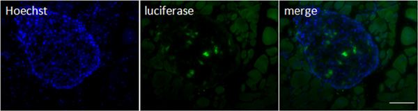
Figure 3. IHC using a firefly luciferase antibody confirms intramuscular implantation of transfected C2C12 cells.
Name of reagent | Company | Catalogue number | Comments (optional) |
C2C12 myoblasts | ATCC | ||
Dulbecco’s Modified Eagle’s Medium | Life Technologies | 12800-017 | |
fetal bovine serum | Life Technologies | 12483020 | |
0.25% Trypsin-EDTA | Life Technologies | 25200-72 | |
Hanks Balanced Salt Solution | Sigma Aldrich | H6648 | |
Lipofectamine 2000 | Life Technologies | 11668-019 | |
Nikon Eclipse TE2000-5 | Nikon Instruments Inc. | ||
Xenolight D-luciferin | PerkinElmer | 122796 | 40 mg/ml in PBS |
Duchenne muscular dystrophy (DMD) is a severe genetic neuromuscular disorder that affects 1 in 3,500 boys, and is characterized by progressive muscle degeneration1, 2. In patients, the ability of resident muscle satellite cells (SCs) to regenerate damaged myofibers becomes increasingly inefficient4. Therefore, transplantation of muscle progenitor cells (MPCs)/myoblasts from healthy subjects is a promising therapeutic approach to DMD. A major limitation to the use of stem cell therapy, however, is a lack of reliable imaging technologies for long-term monitoring of implanted cells, and for evaluating its effectiveness. Here, we describe a non-invasive, real-time approach to evaluate the success of myoblast transplantation. This method takes advantage of a unified fusion reporter gene composed of genes (firefly luciferase [fluc], monomeric red fluorescent protein [mrfp] and sr39 thymidine kinase [sr39tk]) whose expression can be imaged with different imaging modalities9, 10. A variety of imaging modalities, including positron emission tomography (PET), single-photon emission computed tomography (SPECT), magnetic resonance imaging (MRI), optical imaging, and high frequency 3D-ultrasound are now available, each with unique advantages and limitations11. Bioluminescence imaging (BLI) studies, for example, have the advantage of being relatively low cost and high-throughput. It is for this reason that, in this study, we make use of the firefly luciferase (fluc) reporter gene sequence contained within the fusion gene and bioluminescence imaging (BLI) for the short-term localization of viable C2C12 myoblasts following implantation into a mouse model of DMD (muscular dystrophy on the X chromosome [mdx] mouse)12-14. Importantly, BLI provides us with a means to examine the kinetics of labeled MPCs post-implantation, and will be useful to track cells repeatedly over time and following migration. Our reporter gene approach further allows us to merge multiple imaging modalities in a single living subject; given the tomographic nature, fine spatial resolution and ability to scale up to larger animals and humans10,11, PET will form the basis of future work that we suggest may facilitate rapid translation of methods developed in cells to preclinical models and to clinical applications.