1. Zebrafish Husbandry, Embryo Generation, and Treatment
2. Confocal Imaging of Cerebrovascular Structures in Fixed Zebrafish Embryos
3. 3D Reconstruction of Embryonic Zebrafish Cerebrovasculature
3D reconstruction of vascular structures provides a comprehensive and visually interesting perspective of zebrafish development. Figures 1 and 2 show methods as they typically done. Figure 3 shows several angles of vascular structures in a 6 dpf zebrafish embryo that expressed EGFP in endothelial cells. With a solid green or white color it can be difficult to appreciate signal intensity; pseudo-coloring provide image intensity from a look-up-table and allows better depth perception when structures overlap. An example of a pseudo-colored 3D image of the vasculature in a 6 dpf zebrafish is provided in Figure 4. Fluorescence imaging of live embryos can be used to study physiological characteristics that include eye and body movement, and cardiac activity. Figures 3 and 4 show representative results obtained with these methods, using the transgenic zebrafish line described. Imaging resolution depends on microscope characteristics, but the brightness of the EGFP signal is sufficient for good image quality with most commercial systems. Reconstruction and rendering of 3D representations is consistent and options within this open-source software provide consistently good results.
Figure 1. Eye removal. A) A fixed 3 dpf embryo with a tungsten need positioned next to the eye. Tissue is cut around the eye from this position. B) The eye is falls out and the underlying ocular muscles and optic nerve are cut. The empty eye socket is indicated with dashed circle. C) The same embryo is turned over and mounted with methyl-cellulose, with the intact eye facing up. Please click here to view a larger version of this figure.
Figure 2. Step-by-step 3D reconstruction of a confocal image stack. A) Open file (4104.1.ids) loaded within Fiji using Plugins>LOCI>Bioformat to select. B) After finding a slice with the region of interest, threshold adjustment is selected as shown. C) Threshold is adjusted to 214 using the top slider and apply is selected. D) 3D viewer is called as shown. E) The 3D reconstruction is shown of a zebrafish with the eye intact, for orientation. F) The image has been zoomed and rotated. G) A 360 degree rotation movie is made as shown. Please click here to view a larger version of this figure.
Figure 3. Perspectives from 3D reconstruction. A) Medial perspective of 6 dpf embryo imaged with 10x objective, mouth is on the right, not gills inside mouth. B) Lateral of the same embryo, note fin is a loop in the middle. C) Same embryo imaged with 20x objective, medial perspective, note gill resolution. D) Lateral perspective of 20x objective imaging. The fin is on the right edge of the panel. E) Antero-medial view of 20x objective imaging, note gills inside mouth. F) Abdomen of the same embryo imaged with a 20x objective, head it to the right. Note vasculature on the yolk sac at the bottom right. Please click here to view a larger version of this figure.
Figure 4. Intensity differences in 6 dpf embryo. Image of a 6 dpf reconstruction using a pseudocolor look-up-table for signal intensity. Mouth, brain, gills and yolk sac are labeled for orientation. Please click here to view a larger version of this figure.
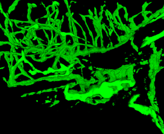
Figure 5. Movie of reconstructed vascular system in a 4 dpf zebrafish. The fish was imaged at 2.5 μm. The images were from imaging one half of the embryo. Compare vascular structures with structures in a GSI-treated zebrafish provided in Figure 6. Note the lower density of blood vessels in the head and larger gills. (See the “Zfish_spin.avi” supplemental file under Downloads)
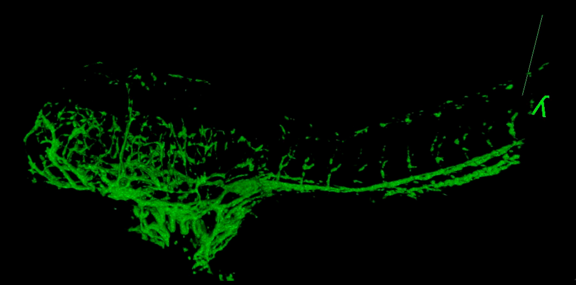
Figure 6. 3D Movie of vascular system in a GSI-treated embryo at 4 dpf. The fish was imaged at 2.5 μm through from lateral to midline. Compare vascular structures with the control 4 dpf fish shown in Figure 5. The arched back and smaller size are typical in embryos treated with this chemical. (See the “GSI-treated_4dpf_fish.avi” supplemental file under Downloads)
N – Phenylthiourea | Alfa Aesar, catalog #41972 | 0.2 M in E3 buffer, kept at 4oC | |
E3 buffer | Sigma | 5 mM NaCl, 0.17 mM KCl, 0.33 mM CaCl2, 0.33 mM MgSO4 | |
Confocal microscope | Nikon | D-EclipseC1 on a Nikon TE-2000U | |
Glass bottom dishes | Mat-Tek | ||
GSI IX/DAPT | N-[N-(3,5-Difluorophenacetyl-L-alanyl)]-S-phenylglycinet-butyl ester EMD Biosciences | ||
24 well plates | Becton-Dickinson, cat# 351147 | BD Falcon | |
Transfer pipettes | VWR, cat #414004-001 | VWR disposable transfer pipets | |
Methyl-cellulose | Alfa Aesar, cat#43146 | 3% in E3 buffer | |
NRD 4/6 Fish food | Brine Shrimp Direct | Dried | |
Brine shrimp | Brine Shrimp Direct | Live | |
Tungsten wire | Small Parts # TW-016-60 | 0.016” OD | |
Tricaine | VWR # 101107-950 | Tricaine methanesulfonate 250 mg/L in E3 buffer |
Zebrafish are a powerful tool to study developmental biology and pathology in vivo. The small size and relative transparency of zebrafish embryos make them particularly useful for the visual examination of processes such as heart and vascular development. In several recent studies transgenic zebrafish that express EGFP in vascular endothelial cells were used to image and analyze complex vascular networks in the brain and retina, using confocal microscopy. Descriptions are provided to prepare, treat and image zebrafish embryos that express enhanced green fluorescent protein (EGFP), and then generate comprehensive 3D renderings of the cerebrovascular system. Protocols include the treatment of embryos, confocal imaging, and fixation protocols that preserve EGFP fluorescence. Further, useful tips on obtaining high-quality images of cerebrovascular structures, such as removal the eye without damaging nearby neural tissue are provided. Potential pitfalls with confocal imaging are discussed, along with the steps necessary to generate 3D reconstructions from confocal image stacks using freely available open source software.