All surgical procedures were subjected to ethical review and approved by the government of Upper Bavaria (reference number: 55.2-1-54-2532.3-13-13 and -2532-136-11). Animals are male C57BL/6 mice with a body weight of approximately 25 g.
1. Animal Preparation
2. SAH Induction
3. End of Experiment
4. Considerations in Case of Survival Surgery (not shown in the video)
Mortality
Once the surgery technique is mastered the procedure does not elicit any intraoperative mortality. Also bleeding can be achieved in virtually all animals. Postoperative mortality is 30-40% with most animals dying on day 1 after surgery (Figure 5).
ICP values after SAH
The ICP before bleeding is around 4 mmHg. Bleeding results in a sharp increase of the ICP up to 120 mmHg. ICP values then stabilize within 5 min at approximately 30 mmHg (Figure 1). At 24 hr after bleeding the ICP is still slightly elevated to 10 mmHg5.
Blood pressure after SAH
Blood pressure rises immediately after bleeding induction (Figure 2). This is due to the Cushing reflex, which is initiated by elevated ICP.
Cerebral perfusion after SAH
After bleeding induction a dramatic decrease of cerebral perfusion can be monitored. Reperfusion to an individually different level occurs within 5 min after the insult (Figure 3).
Blood distributes along brain supplying arteries and cerebellar fissures
We perfused animals 3 hr after SAH transcardially with 20 ml of saline followed by 20 ml of chilled 4% PFA. The brain was carefully removed and blood distribution in the subarachnoid space was observed. Blood distributes along the perivascular space of brain supplying arteries towards the dorsal cortex. In all cases the extravasated blood covered the MCA up to the second branching. The side ipsilateral to the hemorrhage was covered with more blood than the contralateral hemisphere (Figure 4).
Blood distribution does not correlate with ICP rise
In 5 animals we investigated whether the rise of the ICP during SAH has an impact on blood distribution in the subarachnoid space. One hypothesis was that animals that show only a moderate ICP rise during SAH might exhibit less extravasated blood, which then does not distribute to the dorsal parts of the cortex. We found that only the size of the hematoma at the skull base seems to correlate with the ICP rise. Blood distribution along the brain supplying arteries did not differ between animals with different ICP peak values.
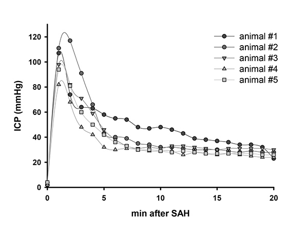
Figure 1. ICP value after SAH. Representative ICP values of 5 animals after SAH. Click here to view larger image.
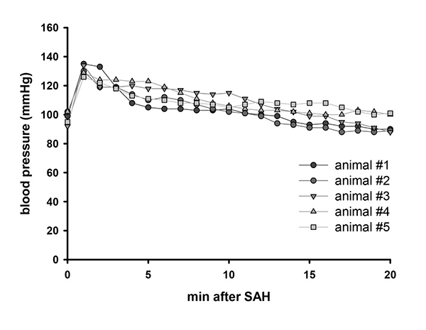
Figure 2. Blood pressure after SAH. Representative blood pressure values of 5 animals after SAH. Click here to view larger image.
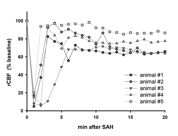
Figure 3. Cerebral perfusion after SAH. Representative laser Doppler flowmeter values of 5 animals after SAH. Click here to view larger image.
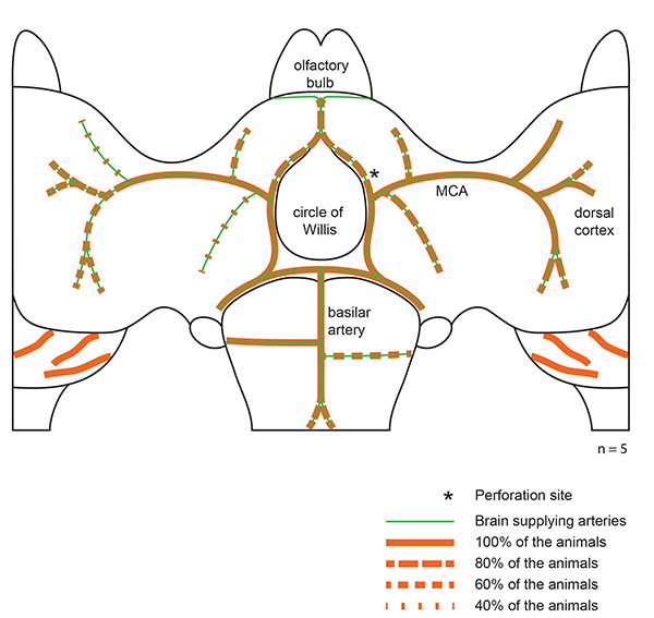
Figure 4. Blood distribution along brain supplying arteries. Representative blood distribution of 5 animals after SAH. Red lines indicate blood distribution along brain supplying arteries. Click here to view larger image.
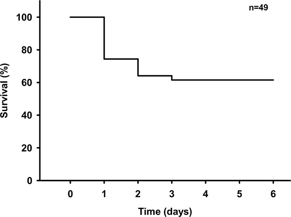
Figure 5. Survival curve after SAH. Survival curve following SAH in 49 male C57BL/6 mice. Click here to view larger image.
Equipment | |||
operation microscope | Leica | KL2500 | |
isoflurane vaporizer | Harvard Instruments | Continuous Flow Vaporizer | |
respirator | Hugo Sachs | Minivent 845 | |
microcapnograph | Hugo Sachs | Type 340 | |
temperature controller | FHC | DC Temperature Controller | |
dental drill | Paggen | Labset- N | |
ICP monitor | Codman | ICP monitor | |
blood pressure monitor | AD Instruments | Bridge Amp FE221 | |
syringe pump | World Precision Instruments | SP101IZ | |
pulsoximeter | Kent Scientific | MouseSTAT | |
LDF | Perimed | Periflux 5000 | |
analog data monitor | AD Instruments | Power Lab 16/35 | |
Material | |||
cement for ICP probe fixation | Speiko | Carboxylate cement | |
glue for LDF probe fixation | Bob Smith Industries | Cyanoacrylate glue (Maxi Cure and Insta Set) | |
venous catheter | Johnson & Johnson | Jelco winged i.v. catheter; REF 4076 | modified intubation tube |
tubing for femoral catheter | Smiths Medical | Fine Bore Polythene Tubing; ID 0.28 mm OD 0.61 mm; REF 800/100/100 | cut to 30 cm length |
filament for vessel perforation | Ethicon | Prolene 5-0 | cut to 12 mm length |
surgical equipment | Fine Scientific Instruments | forceps medical #5, vessel scissors 8 cm, microclip 4 mm jaw |
In this video publication a standardized mouse model of subarachnoid hemorrhage (SAH) is presented. Bleeding is induced by endovascular Circle of Willis perforation (CWp) and proven by intracranial pressure (ICP) monitoring. Thereby a homogenous blood distribution in subarachnoid spaces surrounding the arterial circulation and cerebellar fissures is achieved. Animal physiology is maintained by intubation, mechanical ventilation, and continuous on-line monitoring of various physiological and cardiovascular parameters: body temperature, systemic blood pressure, heart rate, and hemoglobin saturation. Thereby the cerebral perfusion pressure can be tightly monitored resulting in a less variable volume of extravasated blood. This allows a better standardization of endovascular filament perforation in mice and makes the whole model highly reproducible. Thus it is readily available for pharmacological and pathophysiological studies in wild type and genetically altered mice.