Science Education
>

Bacterial Artificial Chromosomes: A Functional Genomics Tool for the Study of Positive-strand RNA Viruses

Instructor Prep
concepts
Student Protocol
JoVE Journal
Immunology and Infection
This content is Free Access.
JoVE Journal Immunology and Infection
Bacterial Artificial Chromosomes: A Functional Genomics Tool for the Study of Positive-strand RNA Viruses

Note: Figure 1 presents a strategy for the construction of a full-length infectious JEV cDNA as a BAC.28 Table 1 provides a list of the oligonucleotides used in this protocol.28

1. Extract Viral RNA from JEV Particles in Cell Culture Supernatants

  1. Start with the cell culture medium containing JEV SA14-14-2, a live JE vaccine virus that requires Biosafety Level 2 containment.
    Note: The viral titer is approximately 1-3 × 10⁶ plaque-forming units/ml.
  2. Take the biosafety training necessary for all standard microbiological practices, safety equipment, and laboratory facilities prior to working with JEV SA14-14-2.
  3. Purify viral RNA from an aliquot of the virus-containing cell culture medium using a monophasic solution of phenol and guanidine isothiocyanate66 (Figure 1A).
    1. Add 600 µl of the monophasic reagent to 200 µl of the culture supernatant in a 1.7 ml microtube. Homogenize the mixture by hand-shaking the tube vigorously for 30 sec and incubating for 5 min at RT.
    2. Add 160 µl of chloroform to the homogenized sample. Mix thoroughly by hand-shaking the tube vigorously for 15 sec and leaving it for 2-3 min at RT.
    3. Centrifuge the total lysate at 13,400 × g for 15 min at 4 °C, which results in a separation of two liquid phases, i.e., a lower organic phase and an upper aqueous phase. Transfer the upper aqueous phase (less than 200 µl) containing the RNA to a new microtube.
    4. Precipitate the RNA by adding 1 µl of 5 µg/µl glycogen and 400 µl of 100% isopropanol and incubating for 10 min at RT.
    5. Centrifuge the mixture at 13,400 × g for 10 min at 4 °C. Discard the supernatant and wash the RNA pellet with 1 mL of 75% ethanol by pulse-vortexing three to five times and centrifuging at 13,400 × g for 5 min at 4 °C.
    6. Air-dry the RNA pellet for 10 min and dissolve it in 40 µl of dH2O. Store the extracted RNA at -80 °C until use.

2. Synthesize a Set of Four Overlapping cDNA Fragments (F1 to F4) Spanning the Entire Viral Genomic RNA by Reverse Transcription (RT)-PCR

  1. Perform a 20 µl RT reaction with a 10 µl aliquot of the purified viral RNA as a template and a modified form of Moloney murine leukemia virus reverse transcriptase.67
    1. Set up a 13 µl mixture containing 10 µl of the purified RNA, 1 µl 10 mM dNTP mix, 1 µl 4 pmol/µl primer, and 1 µl dH2O. Use the fragment-specific primer for each RT reaction: 1RT for F1, 2RT for F2, 3RT for F3, and 4RT for F4.
    2. Incubate the mixture at 65 °C for 5 min, place on ice for 1 min, and then quick-spin to collect the contents at the bottom of the tube.
    3. Add 4 µl 5× RT buffer, 1 µl 0.1 M DTT, 1 µl 40 U/µl RNase inhibitor, and 1 µl 200 U/µl reverse transcriptase. Mix by pipetting up and down three to five times.
    4. Let the reaction proceed at 50 °C for 1 hr, then heat-inactivate the sample at 70 °C for 15 min. Store the synthesized first-strand cDNA at -20 °C until use.
  2. Perform a 100 µl PCR reaction with a 5 µl aliquot of the heat-inactivated RT reaction as a template and a high-fidelity thermostable DNA polymerase68 (Figure 1B).
    1. Set up a 100 µl PCR reaction on ice, containing 5 µl of the RT reaction, 20 µl 5× PCR buffer, 4 µl 10 mM dNTP mix, 5 µl 10 µM forward primer, 5 µl 10 µM reverse primer, 1 µl 2 U/µl DNA polymerase, and 60 µl dH2O. Use the fragment-specific primer pair for each PCR reaction: 1F+1R for F1 (2573 bp), 2F+2R for F2 (4171 bp), 3F+3R for F3 (3922 bp), and 4F+4R for F4 (1798 bp).
    2. Mix gently by finger-flipping the tube three to five times and centrifuge briefly to collect its contents at the bottom.
    3. Begin thermocycling with an initial denaturation step of 30 sec at 98 °C, followed by 25-30 cycles with the following PCR profile: 10 sec at 98 °C, 30 sec at 60 °C, and 1-2 min (30 s/kb) at 72 °C. Store the PCR products at 4 °C until analyzed.
  3. Run a 2-5 µl aliquot of each PCR reaction on a 0.8% agarose gel containing 0.5 µg/ml ethidium bromide (EtBr) (Figure 2).
    CAUTION: EtBr is a potent mutagen and requires lab coats, safety glasses, and gloves to be worn and extreme caution to be observed during its use, storage, and disposal.

3. Subclone Each of the Four cDNA Fragments (F1 to F4) into a BAC Vector to Create pBAC/F1 to pBAC/F4 by Molecular Cloning Techniques

  1. Digest the vector and insert DNAs with two appropriate restriction endonucleases, as follows:
    1. Perform a sequential digestion of pBAC/PRRSV/FL (vector),30 a derivative of the pBeloBAC11 plasmid (7507 bp, GenBank accession number U51113), with Pme I and Not I in a total volume of 60 µl (containing ~500 ng DNA, 10 U enzyme, 1x digestion buffer, and 1× BSA) at 37 °C for 12-15 hr, which yields the 15426-bp vector fragment.
    2. Perform a sequential digestion of each of the four cDNA amplicons (insert) with Sma I and Not I in a total volume of 60 µl (containing ~1 µg DNA, 20 U enzyme, 1× digestion buffer, and 1× BSA) at 25 °C (Sma I) or 37 °C (Not I) for 12-15 hr, which yields the following insert fragments of the desired size: F1 (2559 bp), F2 (4157 bp), F3 (3908 bp), and F4 (1784 bp).
  2. Purify the desired vector and insert DNA fragments by gel extraction.
    1. Separate the doubly digested products on a 1% low-melting point agarose gel containing 0.5 µg/ml EtBr. Cut out a band of the desired DNA fragment with a minimal amount of agarose (usually ~200 µl) under long-wave ultraviolet light.
    2. Add an equal volume of TEM buffer (10 mM Tris-Cl, 1 mM EDTA, and 10 mM MgCl2) to the excised agarose in a 1.7 ml microtube. Incubate the sample at 72 °C for 10-15 min, with vortexing every 2-3 min until the agarose has completely melted.
    3. Add an equal volume of pre-warmed buffer-saturated phenol, vortex vigorously for 1 min, and centrifuge at 13,400 × g for 10 min at RT.
      Note: The mixture separates into a lower organic phase and an upper aqueous phase. Transfer the upper aqueous phase containing the DNA to a new microtube.
    4. Add an equal volume of chloroform:isoamyl alcohol (24:1), vortex for 1 min, and centrifuge at 13,400 × g for 10 min at RT. Transfer the upper aqueous phase to a new microtube.
    5. Add 0.5 µl of 10 µg/µl yeast tRNA, 1/10 volume of 3 M sodium acetate, and 2.5 volumes of 100% ethanol. Keep the mixture on ice for 20 min.
    6. Centrifuge the mixture at 13,400 × g for 10 min at RT. Remove the supernatant and wash the DNA pellet with 1 ml of 70% ethanol by pulse-vortexing three to five times and centrifuging at 13,400 × g for 10 min.
    7. Air-dry the DNA pellet for 10 min and dissolve it in 20 µl of TE buffer (10 mM Tris-Cl and 1 mM EDTA [pH 7.6]).
  3. Ligate the desired vector and insert DNA fragments using T4 DNA ligase.
    1. Set up a 20 µl ligation reaction containing 50-100 ng of the vector DNA, a ~3-fold molar excess of the insert DNA, 400 U T4 DNA ligase, and 1× ligation buffer. Incubate the ligation reaction at 16 °C for 12-15 hr.
      Note: Include a negative control reaction, i.e., vector only without the insert, in parallel.
    2. Perform four separate ligations, each joining the 15426-bp Pme I-Not I fragment of pBAC/PRRSV/FL with the 2559-bp (for F1), 4157-bp (for F2), 3908-bp (for F3), or 1784-bp (for F4) Sma I-Not I fragment of one of the four cDNA amplicons, to generate subclones pBAC/F1 to pBAC/F4.
  4. Transform the ligated DNA into E. coli DH10B by the CaCl2-heat shock method.
    1. Take 100 µl aliquots of the CaCl2-treated competent DH10B cells69 stored at -80 °C and thaw them on ice.
      Note: Use 100 µl of cells per transformation.
    2. Add a 10 µl aliquot of the DNA ligation reaction to 100 µl of the thawed cells in a 1.7 ml microtube, mix gently by tapping the tube, and keep on ice for 30 min.
    3. Heat-shock the DNA-cell mixture for 45 sec in a 42 °C water bath, place on ice for 2 min, and then add 900 µl of LB broth pre-warmed to RT.
    4. Incubate the heat-shocked cells at 35 °C for 1 hr, with shaking at 225-250 rpm.
    5. Spread 50 to 200 µl aliquots of the cultured cells on LB agar plates containing 10 µg/ml chloramphenicol (Cml). Keep the plates at RT, right side up, until they are dry.
    6. Turn the plates upside down and incubate at 35 °C for 15 hr.
  5. Recover the cloned BAC DNA from the host cells by a column-based purification method (Figure 1C).
    1. Pick six to eight bacterial colonies from the LB-Cml agar plates and inoculate them into 3 ml of 2xYT broth containing 10 µg/ml Cml. Incubate the cultures at 35 °C for 10 hr with vigorous shaking (225-250 rpm).
    2. Isolate recombinant BAC DNAs from 1-1.5 ml of the bacterial cultures using spin columns, as directed by the manufacturer.70 Elute the extracted DNA (typically 100-200 ng) in 20 µl of TE buffer.
    3. Perform two analytical restriction enzyme digestions of the isolated BACs for ~6 hr in a total volume of 10 µl, one to identify the presence of the vector with a correct insert using the same enzymes used for cloning (see Protocol 3.1), and the other to test the integrity of the cloned BACs with an appropriate enzyme (e.g., Bgl II, Nco I, or Pst I), generating a unique restriction fragment pattern.
    4. Propagate the correctly cloned BACs by inoculating 500 µl of the positive bacterial cultures (from Protocol 3.5.1) in 500 ml of 2xYT-Cml medium and cultivating the inoculum for 6 hr at 35 °C while shaking at 225-250 rpm. Purify the BAC DNA (typically 10-20 µg) from the 500 ml culture using filter columns, as recommended by the manufacturer.71
      Note: The initial four BAC subclones, each containing a cDNA fragment of JEV genomic RNA, may prove to have one or more unwanted mutation(s) when compared to the consensus sequence of the viral genome42 (which serves as a reference sequence). Any such mutation needs to be corrected by PCR-based site-directed mutagenesis prior to the assembly of a full-length JEV cDNA.27

4. Create a Full-length JEV cDNA with the 5' SP6 Promoter and the 3' Run-off Site

  1. Make three genetic modifications (see below Protocols 4.1.1-4.1.3) in the cloned cDNAs to allow in vitro run-off transcription of genome-length RNAs with the authentic 5' and 3' ends of the viral genome.
    1. Introduce an SP6 promoter directly upstream of the 5' end of the viral genome by overlap extension PCR (Figure 1D, pBAC/F1SP6).
      1. Amplify two overlapping DNA fragments via the first standard PCR of pBAC/F1 with the two primer pairs SP6F+SP6R (product size, 173 bp) and F1F+F1R (product size, 676 bp), each in a 50 µl reaction containing 1 µl template DNA (~200 pg/µl), 10 µl 5× PCR buffer, 2 µl 10 mM dNTPs, 2.5 µl each of 10 µM forward and reverse primers, 0.5 µl 2 U/µl DNA polymerase,68 and 31.5 µl dH2O. Perform the PCR using the following cycling profile: 98 °C for 30 sec, and 25 cycles of 98 °C for 10 sec, 60 °C for 30 sec, and 72 °C for 20 sec.
      2. Gel-purify the two PCR-amplified DNA fragments after running each of the two PCR products on a 1.5% low-melting point agarose gel, as described in Protocol 3.2.
      3. Fuse the two gel-purified DNA fragments via the second fusion PCR using the outermost primers SP6F+F1R (product size, 821 bp) in a 100 µl reaction including 1 µl each of the two purified DNA fragments (~100 pg/µl), 20 µl 5× PCR buffer, 4 µl 10 mM dNTPs, 5 µl each of 10 µM forward and reverse primers, 1 µl 2 U/µL DNA polymerase,68 and 63 µl dH2O. Use the following thermal cycling profile: 98 °C for 30 sec, and 25 cycles of 98 °C for 10 sec, 60 °C for 30 sec, and 72 °C for 30 sec.
      4. Gel-purify the fused PCR amplicon from a 1% low-melting point agarose gel, as described in Protocol 3.2.
      5. Perform the five-step cloning procedure described in Protocols 3.1-3.5 to ligate the 760-bp Pac I-BsiW I fragment of the gel-purified fused PCR amplicon with the 9532-bp Pac I-BsiW I fragment of pBAC/F1 to generate pBAC/F1SP6.
    2. Remove the pre-existing, internal Xba I site at nucleotide 9131 by introducing a silent point mutation (A9134→T) via overlap extension PCR (Figure 1D, pBAC/F3KO).
      1. Amplify two overlapping DNA fragments by the first standard PCR of pBAC/F3 with the two primer pairs X1F+X1R (product size, 746 bp) and X2F+X2R (product size, 316 bp) in a 50 µl reaction under the experimental conditions described in Protocol 4.1.1.1.
      2. Gel-purify the two PCR-amplified DNA fragments after electrophoretic separation on 1.5% low-melting point agarose gels, as detailed in Protocol 3.2.
      3. Fuse the two gel-purified DNA fragments via the second fusion PCR using the outermost primers X1F+X2R (product size, 1033 bp) in a 100 µl reaction under the experimental conditions described in Protocol 4.1.1.3.
      4. Gel-purify the fused PCR amplicon from a 1% low-melting point agarose gel, as detailed in Protocol 3.2.
      5. Perform the five-step cloning procedure described in Protocols 3.1-3.5 to ligate the 949-bp Avr II-Not I fragment of the gel-purified fused PCR amplicon with the 16245-bp Not I-BsiW I and 2141-bp BsiW I-Avr II fragments of pBAC/F3 to produce pBAC/F3KO.
    3. Engineer a new artificial Xba I run-off site just downstream of the 3' end of the viral genome by PCR-based site-directed mutagenesis (Figure 1D, pBAC/F4RO).
      1. Generate one DNA fragment by PCR of pBAC/F4 with primers ROF+ROR (product size, 324 bp) in a 100 µl reaction containing 1 µl template DNA (~200 pg/µl), 20 µl 5× PCR buffer, 4 µl 10 mM dNTPs, 5 µl each of 10 µM forward and reverse primers, 1 µl 2 U/µl DNA polymerase,68 and 64 µl dH2O. Perform the PCR using the following cycling profile: 98 °C for 30 sec, and 25 cycles of 98 °C for 10 sec, 60 °C for 30 sec, and 72 °C for 15 sec.
      2. Gel-purify the PCR-amplified DNA fragment from a 1% low-melting point agarose gel, as detailed in Protocol 3.2.
      3. Perform the five-step cloning procedure described in Protocols 3.1-3.5 to ligate the 283-bp Sfi I-Not I fragment of the gel-purified PCR amplicon with the 16933-bp Sfi I-Not I fragment of pBAC/F4 to create pBAC/F4RO.
  2. Assemble a set of the four modified, overlapping cDNAs into a single full-length SA14-14-2 BAC (pBAC/SA14-14-2) by joining at three natural restriction sites (BsrG I, BamH I, and Ava I) in a sequential manner using the five-step cloning procedures detailed in Protocols 3.1-3.5 (Figure 1E): F1SP6 → F1SP6F2 (by replacing the BsrG I-Not I fragment of pBAC/F1SP6 with that of pBAC/F2) → F1SP6F2F3KO (by replacing the BamH I-Not I fragment of pBAC/F1SP6F2 with that of pBAC/F3KO) → F1SP6F2F3KOF4RO (by replacing the Ava I-Not I fragment of pBAC/F1SP6F2F3KO with that of pBAC/F4RO).

5. Prepare a High-purity Maxi-prep of the Full-length SA14-14-2 BAC

  1. Grow a single colony of E. coli DH10B carrying pBAC/SA14-14-2 in 3 ml of 2xYT broth containing 10 µg/ml Cml for 10 hr at 35 °C with shaking at 225-250 rpm, and then scale up by inoculating 500 µl of the 10 hr bacterial culture into 500 ml of 2xYT-Cml medium and cultivating the inoculum for 6 hr at 35 °C with vigorous shaking.
  2. Centrifuge the bacterial culture in two 250 ml bottles at 3,107 × g for 15 min at 4 °C. Resuspend each pellet in 30 ml of GTE solution (50 mM glucose, 25 mM Tris-Cl, and 10 mM EDTA [pH 8.0]), and then add 500 µl of 60 mg/ml lysozyme. Incubate the two cell suspensions on ice for 10 min.
  3. Add 60 ml of freshly made Lysis solution (0.2 N NaOH and 1% SDS) to each cell suspension, mix well until clear, and keep the lysates at RT for 10 min.
  4. Add 45 ml of Neutralization solution (100 ml, consisting of 60 ml 5 M potassium acetate, 11.5 ml glacial acetic acid, and 28.5 ml dH2O) to each bottle, mix thoroughly by inverting the bottles, and incubate the neutralized lysates on ice for 10 min.
  5. Centrifuge the neutralized lysates at 18,566 × g for 20 min at 4 °C. Transfer the supernatant of both bottles into two new 250 ml bottles; add 0.6 volume of 100% isopropanol to each, and keep them on ice for 20 min.
  6. Spin down the precipitates at 18,566 × g for 20 min at 4 °C. Dissolve each pellet in 5 ml of TE buffer, combine them in a 50 ml tube (10 ml total), and precipitate the RNA by adding an equal volume of 5 M lithium chloride. Incubate the mixture on ice for 10 min.
  7. Centrifuge the RNA precipitate at 14,636 × g for 20 min at 4 °C. Transfer the supernatant to a new 250 ml tube and precipitate DNA by adding 2 volumes of 100% isopropanol. Incubate the mixture on ice for 20 min.
  8. Spin down the DNA precipitate at 18,566 × g for 20 min at 4 °C. Aspirate the supernatant, resuspend the DNA pellet in 9.5 ml of TE buffer (pH 7.6), and add 10 g of cesium chloride (CsCl) and 390 µl of 10 mg/ml EtBr.
  9. Load the DNA-CsCl-EtBr solution into a 16 × 76 mm sealable polypropylene tube using a syringe equipped with an 18 G needle. Spin the sealed CsCl gradient in an ultracentrifuge at 401,700 × g for 16 hr at 20 °C (Figure 3).
  10. Collect a DNA band of BAC plasmid from the CsCl gradient using an 18 G needle to create an air vent at the top of the gradient and a 20 G needle-equipped syringe to retrieve the BAC DNA from the side of the gradient.
  11. Add 2.5 volumes of dH2O-saturated butanol to the EtBr-stained BAC DNA sample and mix by vortexing. Centrifuge the mixture at 13,400 × g for 1 min and transfer the lower aqueous phase to a new 1.7 ml microtube. Repeat this procedure six times.
  12. Precipitate the EtBr-free BAC DNA by adding 1/10 volume of 3 M sodium acetate and 2.5 volumes of 100% ethanol to the butanol-extracted BAC DNA and incubating for 10 min on ice. Centrifuge the precipitate at 13,400 × g for 10 min, wash the DNA pellet with 1 ml of 70% ethanol, and re-pellet it by centrifugation.
  13. Air-dry the DNA pellet for 10 min and dissolve it in 200 µl of TE buffer (pH 7.6).
    Note: Figure 4 shows an overview of the reverse genetics system for JEV SA14-14-2.

6. Transcribe Synthetic RNAs In Vitro from a Linearized Full-length JEV BAC DNA

  1. Perform a large-scale restriction enzyme digestion of pBAC/SA14-14-2 with Xba I in a total volume of 100 µl (containing 3 µg DNA, 60 U enzyme, 1× digestion buffer, and 1× BSA) at 37 °C for 12-15 hr. Examine a 3 µl aliquot of the digestion reaction on a 0.8% agarose gel containing 0.5 µg/ml EtBr.
  2. Incubate the digestion reaction further with 25 U of mung bean nuclease (MBN) at 30 °C for 2 hr (Figure 4A).
  3. Bring the volume of the Xba I-digested, MBN-treated sample up to 300 µl with dH2O. Add an equal volume of phenol:chloroform:isoamyl alcohol (25:24:1) to the diluted sample, vortex vigorously for 1 min, spin at 13,400 × g for 10 min, and then transfer the upper aqueous phase to a new 1.7 ml microtube. Add an equal volume of chloroform and repeat the extraction procedure.
  4. Recover the phenol/chloroform-extracted, linearized BAC by ethanol precipitation: Add 1/10 volume of 3 M sodium acetate and 2.5 volumes of 100% ethanol, and incubate on ice for 20 min. Centrifuge the precipitate at 13,400 × g for 10 min, wash the DNA pellet with 1 ml of 70% ethanol, and then spin it down by re-centrifugation.
  5. Air-dry the DNA pellet for 10 min and dissolve it in 30 µl of dH2O. Examine a 1 µl aliquot of the recovered BAC on a 0.8% agarose gel with 0.5 µg/ml EtBr (Figure 5A).
  6. Perform a run-off transcription of the linearized BAC DNA in a total volume of 25 µl (containing ~200 ng template DNA, 0.8 mM cap analog [m7G(5')ppp(5')A], 1 mM rNTPs, 40 U RNase inhibitor, 20 U SP6 RNA polymerase,72 and 1× transcription buffer) at 37 °C for 1 hr (Figure 4B). Include 0.5 µM [3H]UTP for RNA quantification on the basis of [3H]UTP incorporation, as monitored by adsorption to DE-81 filter paper.69
  7. Run a 1-2 µl aliquot of the run-off transcription reaction on a 0.6% agarose gel containing 0.5 µg/ml EtBr (Figure 5B).

7. Determine RNA Infectivity and Virus Yield

  1. Cultivate BHK-21 cells in 150 mm culture dishes at a density of 3 × 10⁶ cells/dish for 24 hr at 37 °C with 5% CO2.
    Note: Maintain the BHK-21 cells in alpha minimal essential medium supplemented with 10% fetal bovine serum, 2 mM glutamine, vitamins, and penicillin/streptomycin.
  2. Rinse the cell monolayer with 10 ml of cold Solution A (137 mM NaCl, 2.7 mM KCl, 8.1 mM Na2HPO4, and 1.5 mM KH2PO4). Detach the cells from the dishes by treatment with 4 ml of trypsin-EDTA (0.25%), and collect them by centrifugation at 270 × g in a desktop centrifuge for 2 min.
  3. Resuspend the cell pellet with 50 ml of cold Solution A in a 50 ml conical tube and centrifuge the cell suspension at 270 × g for 2 min. Repeat this wash procedure three times; after the last wash, resuspend the cell pellet at a density of 2 × 10⁷ cells/ml in Sol A.
  4. Mix a 400 µl aliquot of cell suspension with 2 µg of synthetic RNA in a 2-mm gap cuvette, and promptly electroporate the mixture with an electroporator under optimal electroporation conditions: 980 V, 99 µsec pulse length, and 5 pulses (Figure 4C).
    Note: Use the 3H-labeled RNA synthesized in Protocol 6.5 directly for electroporation without further purification.
  5. Leave the electroporated cells at RT for 10 min and transfer to a 1.7 ml microtube containing 600 µl of complete culture medium.
  6. Prepare a 10-fold serial dilution of the electroporated cells in 1 ml of complete culture medium and plate a 100 µl aliquot of each dilution on the monolayers of unelectroporated BHK-21 cells (5 × 10⁵) in a 6-well plate.
  7. After 4-6 hr of incubation, overlay the cells with 0.5% agarose in minimal essential medium containing 10% fetal bovine serum. Incubate the plates for 4 days at 37 °C with 5% CO2.
  8. Visualize the infectious centers (plaques) by fixation with 7% formaldehyde and staining with 1% crystal violet in 5% ethanol27 (Figure 6A).
    1. Optional: Examine RNA-electroporated cells at 18-20 hr post-transfection for JEV protein expression by immunofluorescence assays27,73 (Figure 6B), and harvest the supernatants from the RNA-electroporated cells at 22 and 40 hr post-transfection for virus titration by plaque assays27,63 (Figure 6C).

Bacterial Artificial Chromosomes: A Functional Genomics Tool for the Study of Positive-strand RNA Viruses

Learning Objectives

For all positive-strand RNA viruses, the reliability and efficiency of a reverse genetics system depend on the genetic stability of a cloned full-length cDNA, whose sequence is equivalent to the consensus sequence of viral genomic RNA.27 Figure 1 shows a five-step strategy for the construction of a full-length infectious cDNA as a BAC for JEV SA14-14-228: Step 1, purification of viral RNA from the cell culture supernatant of JEV-infected BHK-21 cells (Figure 1A); Step 2, synthesis of four overlapping cDNA amplicons (F1 to F4) spanning the whole viral genome (Figure 1B); Step 3, subcloning of each of the four contiguous cDNA fragments into a BAC vector, creating pBAC/F1 to pBAC/F4 (Figure 1C); Step 4, modification of the cloned cDNAs for in vitro run-off transcription with SP6 RNA polymerase, i.e., placing an SP6 promoter sequence immediately upstream of the viral 5'-end (pBAC/F1SP6), eliminating a pre-existing internal Xba I site at nucleotide 9131 by introducing a silent point mutation, A9134→T (pBAC/F3KO), and inserting a new artificial Xba I run-off site immediately downstream of the viral 3'-end (pBAC/F4RO) (Figure 1D); and Step 5, assembly of a full-length SA14-14-2 cDNA BAC, pBAC/SA14-14-2 (Figure 1E). Table 1 lists the oligonucleotides used in this cloning procedure.28

For the construction of a functional JEV cDNA, the first important step is the synthesis of the four overlapping cDNA fragments using the purified viral RNA as a template for RT-PCR. Figure 2 provides a representative result for the four RT-PCR products that were electrophoresed on a 0.8% agarose gel. This gel demonstrates clearly that a full-length JEV cDNA is amplified into four overlapping cDNA fragments. Occasionally, RT-PCR reactions might yield one or more additional virus-specific or nonspecific products that are mostly smaller than the expected product, because of the nonspecific annealing of primers during cDNA synthesis/amplification. On the other hand, little or no expected RT-PCR product would be amplified because of accidental RNase contamination during the viral RNA isolation or improper RT-PCR performance.

The next key step is the cloning and modification of a partial- or full-length JEV cDNA in BAC, which is a relatively straightforward procedure that uses standard recombinant DNA techniques.69 Figure 3 presents a representative outcome for the purification of the BAC clone containing a full-length cDNA of JEV SA14-14-2 by banding in a CsCl-EtBr gradient. In this experiment, after centrifugation for 16 hr at 401,700 × g, two distinct bands, i.e., the E. coli chromosomal DNA above and the supercoiled BAC plasmid DNA below, are visible in the middle of the tube under long-wave ultraviolet light. A minimal volume (~400 µl) of the lower BAC DNA band was carefully collected by poking a hole with a syringe on the side of the tube. Subsequently, the EtBr was extracted from the BAC DNA by butanol extraction, and the EtBr-free BAC DNA was concentrated by ethanol precipitation.

The final step is the determination of the specific infectivity of the synthetic RNAs transcribed in vitro from the full-length SA14-14-2 BAC (pBAC/SA14-14-2) after RNA transfection into permissive cells (Figure 4). This step involves three sequential steps: Step 1, linearization of the full-length SA14-14-2 cDNA at the 3'-end of the viral genome (Figure 4A); Step 2, production of synthetic RNAs from the linearized cDNA by run-off transcription (Figure 4B); and Step 3, rescue of the recombinant viruses in BHK-21 cells transfected with the synthetic RNAs (Figure 4C). Experimentally, two independent clones of pBAC/SA14-14-2 were linearized with Xba I digestion and treated with MBN to remove the four-base 5' overhang generated by the Xba I digestion. The linearized BACs were cleaned up by phenol-chloroform extraction, followed by ethanol precipitation. The linearization of the two purified BACs was demonstrated on a 0.8% agarose gel (Figure 5A). The phenol-chloroform extraction must be done carefully to ensure that the linearized BACs are RNase-free. Each of the two linearized BACs served as a cDNA template for run-off transcription using SP6 RNA polymerase in the presence of the m7G(5')ppp(5')A cap analog. The integrity of the synthetic RNAs was shown by running aliquots of the two transcription reaction mixtures on a 0.6% agarose gel, along with a reference 1 kb DNA ladder (Figure 5B). In this simple assay, the major prominent RNA band always migrated just below the 3 kb reference DNA band and appeared to be sharp. However, degraded RNA would have a smeared appearance on the same gel.

An infectious center assay is the gold standard for determining the specific infectivity of the synthetic RNAs. This assay was done by electroporating BHK-21 cells with RNA samples, seeding equal aliquots of the 10-fold serially diluted electroporated cells in 6-well plates containing naïve BHK-21 cells (3 × 10⁵ cells/well), and overlaying agarose onto the cell monolayers. After incubation for 4 days, surviving cells were fixed with formaldehyde and stained with a crystal violet solution to quantify the number of infectious centers (plaques), which corresponds to the number of infectious RNA molecules delivered into the cells (Figure 6A). Since the cDNA template used for in vitro transcription has been proven to be non-infectious,27 an aliquot of the transcription reaction mixture was directly used for electroporation. Electroporation is the preferred method for RNA transfection; alternatively, RNAs can be transfected by other methods using DEAE-dextran and cationic liposomes. RNA electroporation is very effective, but "arcing" of the electric pulse occurs rarely if salts are present in the electroporation reaction or if the electroporation cuvette is reused. The expression of viral proteins in RNA-transfected cells was examined by immunofluorescence assays using an anti-NS1 rabbit antiserum (Figure 6B). The production of viral particles accumulated in the supernatants of RNA-transfected cells was analyzed by plaque assays (Figure 6C). The results of these experiments show clearly that the cDNA-derived synthetic RNAs are infectious in permissive BHK-21 cells, generating a high titer of recombinant viruses.

Oligonucleotide Sequencea (5' to 3') Positionb Polarity
1RT TAGGGATCTGGGCGTTTCTG
GCAAAT
2578–2603 Antisense
1F aatcccgggAGAAGTTTATC
TGTGTGAACTT
1–22 Sense
1R attgcggccgcCCACGTCGT
TGTGCACGAAGAT
2532–2553 Antisense
2RT TTCTGCCTACTCTGCCCCTC
CGTTGA
5975–6000 Antisense
2F aatcccgggTCAAGCTCAGT
GATGTTAACAT
1800–1821 Sense
2R attgcggccgcGATGGGTTT
CCGAGGATGACTC
5929–5950 Antisense
3RT ACGGTCTTTCCTTCTGCTGC
AGGTCT
9426–9451 Antisense
3F aatcccgggGAGGATACATT
GCTACCAAGGT
5500–5521 Sense
3R attgcggccgcGTAAGTCAG
TTCAATTATGGCT
9380–9401 Antisense
4RT AGATCCTGTGTTCTTCCTCA
CCACCA
10952–10977 Antisense
4F aatcccgggAGTGGAAGGCT
CAGGCGTCCAA
9200–9221 Sense
4R attgcggccgcAGATCCTGT
GTTCTTCCTCACC
10956–10977 Antisense
SP6F cataccccgcgtattcccac
ta
Sense
SP6R ACAGATAAACTTCTctatag
tgtcccctaaa
1–14 Antisense
F1F aggggacactatagAGAAGT
TTATCTGTGTG
1–17 Sense
F1R TGGATCATTGCCCATGGTAA
GCTTA
638–662 Antisense
X1F CGAATGGATCGCACAGTGTG
GAGAG
8403–8427 Sense
X1R AAAGCTTCAAACTCAAGATA
CCGTGCTCC
9120–9148 Antisense
X2F GGAGCACGGTATCTTGAGTT
TGAAGCTTT
9120–9148 Sense
X2R cacgtggacgagggcatgcc
tgcag
Antisense
ROF CCAGGAGGACTGGGTTACCA
AAGCC
10670–10694 Sense
ROR agggcggccgctctagAGAT
CCTGTGTTCTTCCTCACCAC
10954–10977 Antisense
aJEV sequences are shown in uppercase letters, and BAC sequences are indicated in lowercase letters.
bNucleotide position refers to the complete genome sequence of JEV SA14-14-2 (Genbank accession number JN604986).

Table 1: Oligonucleotides used for cDNA synthesis, PCR amplification, and BAC mutagenesis.

Figure 1
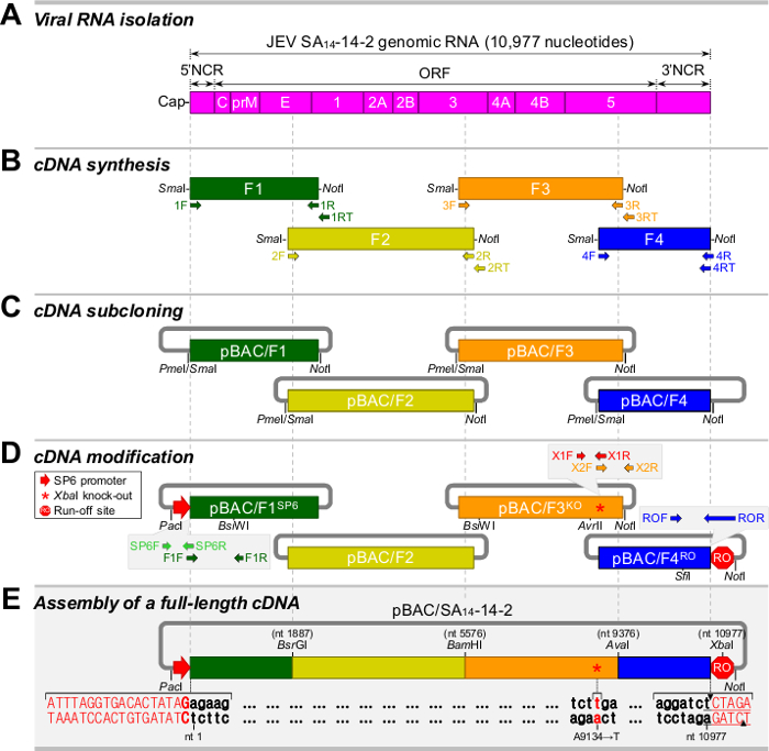
Figure 1. Strategy for the construction of a full-length cDNA of JEV SA14-14-2 as a BAC. (A) Isolation of viral RNA from JEV particles. Shown is a schematic diagram of the genomic RNA of JEV SA14-14-2. (B) Synthesis of four overlapping cDNA fragments (F1 to F4) covering the entire viral genome. (C) Subcloning of four overlapping cDNA fragments into a BAC vector, creating pBAC/F1 to pBAC/F4. (D) Modification of the cloned cDNAs for run-off transcription in vitro. pBAC/F1SP6 is a derivative of pBAC/F1 that contains the SP6 promoter sequence upstream of the viral 5'-end. pBAC/F3KO is a derivative of pBAC/F3 that contains a silent point mutation (A9134→T, asterisk). pBAC/F4RO is a derivative of pBAC/F4 that contains an artificial Xba I run-off site downstream of the viral 3'-end. (E) Assembly of a full-length SA14-14-2 BAC (pBAC/SA14-14-2). Please click here to view a larger version of this figure.

Figure 2
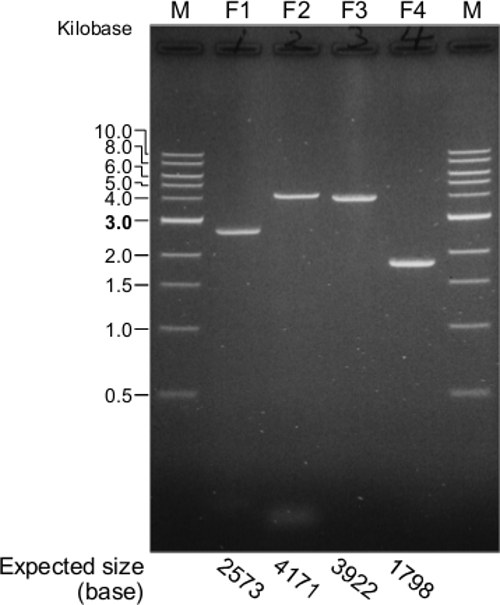
Figure 2. Synthesis of four overlapping cDNA fragments (F1 to F4) spanning the full-length genomic RNA of JEV SA14-14-2. The four RT-PCR products are evaluated by electrophoresis in a 0.8% agarose gel. M, 1 kb DNA ladder. The expected sizes of the four cDNA fragments are indicated at the bottom of the gel image. Please click here to view a larger version of this figure.

Figure 3
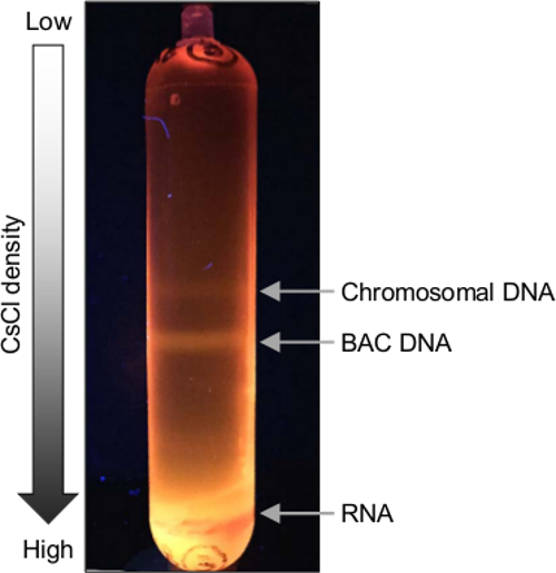
Figure 3. Purification of the BAC containing a full-length cDNA of JEV SA14-14-2. The BAC plasmid is isolated from E. coli DH10B by the SDS-alkaline lysis method and further purified by banding in a CsCl-EtBr gradient. Presented is an example of the CsCl-EtBr gradient using a 16 × 76 mm sealable polypropylene tube. Please click here to view a larger version of this figure.

Figure 4
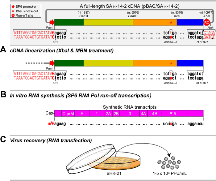
Figure 4. Overview of the recovery of infectious viruses from a full-length JEV SA14-14-2 cDNA assembled in a BAC. (A) Linearization of the cDNA template. The full-length JEV BAC is cut with Xba I and treated with MBN. (B) Synthesis of the RNA transcripts. The linearized cDNA is transcribed by SP6 RNA polymerase in the presence of the m7G(5')ppp(5')A cap analog. (C) Recovery of the synthetic JEVs. The in vitro transcribed RNAs are transfected into BHK-21 cells by electroporation, which generates a high titer of synthetic virus. Please click here to view a larger version of this figure.

Figure 5
Figure 5. Synthesis of the RNAs by in vitro transcription using a full-length JEV BAC as a cDNA template. (A) Generation of the linearized full-length JEV BAC, pBAC/SA14-14-2. Two independent clones of pBAC/SA14-14-2 (Cl.1 and Cl.2) are linearized by digestion with Xba I and subsequent treatment with MBN. The linearized BACs are examined by electrophoresis in a 0.8% agarose gel. (B) Production of the synthetic RNAs by run-off transcription. Each of the two linearized BACs is used as a template for SP6 RNA polymerase run-off transcription. Aliquots of the two transcription reactions are run on a 0.6% agarose gel. M, 1 kb DNA ladder. Please click here to view a larger version of this figure.

Figure 6
Figure 6. Specific infectivity of the synthetic RNAs transcribed from a full-length JEV BAC and the recovery of synthetic virus. BHK-21 cells are mock-electroporated (Mock) or electroporated with the RNA transcripts derived from each of the two independent clones of the full-length JEV BAC (Cl.1 and Cl.2). (A) RNA infectivity. The cells are overlaid with agarose and stained with crystal violet at 4 days post-transfection. RNA infectivity is determined by infectious center assays to estimate the amount of infectious RNA electroporated into the cells (left panel). Also, representative images of infectious centers are shown (right panel). (B) Protein expression. The cells are cultured in 4-well chamber slides. Viral protein expression in RNA-electroporated cells at 20 hr post-transfection (hpt) is analyzed by immunofluorescence assays using a primary anti-NS1 rabbit antiserum and a secondary Cy3-conjugated goat anti-rabbit IgG (red). The nuclei are counterstained with 4',6-diamidino-2-phenylindole (blue). The immunofluorescence images are overlaid on their corresponding differential interference contrast images. (C) Virus yield. The cells are cultured in 150 mm culture dishes. The production of infectious virions accumulated in the culture supernatants of RNA-electroporated cells at 22 and 40 hpt is examined by plaque assays. Please click here to view a larger version of this figure.

List of Materials

1. Molecular Cloning
2xYT Broth Sigma-Aldrich Y2377
[3H]UTP PerkinElmer NET380250UC Radioactive
50-mL Tube Thermo Scientific (Nalgene) 3114-0050
250-mL Bottle Beckman Coulter 356011
Agarose Lonza 50004
Agarose (Low Melting Point) Life Technologies (Invitrogen) 16520-100
AvaI New England BioLabs R0152S
AvrII New England BioLabs R0174S
BamHI New England BioLabs R0136S
BsiWI New England BioLabs R0553S
BsrGI New England BioLabs R0575S
Butanol Fisher Scientific A399-1 
Cap Analog [m7G(5ʹ)ppp(5ʹ)A] New England BioLabs S1405S
Cesium Chloride Fisher Scientific BP1595-1
Chloramphenicol Sigma-Aldrich C0378
Chloroform Sigma-Aldrich C2432 Carcinogenic
DE-81 Filter Paper GE Healthcare Life Sciences 3658-023
dNTP mix Life Technologies (Invitrogen) 18427-088
E. coli DH10B  Life Technologies (Invitrogen) 18297-010
EDTA Sigma-Aldrich E5134
Ethanol  Sigma-Aldrich E7023
Ethidium Bromide Sigma-Aldrich E7637 Toxic and highly mutagenic
Filter Column (Plasmid Maxiprep Kit) Life Technologies (Invitrogen) K2100-26
Glacial Acetic Acid Sigma-Aldrich A6283 Irritating
Glucose Sigma-Aldrich G5400
Glycogen  Roche 10901393001
High-Fidelity DNA Polymerase New England BioLabs M0491S
Isoamyl Alcohol Sigma-Aldrich I9392 Flammable
Isopropanol  Amresco 0918 Flammable
LB Broth Life Technologies (Invitrogen) 12795-027
Lithium Chloride Sigma-Aldrich L9650
Lysozyme Amresco 0663
Magnesium Chloride Sigma-Aldrich M8266
M-MLV Reverse Transcriptase Life Technologies (Invitrogen) 18080-044
Mung Bean Nuclease New England BioLabs M0250S
Needle (18G, 20G) BD 305196, 305175 Biohazardous (Sharps waste)
NotI New England BioLabs R0189S
Oligonucleotide Integrated DNA Technologies Custom Oligonucleotide Synthesis
PacI New England BioLabs R0547S
pBeloBAC11 New England BioLabs ER2420S (E4154S)
Phenol (Buffer-Saturated) Life Technologies (Invitrogen) 15513-039 Toxic and highly corrosive
Phenol:Chloroform:Isoamyl Alcohol Life Technologies (Invitrogen) 15593-031 Toxic and highly corrosive
Phenol:Guanidine Isothiocyanate Life Technologies (Ambion) 10296-010 Toxic, corrosive, and irritating
Pme I New England BioLabs R0560S
Potassium Acetate Amresco 0698
RNase Inhibitor Life Technologies (Invitrogen) 10777-019
rNTP Set GE Healthcare Life Sciences 27-2025-01
Sealable Polypropylene Tube (16 × 76 mm)  Beckman Coulter 342413
SfiI New England BioLabs R0123S
Sma I New England BioLabs R0141S
Sodium Acetate Sigma-Aldrich S2889
Sodium Dodecyl Sulfate Amresco 0227
Sodium Hydroxide Sigma-Aldrich S5881
SP6 RNA Polymerase New England BioLabs M0207S
Spin Column (Plasmid Miniprep Kit) Life Technologies (Invitrogen) K2100-11
Syringe HSW NORM-JECT 4200.000V0
T4 DNA Ligase New England BioLabs M0202S
Tris Amresco 0826
tRNA (yeast) Life Technologies (Invitrogen) 15401-011
XbaI  New England BioLabs R0145S
Name Company Catalog Number Comments
2. Cell Culture
Alpha Minimal Essential Medium Life Technologies (Gibco) 12561-049
Conical Tube (50 mL) VWR 21008-242
Crystal Violet  Sigma-Aldrich C0775
Culture Dish (150 mm)  TPP 93150 
Cuvette (2-mm Gap) Harvard Apparatus 450125 
Fetal Bovine Serum  Life Technologies (Gibco) 16000-044
Formaldehyde Sigma-Aldrich F1635 Toxic and carcinogenic
Glutamine Life Technologies (Gibco) 25030-081
Minimal Essential Medium Life Technologies (Gibco) 61100-061
Penicillin/Streptomycin Life Technologies (Gibco) 15070-063
Potassium Chloride Sigma-Aldrich P3911
Potassium Phosphate Monobasic Sigma-Aldrich P9791
Six-Well Plate TPP 92006
Sodium Chloride Sigma-Aldrich S3014
Sodium Phosphate Dibasic Sigma-Aldrich S3264
Trypsin-EDTA (0.25%) Life Technologies (Gibco) 25200-056
Vitamins Sigma-Aldrich M6895
Name Company Catalog Number Comments
3. Equipment
Agarose Gel Electrophoresis System Mupid MPDEXU-01
CO2 Incubator Thermo Scientific Heracell 150i
Desktop Centrifuge  Thermo Scientific ST16R
Electroporator Harvard Apparatus ECM 830
Longwave Ultraviolet Lamps (Handheld) UVP UVGL-58
Tabletop Centrifuge Beckman Coulter 368826
Thermocycler Life Technologies (Applied Biosystems) GeneAmp PCR System 9700
Vortexer Scientific Industries G-560
Water Bath Jeio Tech WB-10E

Lab Prep

Reverse genetics, an approach to rescue infectious virus entirely from a cloned cDNA, has revolutionized the field of positive-strand RNA viruses, whose genomes have the same polarity as cellular mRNA. The cDNA-based reverse genetics system is a seminal method that enables direct manipulation of the viral genomic RNA, thereby generating recombinant viruses for molecular and genetic studies of both viral RNA elements and gene products in viral replication and pathogenesis. It also provides a valuable platform that allows the development of genetically defined vaccines and viral vectors for the delivery of foreign genes. For many positive-strand RNA viruses such as Japanese encephalitis virus (JEV), however, the cloned cDNAs are unstable, posing a major obstacle to the construction and propagation of the functional cDNA. Here, the present report describes the strategic considerations in creating and amplifying a genetically stable full-length infectious JEV cDNA as a bacterial artificial chromosome (BAC) using the following general experimental procedures: viral RNA isolation, cDNA synthesis, cDNA subcloning and modification, assembly of a full-length cDNA, cDNA linearization, in vitro RNA synthesis, and virus recovery. This protocol provides a general methodology applicable to cloning full-length cDNA for a range of positive-strand RNA viruses, particularly those with a genome of >10 kb in length, into a BAC vector, from which infectious RNAs can be transcribed in vitro with a bacteriophage RNA polymerase.

Reverse genetics, an approach to rescue infectious virus entirely from a cloned cDNA, has revolutionized the field of positive-strand RNA viruses, whose genomes have the same polarity as cellular mRNA. The cDNA-based reverse genetics system is a seminal method that enables direct manipulation of the viral genomic RNA, thereby generating recombinant viruses for molecular and genetic studies of both viral RNA elements and gene products in viral replication and pathogenesis. It also provides a valuable platform that allows the development of genetically defined vaccines and viral vectors for the delivery of foreign genes. For many positive-strand RNA viruses such as Japanese encephalitis virus (JEV), however, the cloned cDNAs are unstable, posing a major obstacle to the construction and propagation of the functional cDNA. Here, the present report describes the strategic considerations in creating and amplifying a genetically stable full-length infectious JEV cDNA as a bacterial artificial chromosome (BAC) using the following general experimental procedures: viral RNA isolation, cDNA synthesis, cDNA subcloning and modification, assembly of a full-length cDNA, cDNA linearization, in vitro RNA synthesis, and virus recovery. This protocol provides a general methodology applicable to cloning full-length cDNA for a range of positive-strand RNA viruses, particularly those with a genome of >10 kb in length, into a BAC vector, from which infectious RNAs can be transcribed in vitro with a bacteriophage RNA polymerase.

Procedure

Reverse genetics, an approach to rescue infectious virus entirely from a cloned cDNA, has revolutionized the field of positive-strand RNA viruses, whose genomes have the same polarity as cellular mRNA. The cDNA-based reverse genetics system is a seminal method that enables direct manipulation of the viral genomic RNA, thereby generating recombinant viruses for molecular and genetic studies of both viral RNA elements and gene products in viral replication and pathogenesis. It also provides a valuable platform that allows the development of genetically defined vaccines and viral vectors for the delivery of foreign genes. For many positive-strand RNA viruses such as Japanese encephalitis virus (JEV), however, the cloned cDNAs are unstable, posing a major obstacle to the construction and propagation of the functional cDNA. Here, the present report describes the strategic considerations in creating and amplifying a genetically stable full-length infectious JEV cDNA as a bacterial artificial chromosome (BAC) using the following general experimental procedures: viral RNA isolation, cDNA synthesis, cDNA subcloning and modification, assembly of a full-length cDNA, cDNA linearization, in vitro RNA synthesis, and virus recovery. This protocol provides a general methodology applicable to cloning full-length cDNA for a range of positive-strand RNA viruses, particularly those with a genome of >10 kb in length, into a BAC vector, from which infectious RNAs can be transcribed in vitro with a bacteriophage RNA polymerase.

Tags