Mice were housed in pathogen-free conditions in compliance with the institutional guidelines until euthanization (The Institutional Animal Care and Use Committee (IACUC) guidelines at the National University of Singapore and the National Advisory Committee for Laboratory Animal Research (NACLAR) guidelines).
1. Mice
2. Preparation of Complete Medium
3. Preparation of Enzyme Solutions
4. Preparation of Collagenase D-pronase Mix (≤2 Tails)
Note: Perform the subsequent steps in a sterile cell culture hood.
5. Extraction of Fibroblasts from Ear and Tail Tissues
6. Culturing of Cell Mixture
7. Sub-culture of Fibroblasts
8. Preparation of Ears for Shipment
Note: Perform the subsequent steps in a sterile cell culture hood.
Extraction of fibroblasts from tissue results in a significant amount of tissue debris (Figure 1). In contrast to tissue debris, fibroblasts adhere to tissue culture plastic surfaces between day 1 and 3 of culture. The medium of fibroblast cultures can be safely changed on day 3 of culture, which should significantly decrease the levels of debris present in the culture (Figure 2). Fibroblasts display an elongated morphology and a clearly visible cytoplasm (Figures 1 and 2). Mitotic cells should be present from day 3 of culture onwards and cells should reach 70-80% of cell confluency within 3-4 days of culture. The yield from ear and tail tissues in a 10 cm culture dish ranges from 4 to 5 x 10⁵ (ears) and 5 to 6 x 10⁵ cells (tail) on day 3 of culture. Following the third day of fibroblasts isolation, the cells can be passaged and seeded at 2 x 10⁵ cells per 10 cm culture dish.
Extraction of fibroblasts from ears stored at RT for 10 days should result in 70-80% cell confluency within 5 to 6 days of culture (Figure 3). Seeding the cells at 2 x 10⁵ cells per 10 cm culture dish after day 5 should also give rise to approximately 1 x 10⁶ cells within 3-4 days of culture. Long-term storage does not affect the time it takes for fibroblasts to enter senescence in our experience (data not shown).
To verify the identity of cells after 3 days of culture, cells can be labelled for the fibroblasts marker vimentin14. Using the above protocol, we routinely obtain pure fibroblast cultures as indicated by vimentin staining (Figure 4).
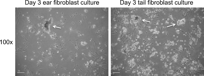
Figure 1. Fibroblast culture on day 3 post extraction prior to change of medium. Representative bright field images of cell debris (white arrows) present on day 3 of culture. Images were captured at 100× magnification using a light microscope. The scale bar represents 100 µm. Please click here to view a larger version of this figure.
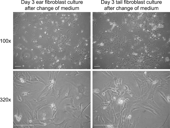
Figure 2. Fibroblast culture on day 3 post extraction after addition of new medium. Representative bright field images of fibroblasts on day 3 in culture. Images were captured at 100× and 320× magnification using a light microscope. The scale bar represents 100 µm. Please click here to view a larger version of this figure.
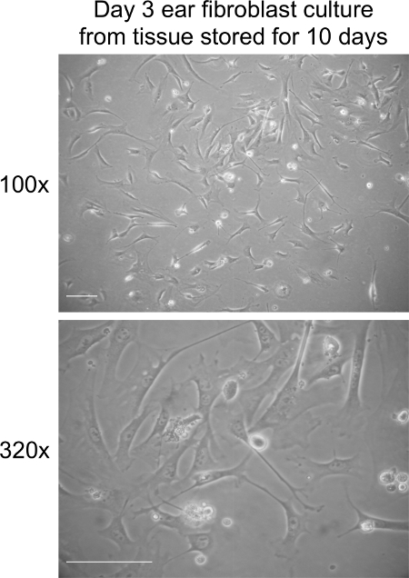
Figure 3. Fibroblast culture on day 3 post extraction from ear tissues stored at RT for 10 days. Representative bright field images of fibroblasts at day 3 in culture. Images were captured at 100× and 320× magnification using a light microscope. The scale bar represents 100 µm. Please click here to view a larger version of this figure.
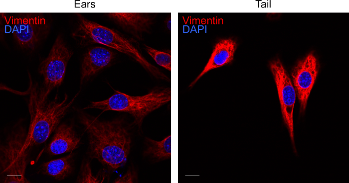
Figure 4. Labelling of fibroblast cultures for vimentin. Representative confocal image of ear and tail fibroblasts on day 3 of culture. Fibroblasts extracted from the tissues were labelled for vimentin (red) and DAPI (blue). The fluorescent images were acquired by confocal microscopy. The scale bar represents 10 µm. Please click here to view a larger version of this figure.
RPMI-1640 | HyClone | SH30027.01 | |
Fetal Calf Serum | HyClone | SV30160.03 | |
2-mercaptoethanol | Sigma-Aldrich | M3148 | |
Asparagine | Sigma-Aldrich | A4159 | |
Glutamine | Sigma-Aldrich | G8540 | |
Penicillin/Streptomycin | HyClone | SV30010 | |
Ethanol | Merck Millipore | 107017 | Absolute for analysis |
Collagenase D | Roche Diagnostics | 11088866001 | From Clostridium histolyticum, lyophilized, non-sterile |
Pronase protease | Merck Millipore | 53702 | From Streptomyces griseus |
Tris buffer (pH 8) | 1st BASE | 1415 | Ultra pure grade |
0.5M EDTA (pH 8) | 1st BASE | BUF-1053 | Biotechnology grade |
10X Phosphate Buffered Saline (PBS) | 1st BASE | BUF-2040-10X4L | Ultra pure grade |
Trypsin-EDTA solution 10X | Sigma-Aldrich | 59418C-100ML | 0.5% trypsin, 0.2% EDTA, trypsin gamma irradiated by SER-TAIN process, without phenol red, in saline |
Amphotericin B | Sigma-Aldrich | A2492-20ml | 250 μg/ml in deionized water, sterile-filtered |
Scissors | Aesculap | ||
Forcep | Aesculap | AE-BD312R | |
0.2 μM syringe filter | Sartorius Stedim | 16534 | |
70 μM cell strainer | SPL | 93070 | |
Syringe plunger | Terumo | SS+10L | |
Cryovial tube | NUNC | 368362 | |
1.7 ml microcentrifuge tube | Axygen | MCT-175-C | |
10 cm cell culture dish | Greiner | 664160 | Cell culture treated dish |
15 ml conical bottom tube | Greiner | 188271 | |
50 ml conical bottom tube | Greiner | 227261 | |
Water bath | GFL | 1002 | |
Centrifuge | Eppendorf | 5810R | |
Incubation shaker | Sartorius Stedim | Certomat-BS1 | |
Zeiss Axiovert 25 light microscope | Carl Zeiss AG |
Primary cells are derived directly from tissue and are thought to be more representative of the physiological state of cells in vivo than established cell lines. However, primary cell cultures usually have a finite life span and need to be frequently re-established. Fibroblasts are an easily accessible source of primary cells. Here, we discuss a simple and quick experimental procedure to establish primary fibroblast cultures from ears and tails of mice. The protocol can be used to establish primary fibroblast cultures from ears stored at RT for up to 10 days. When the protocol is carefully followed, contaminations are unlikely to occur despite the use of non-sterile tissue stored for extended time in some cases. Fibroblasts proliferate rapidly in culture and can be expanded to substantial numbers before undergoing replicative senescence.