The setup of the transcutaneous measurement is very simple and fast: the device is placed on the double-sided adhesive patch (Figure 1) and adjusted in size if necessary (Figure 2), the battery is prepared (Figure 3) and connected (Figure 4).
This method to assess renal function has been already validated in different species by comparison with the traditional approach of plasma clearance9,11,12. Following the protocol exposed here, Schreiber et al. demonstrated the validity of the technique in different mouse models, showing highly comparable results among the transcutaneous measurement and plasma clearance for all the groups studied (Table 1)9. In this work the obtained t1/2 was converted to GFR using a mouse specific semi-empirical conversion factor.
The consistency of the transcutaneous assessment of renal function has been also proved using different strains of mice. Sequential measurements within 3 days in the same animal showed a coefficient of variances of 3.0-6.2%13. In this study there was no conversion to GFR but the results were expressed and interpreted directly in terms of t1/2.
The possibility to have the results almost in real-time is one of the great advantages of this method. After the recording period, the results are immediately available for analysis and the provided software displays the excretion curve of FITC-sinistrin instantaneously (Figure 6). Within the same software the t1/2 of FITC-sinistrin can be obtained, which can be directly used as the parameter to evaluate renal function of converted in terms of GFR. Figure 7 shows how it looks the FITC-sinistrin excretion curve measured transcutaneously in an animal with renal impairment. When renal function is affected, FITC-sinistrin t1/2 increases due to the reduced excretion of the substance and the appearance of the curve changes. Typically, the measured curve does not come back to background level and presents an increased area under the curve. In the same animal, measurements from pre- to post-injury can experience an increase in the maximum fluorescence intensity due to accumulation of the marker caused by its reduced excretion. In presence of kidney failure, the transcutaneously measured curve of FITC-sinistrin can show a steady state due to severely impaired function (Figure 7B).
The use of the miniaturized device in several strains of rodents with different health status has shown that this technique is suitable and sensitive enough to detect changes due to renal disease and ageing. Table 2 shows a summary of the murine models studied to date with this method.
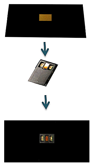
Figure 1. Placement of the device on the double-sided adhesive patch. The device is mounted on the adhesive patch positioning its optical part in the transparent window.
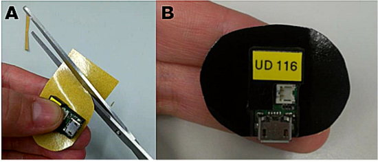
Figure 2. Adapting the adhesive patch for small-sized rodents. For use in small animals it is recommended to reduce the size of the patch. A: Use the device as a guide to cut the patch properly. B: Once the desired size has been obtained, the device can be placed on the sticky surface of the patch. Please click here to view a larger version of this figure.
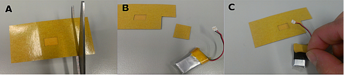
Figure 3. Preparation of the battery. To attach the battery to the device, cut a small piece of double-sided adhesive patch (A and B) and place it on the surface of the battery (C). Please click here to view a larger version of this figure.
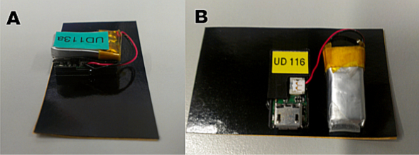
Figure 4. Connection and placement of the battery during the measurement. (A) In small rodents such as mice, due to the reduced space, the battery must be placed on top of the device. (B) In larger animals the battery can be placed next to the device. Please click here to view a larger version of this figure.
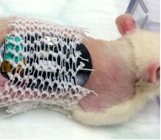
Figure 5. Placement of the tubular elastic gauze bandage in a rat. The tubular bandage should cover the double-sided adhesive patch without interfering with the free movement of the limbs for the comfort of the animal. Please click here to view a larger version of this figure.
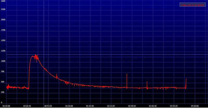
Figure 6. Representative image of an elimination curve of FITC-sinistrin. The signal generated by FITC-sinistrin is detected transcutaneously and stored in the internal memory of the device. When the recorded data are downloaded onto a PC, the software generates a curve comparable to the one presented in the image. Y-axis shows the recorded fluorescence intensity [AU], emitted by the injected FITC-sinistrin marker, while X-axis represents the duration of the measurement in time [min]. Please click here to view a larger version of this figure.
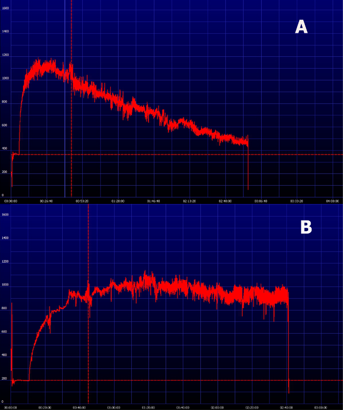
Figure 7. Representative image of an elimination curve of FITC-sinistrin in animals with impaired renal function. (A) FITC-sinistrin elimination curve in animals with reduced renal function typically shows an increased area under the curve and inability to reach the baseline within the normal measurement period. (B) In severely impaired animals the transcutaneously measured curve can show a steady state which indicates renal failure. Please click here to view a larger version of this figure.
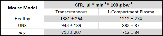
Table 1. Validation of the transcutaneous measurement by comparing with the traditional plasma clearance. The transcutaneous measurement of renal function has been validated in different mouse models (healthy and unilaterally nephrectomized (UNX) C57BL/6-129 SV mice and mouse model of nephronophthisis (pcy)) by comparison with plasma clearance. Values are means ± SD. GFR, glomerular filtration rate9.
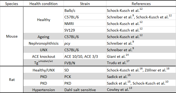
Table 2. Murine models studied using the transcutaneous measurement. UNX, unilaterally nephrectomized; ACE, angiotensin-converting enzyme; Tgumodwt/wt, transgenic uromodulin knockout, SD, Sprague Dawley rat; PKD, polycystic kidney disease.