1. DNA Biotinylation
2. Functionalized Surface Derivatization
Note: In order to tether an end of a DNA molecule on glass surface, a primary amine group is silanized on a coverslip followed by biotin-PEG coating as shown in Figure 1. This PEGylation process is important for single-molecule DNA imaging because it can significantly decrease random noise created by attachment of undesired molecules on the surface.
3. Assembling a Flow Chamber
4. Sample Loading into Flow Chamber
Note: Neutravidin can be replaced with other avidin proteins such as streptavidin. All of the reactions can be performed at room temperature, unless it is mentioned. Take sample solution in a yellow tip, and install the tip with the solution onto the hole of acrylic holder (Figure 2b).
Figure 1 shows two different DNA tethering methods depending on terminal structures of the DNA molecule. Figure 1a illustrates how the sticky ended DNA molecules are hybridized with complementary biotinylated oligonucleotides, which are immobilized on the avidin-coated PEG surface. Figure 1b shows the addition of biotinylated ddNTP or dNTP to the 3' hydroxyl group of a blunt ended DNA by terminal transferase. We added flexible linkers between DNA molecules and biotin. This increased the ligation and avidin-biotin binding efficiency to provide sufficient space for the reactions. Figure 1c demonstrates the overall schematic representation for DNA tethering on the avidin-coated PEG surface.
Figure 2 depicts the assembly of the flow chamber along with an acrylic holder. Compared to glass/quartz slide glasses, a custom-made acrylic holder simplifies the preparation of the flow chamber for epi-fluorescent microscopy.12 The flow chamber enables instant changing of buffer conditions for enzyme reactions,6 and stretching DNA molecules.
Figure 3 demonstrates single-tethered DNA and T4 λDNA stained with FP-DBP. Microfluidic shear flows elongated single DNA molecules up to full contour lengths such as 16.5 μm (48.5 kb λ DNA x 0.34 nm) and 56.4 μm (166 kb T4 DNA x 0.34 nm). We were able to repeat DNA stretching and relaxation multiple times during an extended period (e.g., half-hour), since we utilized FP-DBP that does not cause photo-cleavage.
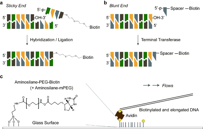
Figure 1. Schematic illustration of DNA tethering. a) A sticky ended DNA is hybridized with a biotinylated complementary oligonucleotide. Triethylene glycol is a flexible linker between the oligonucleotide and biotin. b) Biotinylated dNTP is added to a blunt end of double stranded DNA, by terminal transferase. Polycarbon spacer (11-atoms) is a flexible linker between dNTP and biotin. c) Biotinylated DNA is anchored to an avidin protein, which is linked to a biotinylated PEG covalently bonded to the glass surface. The ratio of biotinylated PEG to normal PEG was 0.025 (1:40). Please click here to view a larger version of this figure.
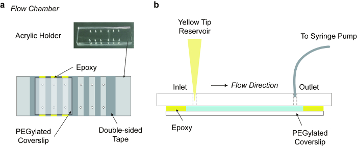
Figure 2. Flow chamber. a) Flow chamber consisting of an acrylic acid resin holder, strips of double-sided tapes and a PEGylated cover slip. The acrylic holder of dimensions 76 mm x 26 mm x 5 mm (L x W x H), was fabricated with laser cutting. b) Side view of the Flow chamber: punched holes are an inlet port on which a yellow tip is installed for a buffer reservoir, and an outlet port that is connected to a tubing controlled by a syringe-pump. Please click here to view a larger version of this figure.
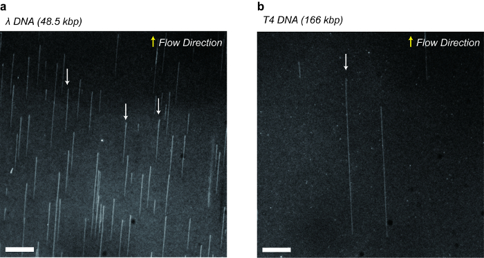
Figure 3. Fluorescent microscopic images of stretched DNA molecules stained with FP-DBP. a) Bacteriophage λ DNA (48.5 kbp) tethered on the surface after ligation with biotinylated complementary oligonucleotides. b) Bacteriophage T4 DNA (166 kbp) tethered on the surface after adding biotin with terminal transferase. Indicated arrows show DNA molecules stretched up to their full contour lengths such as 16.5 µm for λ DNA and 56.4 µm for T4 DNA. Scale bars: 10 µm. Please click here to view a larger version of this figure.
1. DNA Biotinylation | |||
1.1) Biotinylation of blunt-end DNA using Terminal Transferase (TdT) | |||
Terminal Transferase | New England Biolabs | M0315S | Provided with 10x reaction buffer, 2.5 mM Cobalt chloride |
Biotin-11-dUTP | Invitrogen | R0081 | Biotin-ddNTP is also available |
T4GT7 Phage DNA | Nippon Gene | 318-03971 | |
Ethylenediaminetetraacetic acid | Sigma-Aldrich | E6758 | EDTA |
1.2) Biotinylation of sticky end DNA Using DNA Ligase | |||
T4 DNA Ligase | New England Biolabs | M0202S | Provided with 10x reaction buffer |
Lambda Phage DNA | Bioneer | D-2510 | Also available at New England Biolabs |
2. Functionalized Surface Derivatization | |||
2.1) Piranha Cleaning | |||
Coverslip | Marienfeld-Superior | 0101050 | 22×22 mm, No. 1 Thickness |
Teflon rack | Custom Fabrication | ||
PTFE Thread Seal Tape | Han Yang Chemical Co. Ltd. | 3032292 | Teflon™ tape |
Sulferic acid | Jin Chemical Co. Ltd. | S280823 | H2SO4, 95 % Purity |
Hydrogen peroxide | Jin Chemical Co. Ltd. | H290423 | H2O2, 35 % in water |
Sonicator | Daihan Scientific Co. Ltd. | WUC-A02H | Table-top Ultrasonic Cleaner |
2.2) Aminosilanization on Glass Surface | |||
N-[3-(Trimethoxysilyl)propyl] ethylenediamine |
Sigma-Aldrich | 104884 | |
Glacial Acetic Acid | Duksan Chemicals | 414 | 99 % Purity |
Methyl Alcohol | Jin Chemical Co. Ltd. | M300318 | 99.9 % Purity |
Polypropylene Container | Qorpak | PLC-04907 | |
Ethyl Alcohol | Jin Chemical Co. Ltd. | A300202 | 99.9 % Purity |
2.3) PEGylation of the coverslip | |||
Sodium Bicarbonate | Sigma-Aldrich | S5761 | |
Syringe Filter | Sartorius | 16534———-K | |
Biotin-PEG-SC | Laysan Bio | Biotin-PEG-SC-5000 | |
mPEG-SVG | Laysan Bio | MPEG-SVA-5000 | |
Acetone | Jin Chemical Co. Ltd. | A300129 | 99 % Purity |
Microscope Slides | Marienfeld-Superior | 1000612 | ~76x26x1 mm |
3. Assembling a Flow Chamber | |||
Acrylic Support | Custom Fabrication | ||
Double-sided Tape | 3M | Transparent type | |
Quick-dry Epoxy | 3M | ||
Polyethylene Tubing | Cole-Parmer | 06417-11, 06417-21 | |
Gas Tight 250 µl Syringe | Hamilton | 81165 | |
Syringe Pump | New Era Pump Systems Inc. | NE-1000 | |
4. Sample Loading into Flow Chamber | |||
Neutravidin | Thermo Scientific | 31000 | |
Tris base | Sigma-Aldrich | T1503-5KG | Trizma base |
Microscope | Olympus | IX70 | |
EMCCD Camera | Q Imaging | Rolera EM-C2 | |
Solid-state Laser (488 nm) | Oxxius | LBX488 | |
Alconox | Alconox Inc. |
Large DNA molecules tethered on the functionalized glass surface have been utilized in polymer physics and biochemistry particularly for investigating interactions between DNA and its binding proteins. Here, we report a method that uses fluorescent microscopy for visualizing large DNA molecules tethered on the surface. First, glass coverslips are biotinylated and passivated by coating with biotinylated polyethylene glycol, which specifically binds biotinylated DNA via avidin protein linkers and significantly reduces undesirable binding from non-specific interactions of proteins or DNA molecules on the surface. Second, the DNA molecules are biotinylated by two different methods depending on their terminals. The blunt ended DNA is tagged with biotinylated dUTP at its 3′ hydroxyl terminus, by terminal transferase, while the sticky ended DNA is hybridized with biotinylated complimentary oligonucleotides by DNA ligase. Finally, a microfluidic shear flow makes single DNA molecules stretch to their full contour lengths after being stained with fluorescent protein-DNA binding peptide (FP-DBP).