1. Bacterial Culture
2. Host Cell Culture
3. Host Cell Infection with Permeable Membrane Insert System
4. Sample Collection
5. Suggested Applications
The permeable membrane insert-based infection system protocol developed for the study of secreted bacterial factors is detailed in Figure 1. This system relies on the separation of bacteria and host cells via a porous membrane to assess the effects of secreted bacterial factors, in this case Streptolysin S (SLS), on host responses such as host membrane integrity, cellular viability, cellular signal transduction, and secreted host cell factors. Figure 2 provides representative Western Blot data demonstrating that this system may be used to assess changes in the activation of contact-independent host signaling proteins. Specifically, the representative data show significantly enhanced p38 MAPK activation in the presence of SLS-producing GAS strains. This system can also be applied to visualize the effects of secreted bacterial factors on host protein localization by immunofluorescence microscopy (Figure 3). The data show SLS-dependent activation of the key inflammatory mediator Nuclear Factor kappa B (NFκB), which translocates from the cytoplasm to the nucleus upon activation21. The results in Figures 2 and 3 indicate that both SLS-dependent inflammatory signaling responses do not require direct contact between the bacteria and host cells. As previous experiments have already demonstrated SLS-dependent p38 and NFκB activation in direct infection models21, similar levels of p38 or NFκB activation among the three GAS strains in the permeable membrane insert-based system would have indicated that the response required direct contact between the bacteria and host cells. Figure 4 demonstrates that this infection system may be applied to assess toxin-dependent changes in host cytotoxicity via ethidium homodimer and LDH release assays. This is evidenced by the significant increase in both membrane permeabilization and in the release of LDH from host cells exposed to GAS strains containing SLS compared to uninfected cells or cells exposed to the SLS-deficient strain. These changes in cytotoxicity are not immediate, as significant effects are not evident until 12 hr post infection in the case of membrane permeabilization and 16 hr in the case of LDH release. The representative data illustrate the importance of selecting appropriate time points and infection conditions for the evaluation of these host responses. In addition to allowing for the assessment of host signaling changes and cytotoxicity, the permeable membrane insert-based infection system is applicable to the study of metabolic changes such as accurate determination of host ATP levels by preventing bacterial contamination of host cell lysates (Figure 5). These data show a significant loss of keratinocyte ATP in response to GAS infection by 16 hours post infection, with enhanced ATP loss in the presence of SLS. These results are consistent with the observed toxin-dependent increases in host stress-response signaling and cytotoxicity.
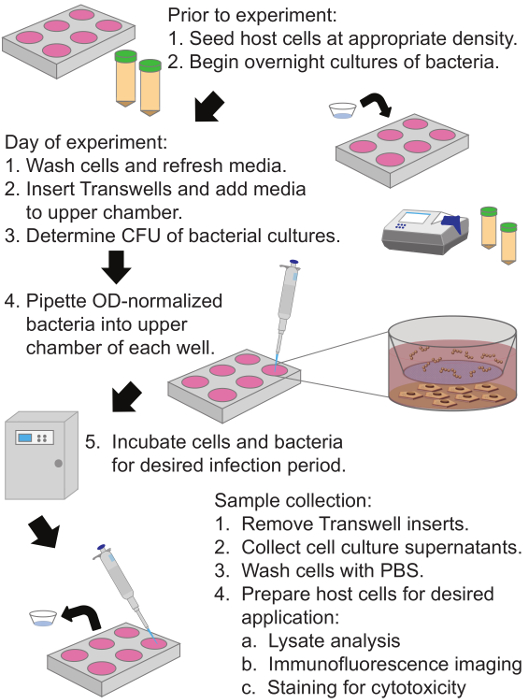
Figure 1: Diagram of Permeable Membrane Insert-Based Infection System Protocol to Assess the Effects of Secreted Bacterial Factors on Host Cells. Human keratinocytes are plated in the bottom compartment and grown to 90% confluence. A collagen coated membrane with 0.4 µm pores separates the upper and lower chambers, and bacteria are added to the upper chamber of the permeable membrane insert system for the desired infection period. Cell culture supernatants and host cells may be collected following the infection period and utilized for a variety of analyses. Please click here to view a larger version of this figure.
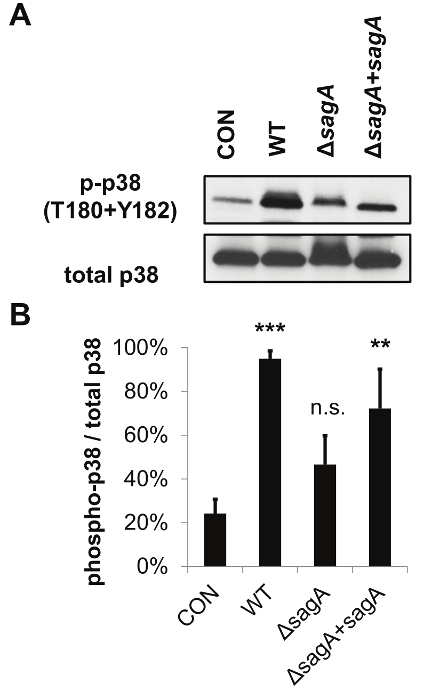
Figure 2: The Permeable Membrane Insert-Based Infection System can be Utilized for Host Cell Lysate Analysis by SDS-PAGE and Western Blotting. The representative data demonstrate that SLS enhances activation of the p38 MAPK pathway in infected keratinocytes. (A) HaCaTs were infected with GAS for 7 hr via the permeable membrane insert infection system (at MOI = 10) and lysates were assessed for activation of p38. (B) Densitometry from three independent Western Blots was performed to quantify the relative activation of p38 in response to GAS'infection. Averages from three biological replicates are shown, with error bars representing standard deviation. Relative activation of p38 is represented as phosphorylated/total protein level. Statistical significance was determined compared to uninfected cells. The overall p-value was determined by ANOVA (p = 0.0063). Dunnett's tests were performed post hoc to compare each condition to the corresponding uninfected control mean. *, p = 0.01-0.05; **, p = 0.001-0.01; ***, p = 0.0001-0.001; ****, p <0.0001. This figure has been modified from Flaherty et al. 201521. Please click here to view a larger version of this figure.
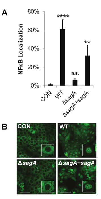
Figure 3: The Permeable Membrane Insert-Based Infection System Allows for the Visualization of Host Signaling Changes by Immunofluorescence Microscopy. The representative data show that Streptolysin S enhances pro-inflammatory signaling through activation of NFκB. HaCaT human keratinocytes were infected with GAS at an MOI of 10 for 8 hr using the permeable membrane insert-based infection system. (A and B) Nuclear localization of NFκB was assessed by immunofluorescence imaging. The percentage of nuclear localized cells was calculated by counting the number of cells in which NFκB had translocated from the cytoplasm to the nucleus for a given field and dividing that number by the total number of cells for the same field. Scale bars indicate 100 µm. (A) The average of three biological replicates are represented for each condition with error bars representing standard deviation. The overall p-value was determined by ANOVA; p <0.0001. Dunnett's tests were performed to compare each condition to the corresponding uninfected control condition. *, p = 0.01-0.05; **, p = 0.001-0.01; ***, p = 0.0001-0.001; ****, p <0.0001. This figure has been modified from Flaherty et al. 201521. Please click here to view a larger version of this figure.
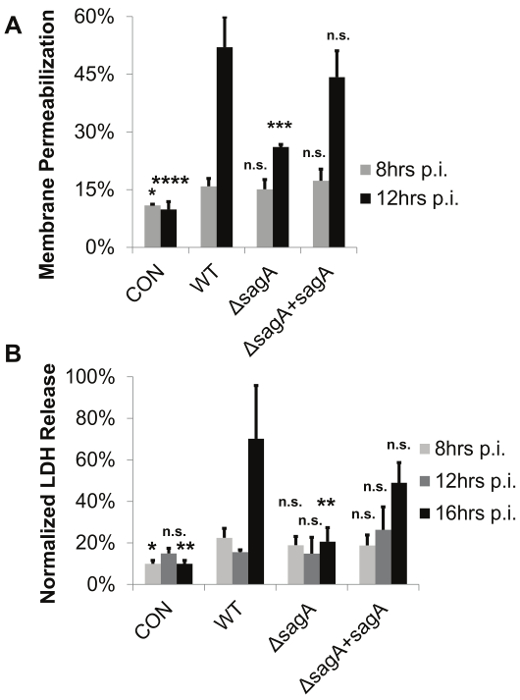
Figure 4: The Permeable Membrane Insert-Based Infection System Allows for the Determination of Bacterial-Mediated Membrane Permeabilization and Cytotoxicity in the Absence of Direct Contact between Bacteria and Host Cells. The representative data demonstrate that keratinocyte viability decreases in the presence of active SLS toxin. GAS–induced cell death was assessed in HaCaT cells in the presence of WT, SLS-deficient or sagA complemented GAS. Keratinocytes were exposed to GAS using the permeable membrane insert-based infection system for 8-16 hr at an MOI of 10. (A) Viability was assessed by ethidium homodimer assay or (B) LDH release assay. In both panels, 3 replicates are averaged and error bars represent standard deviation. Significance for each time point was determined by ANOVA (A) 8 hr, p = 0.0241; 12 hr, p <0.0001 (B) 8 hr, p = 0.0287; 12 hr, p = 0.1977; 16 hr, p = 0.0031. Dunnett's tests were performed to compare means from each condition to the wild-type infection for the corresponding time point. *, p = 0.01-0.05; **, p = 0.001-0.01; ***, p = 0.0001-0.001; ****, p <0.0001. This figure has been modified from Flaherty et al. 201521. Please click here to view a larger version of this figure.
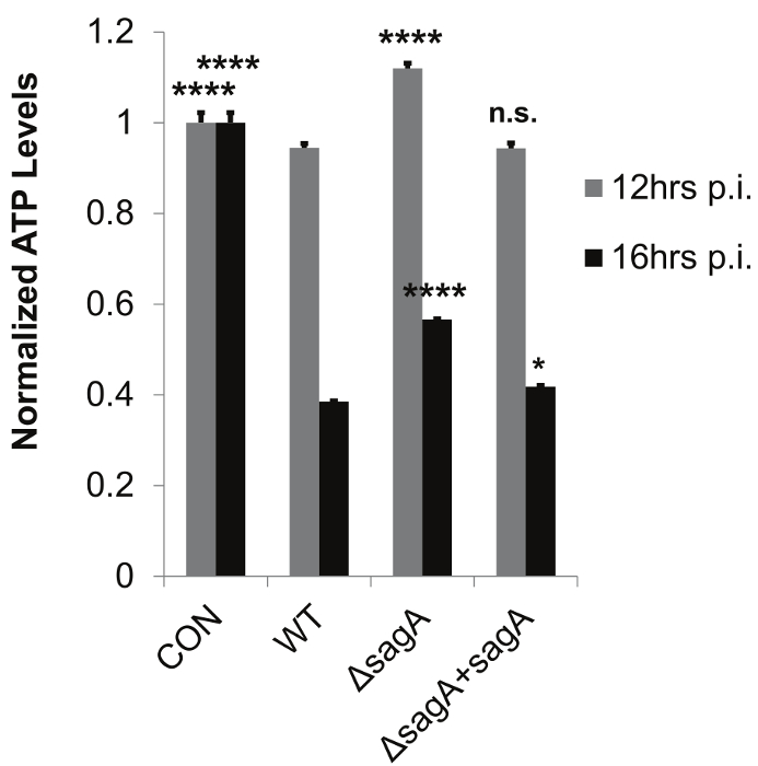
Figure 5: The Permeable Membrane Insert-Based Infection System Allows for Accurate Determination of Host ATP Levels by Preventing Bacterial Contamination of Host Cell Lysates. The representative data demonstrate SLS-dependent loss of ATP during Group A Streptococcal infection. HaCaT cells were infected with GAS for 8-16 hr using the permeable membrane insert-based infection system. Technical replicates (n = 3) from one representative biological replicate (2 x 10⁶ cells per sample) were averaged for each condition, with error bars representing standard deviation. The overall p-values were determined by ANOVA (12 hr, p <0.0001; 16 hr, p <0.0001). Dunnett's tests were performed to compare each condition with wild-type infection for the corresponding time point. *, p = 0.01-0.05; **, p = 0.001-0.01; ***, p = 0.0001-0.001; ****, p <0.0001. This figure has been modified from Flaherty et al. 201521. Please click here to view a larger version of this figure.
Todd-Hewitt broth | Acumedia | 7161A | Use appropriate broth for bacterial species of interest. |
BioSpectrometer | Eppendorf | basic model | Any UV-Vis spectrophotometer is suitable. |
Petri Dishes | Fisher Scientific | FB0875713 | Other brands are suitable. |
Agar | Sigma | A1296-1kg | Other brands are suitable. |
HaCaT human epithelial keratinocytes | Gift from lab of Victor Nizet. | ||
Dulbecco's Modified Eagle's Medium (DMEM) | Life Technologies | 11995-073 | Use appropriate media for cell line of interest. |
Fetal Bovine Serum (FBS) | Biowest | S162H | Use appropriate media additives for cell line of interest. |
10cm tissue culture dishes | Nunc | 150350 | Other brands are suitable. |
6 well tissue culture dishes | CytoOne | cc7682-7506 | Other brands are suitable but need to be compatible with the Corning insert. |
24 well tissue culture dishes | CytoOne | cc7682-7524 | Other brands are suitable but need to be compatible with the Corning insert. |
Glass coverslips | Fisherbrand | 12-541-B | These must be autoclaved prior to use for cell plating. |
Phosphate Buffered Saline (PBS) | Gibco | 10010-023 | |
Phenol Red Free DMEM | Life Technologies | 31053028 | Phenol Red Free media is only required for the LDH release assay. |
Bovine Serum Albumin (BSA) | Fisher Scientific | BP1600-100 | |
L-glutamine | Gibco | 25030149 | Only required to supplement cell culture media for LDH release assay. |
Sodium pyruvate | Gibco | BP356100 | Only required to supplement cell culture media for LDH release assay. |
Collagen-coated 0.4μm Transwell inserts (6 well) | Corning | 3540 | |
Collagen-coated 0.4μm Transwell inserts (24 well) | Corning | 3595 | |
Forceps | Fisher | 3120018 | These must be sterilized with ethanol prior to handeling Transwell inserts. |
Cell scrapers | Fisherbrand | 08-100-241 | These are only required for the collection of cell lysates. |
Paraformaldehyde | Fisher Scientific | 04042-500 | TOXIC. This is only required for immunofluorescence imaging. |
Bicinchoninic acid (BCA) assay kit | Pierce | 23227 | Other protein quantification assays may be used. |
Tris-glycine 4-15% polyacrylamide gels | BioRad | 4561083 | Buffer system and percent polyacrylamide should be selected based on proteins of interest. |
Electrophoresis cassette supplies | BioRad | 1658063 | Only required for Western Blotting. |
Electrophoresis power supply | BioRad | 1645050 | Only required for Western Blotting. |
Western blot transfer cassette | BioRad | Only required for Western Blotting. | |
Polyvinylidene (PVDF) Membrane | EMD Millipore | IPVH00010 | Only required for Western Blotting. |
Tween 20 | Sigma | P1379-500mL | |
Rabbit IgG-HRP secondary antibody | Santa Cruz Biotechnology | sc2030 | The secondary antibody should be determined based on the selected method of detection. |
Mouse IgG-HRP secondary antibody | Santa Cruz Biotechnology | sc2031 | The secondary antibody should be determined based on the selected method of detection. |
Phospho-p38 (T180+T182) MAPK antibody | Cell Signaling | 4511 | Select appropriate primary antibodies based on proteins of interest. |
Total p38 MAPK antibody | Cell Signaling | 8690 | Select appropriate primary antibodies based on proteins of interest. |
Goat anti-rabbit IgG Alexafluor488 | Molecular Probes | A-11034 | Other secondary antibodies may be used for immunofluoresence imaging detection. |
LumiGLO Detection Reagent | KPL | 54-61-00 | Only required for Western Blotting with detection on film. |
Detection film | Biodot | BDB57 | Only required for Western Blotting with detection on film. |
DeltaVision Nikon 90i fluoresence microscope | Nikon | Other fluorescence microscopes are suitable for these analyses. | |
Normal goat serum | Thermo Scientific | 31873 | |
Triton X-100 | Sigma | T9284-500mL | |
DAPI nuclear stain | Cell Signaling | 4083 | Select stains based on specific immunofluorescence applications of interest. |
Rhodamine-phalloidin actin stain | Molecular Probes | R415 | Select stains based on specific immunofluorescence applications of interest. |
Fluoromount-G | Southern Biotech | 0100-01 | Only required for immunofluorescence imaging. |
Ethidium homodimer 1 | Molecular Probes | E1169 | Only required for membrane permeabilization cytotoxicity assay. |
Spectramax M5 Microplate Reader | Molecular Devices | Other microplate readers capable of detecting UV-vis, fluorescence and luminescence may be suitable. | |
Saponin | Sigma | 47036-50G-F | Only required for membrane permeabilization cytotoxicity assay. |
Molecular Probes ATP Determination Kit | Life Technologies | A22066 | Other kits are likely to be suitable. |
Cytotoxicity Detection Kit for LDH release | Roche | 11644793001 | Other kits are likely to be suitable. |
Nonidet P40 Substitute | Sigma | 74385-1L | Other suppliers are suitable. |
HALT Phosphatase Inhibitor Cocktail | Thermo Scientific | 78420B | Other cocktails are likely to be suitable. |
SIGMAFAST Protease Inhibitor Cocktail Tablets, EDTA-free | Sigma | S8830-2TAB | Other cocktails are likely to be suitable. |
Many bacterial pathogens secrete potent toxins to aid in the destruction of host tissue, to initiate signaling changes in host cells or to manipulate immune system responses during the course of infection. Though methods have been developed to successfully purify and produce many of these important virulence factors, there are still many bacterial toxins whose unique structure or extensive post-translational modifications make them difficult to purify and study in in vitro systems. Furthermore, even when pure toxin can be obtained, there are many challenges associated with studying the specific effects of a toxin under relevant physiological conditions. Most in vitro cell culture models designed to assess the effects of secreted bacterial toxins on host cells involve incubating host cells with a one-time dose of toxin. Such methods poorly approximate what host cells actually experience during an infection, where toxin is continually produced by bacterial cells and allowed to accumulate gradually during the course of infection. This protocol describes the design of a permeable membrane insert-based bacterial infection system to study the effects of Streptolysin S, a potent toxin produced by Group A Streptococcus, on human epithelial keratinocytes. This system more closely mimics the natural physiological environment during an infection than methods where pure toxin or bacterial supernatants are directly applied to host cells. Importantly, this method also eliminates the bias of host responses that are due to direct contact between the bacteria and host cells. This system has been utilized to effectively assess the effects of Streptolysin S (SLS) on host membrane integrity, cellular viability, and cellular signaling responses. This technique can be readily applied to the study of other secreted virulence factors on a variety of mammalian host cell types to investigate the specific role of a secreted bacterial factor during the course of infection.