The engineering scheme of HDL-mimicking, α-tocopherol-coated NGF NPs prepared by an ion-pair strategy is shown in Figure 1. To neutralize the surface charges of NGF, protamine USP was used as an ion-pair agent to form a complex with NGF. To protect the bioactivity, prototype HDL-mimicking NPs were engineered, first using homogenization; then, the NGF/protamine complex was encapsulated into the prototype NPs. Homogenization provided sufficient energy and successfully promoted the mixing of the excipients. After a 3 min homogenization, consistent particle sizes (around 170 nm) were obtained for the prototype NPs (Figure 2). Apo A-I was incubated with the prototype NPs in different conditions, including 2 h of stirring at room temperature; 4 h of stirring at room temperature; 4 h of stirring at room temperature, followed by overnight incubation at 4 °C; and stirring at room temperature overnight. Over 26% of Apo A-I was incorporated in the NPs when stirred at room temperature overnight. To add the NGF-protamine complex, the complex was incubated with the prototype NPs for 30 min at 37 °C and then Apo A-I was added to the mixture to finish the final coating of Apo A-I on the surface of the NPs. Using the procedure described here, the final NGF HDL-mimicking NPs had particle sizes of 171.4 ±6.6 nm (n = 3), with 65.9% of NGF entrapment efficiency (Table 1). NGF HDL-mimicking NPs had a slight negative charge (Table 1). The NPs had a narrow size distribution, and incorporating NGF into the NPs did not affect the particle size (Figure 3).
To measure the entrapment efficiency of NGF, various methods were evaluated to separate unloaded NGF from NGF HLD-mimicking NPs. Unexpectedly, NGF cannot pass a separation filter (molecular weight cutoff: 100 kDa). Gel filtration columns, including Sephadex G-50, Sephadex G-100, and Sephacryl S-100, cannot separate unloaded NGF and NGF HDL-mimicking NPs, since both of them come out in the same fractions after elution. A Sepharose CL-4B column performed the separation with the optimized sample loading, elution buffer, and elution rate. As shown in Figure 4, unloaded NGF and NGF HDL-mimicking NPs were completely separated on a Sepharose CL-4B column.
A dialysis method was used to study the in vitro release of NGF HDL-mimicking NPs in PBS with 5% BSA, which was included to mimic the physiological conditions in blood. By increasing the size of the dialysis device (molecular weight cutoff: 300 kDa) and by adding PBS and BSA to the release medium, NGF did not bind with the dialysis membrane and freely passed through. As a result, the recovery of free NGF in this dialysis method was over 85% (Figure 5). NGF HDL-mimicking NPs showed a slow release profile, and about 10% of the NGF was released from the NPs over 72 h (Figure 5).
To test the bioactivity of NGF HDL-mimicking NPs, the subculture of PC12 cells that had a strong adhesion property was selected to conduct a neurite outgrowth assay. Figure 6 represents the imaging of neurite outgrowth when the cells were treated with 50 ng/mL free NGF (Figure 6A) and NGF HDL-mimicking NPs (Figure 6B). When the treatment concentration was higher than 10 ng/mL, neurite outgrowth was clearly observed by the microscope. At these high concentrations, free NGF and NGF HDL-mimicking NPs did not show a significant difference on the effect of neurite outgrowth. When the concentration of NGF was lower than 10 ng/mL, neurite outgrowth could not be observed clearly, both for free NGF and for NGF HDL-mimicking NPs. Biodistribution studies were performed to compare the in vivo behaviors of free NGF and NGF HDL-mimicking NPs. As shown in Figure 7, NGF HDL-mimicking NPs significantly increased the plasma concentration and decreased the uptake in the liver, kidney, and spleen.
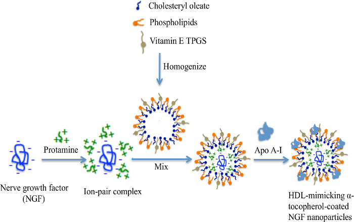
Figure 1: The engineering scheme of NGF HDL-mimicking nanoparticles prepared by an ion-pair strategy. NGF is a negatively charged hydrophilic molecule. A cationic peptide, protamine, was used to neutralize the charges and formed an ion-pair complex with NGF. Cholesteryl oleate, phospholipids, and TPGS formed self-assembly prototype NPs by homogenization. The NGF/protamine complex was incorporated into the prototype NPs. Finally, Apo A-I was coated on the NP surface after overnight incubation. This figure has been modified from Prathipati et al.16. Please click here to view a larger version of this figure.
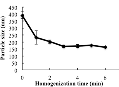
Figure 2: The influence of homogenization on the particle size of the prototype nanoparticles. The excipients, PC, SM, PS, CO, and TPGS, which were dissolved in ethanol, were added to glass vials, and the solvent was evaporated under N2 stream. 1 mL of water was added and homogenized at 9,500 rpm for different times. The particle sizes of subsequent nanoparticles were measured. Data are presented as the mean ± standard deviation (n = 4). This figure has been modified from Prathipati et al.16. Please click here to view a larger version of this figure.

Figure 3: Particle size and size distribution of blank HDL-mimicking nanoparticles and NGF HDL-mimicking nanoparticles. The particle sizes and distributions were measured using a particle analyzer. This figure has been modified from Prathipati et al.16. Please click here to view a larger version of this figure.
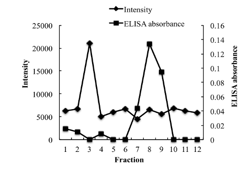
Figure 4: Chromatograms of free NGF and NGF HDL-mimicking nanoparticles on a Sepharose CL-4B column eluted by PBS. 200 µL of free NGF solution (10 µg/mL) and NGF NP solution were loaded onto the gel filtration column and eluted with 1x PBS. A total of twelve fractions (1 mL for each fraction) were collected for both samples. The NP intensity in each fraction was measured by a particle analyzer, and the concentration of NGF in each fraction was measured using a Sandwich ELISA kit. This figure has been modified from Prathipati et al.16. Please click here to view a larger version of this figure.
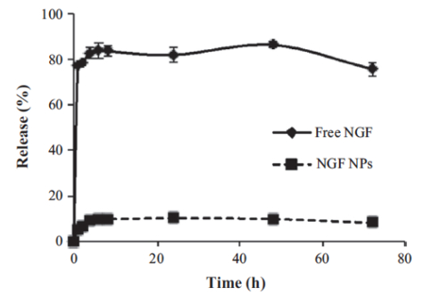
Figure 5: In vitro release of NGF HDL-mimicking nanoparticles measured by a dialysis method. PBS with 5% BSA was used as a release medium. 200 µL of free NGF solution (10 µg/mL) or NGF NPs were added to a dialysis tube supplemented with 400 µL of the release medium. The dialysis tube was put into 30 mL of pre-warmed release medium. The study was performed at 37 °C with 135 rpm shaking. At 1, 2, 4, 6, 8, 24, 48, and 72 h, 100 µL of the release medium was withdrawn and replaced with 100 µL of fresh medium. The NGF concentration in each sample was measured using a Sandwich ELISA kit. Data are presented as the mean ± standard deviation (n = 4). This figure has been modified from Prathipati et al.16. Please click here to view a larger version of this figure.
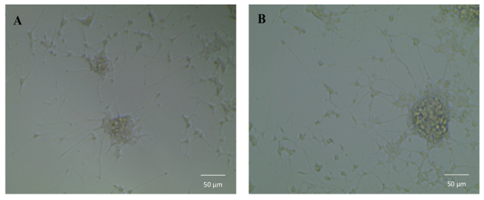
Figure 6: The influence of NGF HDL-mimicking nanoparticles on neurite outgrowth in PC12 cells. Cells were treated with 50 ng/mL free NGF (A) and NGF HDL-mimicking nanoparticles (B) for 7 days. The neurite was imaged using an inverted light microscope under 10X magnification. Please click here to view a larger version of this figure.
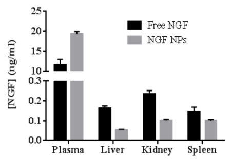
Figure 7: The comparison of biodistribution between free NGF and NGF HDL-mimicking nanoparticles in mice (n = 3). The mice were administered with 40 mg/kg of NGF by tail-vein injection and were sacrificed at 30 min after administration. The blood, liver, spleen, and kidney were collected, and the concentration of NGF in each sample was measured using a Sandwich ELISA kit. Data are shown as the mean ± standard deviation. This figure has been modified from Prathipati et al.16. Please click here to view a larger version of this figure.
| Sample | Particle size (nm) | P.I. | EE% of NGF | Zeta potential (mV) |
| NGF HDL-mimicking NPs | 171.4 ±6.6 | 0.239 ±0.01 | 65.9 ±1.4 | -12.5 ±1.9 |
Table 1: Characterization of NGF HDL-mimicking nanoparticles (n = 3). Data are shown as the mean ± standard deviation. This table has been modified from Prathipati et al.16.