This method describes a synthesis of the mitochondrial calcium uptake inhibitor [(OH2)(NH3)4Ru(µ-O)Ru(NH3)4(OH2)]Cl5 starting from [Ru(NH3)5Cl]Cl2, a well known ruthenium(III) starting material. [Ru(NH3)5Cl]Cl2 is characterized by IR spectroscopy, with vibrational modes at 3200 cm-1, 1608 cm-1, 1298 cm-1, and 798 cm-1 (Figure 1). A minor impurity at 2069 cm-1 can be attributed to [Ru(NH3)5N2]Cl3. The reaction of this Ru(III) species with 1 M NH4OH affords [(OH2)(NH3)4Ru(µ-O)Ru(NH3)4(OH2)]Cl5. The progress of this reaction is evidenced by a dramatic change in color of the solution from yellow to green. The final reaction solution is dark green in color. Acidification of this solution with concentrated HCl results in a color change to brown; a byproduct of this neutralization is ammonium chloride, which can contaminate the final product if care is not exercised. Purification of the compound proceeds via cation-exchange chromatography using strongly acidic mesh (H+ form) resin. The resin is equilibrated first with 0.10 M HCl, and the solution of [(OH2)(NH3)4Ru(µ-O)Ru(NH3)4(OH2)]Cl5 is loaded on the column. The ammonium chloride byproduct elutes with the 1 M HCl washings. Ruthenium-containing compounds elute at higher HCl concentrations. A series of yellow fractions, which contain unreacted [Ru(NH3)5Cl]Cl2 starting material, come off of the column when the HCl concentration is 1.5 M. The desired product elutes between 2.5-3.0 M HCl, and the resulting fractions appear green to green-brown in color.
Prior to pooling the fractions of the product, their purity should be verified. Because the desired compound exhibits some pH-dependence in its UV-vis spectrum, we suggest adding small aliquots of the fractions to highly basic 3 M NH3 solutions to ensure that the spectral features are the same for all of the fractions. Pure fractions should only display an intense peak at 360 nm and a weak peak at 600 nm (Figure 2). Peaks near 480 and 533 nm indicate the presence of ruthenium brown and red, respectively, and a significant peak at 260 nm, with a shoulder at 290 nm, signifies the presence of [Ru(NH3)5Cl]Cl2 starting material (Figure 3). Rotary evaporation of pure fractions affords the desired compound as a green-brown solid.
The isolated compound can further be characterized by both UV-vis and IR spectroscopy. The UV-vis spectrum (Figure 2) displays the 360 nm and 600 nm absorbance bands as described above. The extinction coefficient for the 600 nm band is 850 M-1cm-1 and for the 360 nm band is 27000 M-1cm-1. The ratio of the intensity of the 360 nm band compared to the 600 nm band should be 31 in pH 7.4 PBS, and this metric can effectively be used to gauge the purity of the compound. The IR spectrum should appear as shown in Figure 4. The Ru-O-Ru stretching frequency, for example, is diagnostic at 850 cm-1. The IR spectrum was employed to determine the Ru-O-Ru stretch and to ensure no ammonium chloride was present in the final product. Ammonium chloride, a common impurity, has vibrational modes at 1762 cm-1 (very weak) and 1400 cm-1 (strong) that can be easily discerned from the IR spectrum (Figure 5). Ruthenium red is a common byproduct of the reaction and can be identified in the IR spectrum with stretches at 1404 cm-1, 1300 cm-1, 1037 cm-1 and 800 cm-1, although some overlap with the desired product will occur (Figure 6).
The calcium uptake response in digitonin-permeabilized HeLa cells is observed (Figure 7). The asterisks indicate the addition of a CaCl2 bolus. In the presence of [(OH2)(NH3)4Ru(µ-O)Ru(NH3)4(OH2)]Cl5 an increase in emission intensity is observed upon calcium addition, but no decay due to mitochondrial calcium uptake is observed.
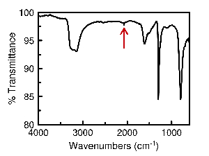
Figure 1: Infrared spectra of [Ru(NH3)5Cl]Cl2. The infrared spectra of [Ru(NH3)5Cl]Cl2 with a very small [Ru(NH3)5N2]Cl3 impurity. The red arrow indicates the impurity at 2,069 cm-1. Please click here to view a larger version of this figure.
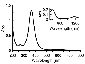
Figure 2: Representative UV-vis spectra for pure material. [(OH2)(NH3)4Ru(µ-O)Ru(NH3)4(OH2)]Cl5 UV-vis absorbance spectra and near infrared (inset) taken in pH 7.4 PBS. The extinction coefficient for the major absorbance at 360 nm is 27,000 M-1 cm-1. Please click here to view a larger version of this figure.
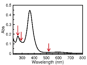
Figure 3: UV-vis spectra of the crude reaction mixture. [(OH2)(NH3)4Ru(µ-O)Ru(NH3)4(OH2)]Cl5 UV-vis absorbance spectra of the crude reaction mixture before purification taken in pH 7.4 PBS. The red arrows indicate common impurities. Please click here to view a larger version of this figure.
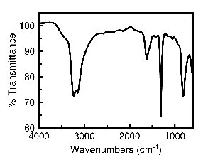
Figure 4: Representative infrared spectra for pure material. The infrared spectra of [(OH2)(NH3)4Ru(µ-O)Ru(NH3)4(OH2)]Cl5. The Ru-O-Ru stretch is at 850 cm-1, the other stretches are from the NH3 oscillators. Please click here to view a larger version of this figure.
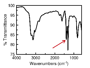
Figure 5: Infrared spectra containing NH4Cl impurities. The infrared spectra of [(OH2)(NH3)4Ru(µ-O)Ru(NH3)4(OH2)]Cl5 with ammonium chloride impurity. The red arrow indicates ammonium chloride at 1,400 cm-1. Please click here to view a larger version of this figure.
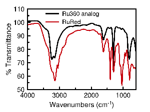
Figure 6: Infrared spectra of commercial ruthenium red. The infrared spectrum of [(OH2)(NH3)4Ru(µ-O)Ru(NH3)4(OH2)]Cl5 (black trace) and commercial ruthenium red (red trace). Please click here to view a larger version of this figure.
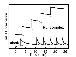
Figure 7: Representative calcium uptake results. The fluorescence increase due to the addition of calcium to a cocktail of digitonin-permeabilized HeLa cells, Calcium Green-5N and [(OH2)(NH3)4Ru(µ-O)Ru(NH3)4(OH2)]Cl5 in BGSS. The blank contains no [(OH2)(NH3)4Ru(µ-O)Ru(NH3)4(OH2)]Cl5 and fluorescence decrease due to Ca2+ uptake into the mitochondria can be seen. Please click here to view a larger version of this figure.